ICU · Procedural
Tracheostomy in ICU: timing, technique, and weaning
Also known as Tracheostomy · Percutaneous tracheostomy · Surgical tracheostomy · Tracheostomy timing · Early vs late tracheostomy · Trach weaning
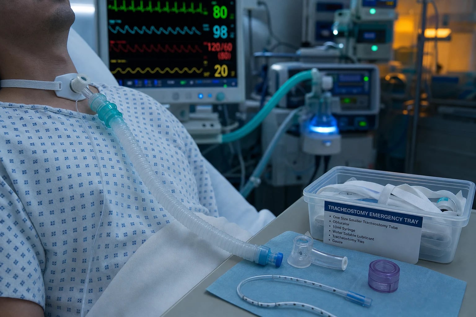
Tracheostomy in ICU: for patients requiring prolonged mechanical ventilation (7-14 days). BENEFITS: patient comfort, reduced sedation, easier secretion clearance, allows oral intake, potential for speech, easier weaning. TIMING: controversial — early (<10 days) vs late (10-21 days). TracMan, SETT2, TracMan trials: NO mortality benefit of early tracheostomy. Current: individualise — consider at day 7-14 if prolonged ventilation expected. TECHNIQUE: percutaneous dilatational (PDT — bedside, intensivist-performed, bronchoscopy-guided) vs surgical (OR — ENT/thoracic). PDT preferred (cheaper, faster, similar outcomes). COMPLICATIONS: bleeding, infection, displacement, tracheal stenosis, tracheo-oesophageal fistula.
On this page & tools
Your progress
Saved locally on this device.
Target exams
Red flags
Percutaneous vs surgical tracheostomy
| Feature | Percutaneous (PDT) | Surgical |
|---|---|---|
| Location | ICU bedside | Operating theatre |
| Operator | Intensivist | ENT/thoracic surgeon |
| Technique | Seldinger (guidewire + dilators) | Surgical incision + dissection |
| Bronchoscopy | Guided (recommended) | Optional |
| Time | 20-40 min | 30-60 min |
| Bleeding | Less (smaller incision) | More (larger dissection) |
| Infection | Similar | Similar (or slightly more) |
| Stomal maturation | Slower (tract takes 7-10 days) | Faster (sutured stoma) |
| Scar/cosmesis | Smaller scar | Larger scar |
| Cost | Cheaper (no OR) | More expensive (OR, surgeon) |
| Outcomes | Similar (multiple RCTs) | Similar |
| Preferred | YES (for most ICU patients) | Selected (coagulopathy, difficult anatomy, previous trach/neck surgery) |
Early vs late tracheostomy — timing strategies
| Feature | Early (<4-7 days) | Late (>10-14 days) |
|---|---|---|
| Definition | Tracheostomy within first week of ventilation | Tracheostomy after 10-14 days of ventilation |
| TracMan trial | Within 4 days | After 10 days (if still ventilated) — only 45% actually needed trach |
| Mortality | NO benefit (30% vs 31% at 30 days) | Same — trial showed no difference |
| ICU length of stay | Shorter (~2 days) | Longer |
| Sedation/analgesia exposure | LESS (patient more comfortable sooner) | More (prolonged ETT discomfort) |
| Mechanical ventilation days | Marginally less | Slightly more |
| Trach performed (avoided) | ~92% received trach | ~45% received — many extubated before day 10 |
| Anticipated benefit | Comfort, easier weaning, less sedation | Wait — patient may extubate, avoid unnecessary trach |
| Risk of unnecessary procedure | HIGH (patient may have extubated by day 7-10) | LOW (only if clearly needing prolonged ventilation) |
| 2025 recommendation | Individualise — consider at day 7-10 if prolonged course still expected | Default unless clearly prolonged course |
| Selected early | Severe brain injury (low GCS, prolonged coma), high spinal cord injury, neuromuscular weakness expected to need prolonged ventilation | — |
Percutaneous dilatational tracheostomy (PDT) techniques compared
| Technique | Mechanism | Key feature | Evidence |
|---|---|---|---|
| Ciaglia Blue Rhino (single-dilator) | Seldinger → single tapered hydrophilic-coated dilator (rhino-shaped) curved over guidewire → trach tube loaded over dilator | Single dilator (no serial dilation) — faster, less bleeding; MOST COMMON worldwide | Byhahn 2000 — modified standard Ciaglia; equivalent/better outcomes than serial; favoured by 2025 practice |
| Ciaglia multiple-dilator (original) | Seldinger → serial dilators (progressively larger, 12-36 Fr) → trach tube | Original technique (1985); more passes → more bleeding/trauma | Largely superseded by Blue Rhino |
| Griggs (guide-wire dilating forceps) | Seldinger → specially modified Howard-Kelly forceps advanced over guidewire → forceps opened to dilate tract → trach tube | Single-step forceps dilation; fast; risk of posterior wall injury if forceps advances too deep | Griggs 1990; widely used (esp. UK/Australasia); comparable to Ciaglia |
| Fantoni translaryngeal (TLT) | Retrograde technique — needle/needle-catheter introduced from within trachea (via bronchoscope), pushed OUT through neck → wire pulled up → trach tube advanced from inside out, then rotated down | 'Inside-out' — minimal skin incision; lowest bleeding/paratracheal injury; more complex; tube rotation risks posterior wall trauma | Fantoni 1996; niche — for coagulopathy/difficult anatomy |
| PercuTwist (screw-dilator) | Seldinger → self-cutting single screw dilator rotated into trachea → trach tube | Single rotating screw; designed to minimise shear/bleeding | Less common; case reports of ring fracture |
| Balloon dilatation | Seldinger → high-pressure balloon dilates tract in single inflation → trach tube | Single-step radial dilation; minimal longitudinal force (less posterior wall trauma) | Growing use; emerging evidence |
Tracheostomy tube types — choosing the right tube
| Tube type | Description | Indication | Notes |
|---|---|---|---|
| Cuffed (standard) | Inflatable cuff seals trachea — protects from aspiration, allows positive-pressure ventilation | Most newly tracheostomised ICU patients on ventilation | Check cuff pressure 20-25 cmH2O daily |
| Uncuffed | No cuff — air leaks around tube | Weaning, decannulation pathway, paediatric | Cannot deliver positive pressure reliably |
| Fenestrated | Opening (fenestration) in the cannula above cuff → airflow up through larynx → speech (with cuff deflated) | Patients who can tolerate cuff deflation, want to speak, on minimal/no ventilator support | Cap outer lumen + deflate cuff → airflow through fenestration + around tube → speech |
| Non-fenestrated | Solid cannula wall — no opening | Patients on positive-pressure ventilation, high aspiration risk | Default for early ICU use |
| Single cannula | One-piece tube (no inner liner) | Most PVC single-use tubes | Cannot be cleaned internally — must suction/change |
| Double cannula | Outer tube + removable inner cannula (can be removed for cleaning) | Long-term trach patients; reduces blockage from secretions | Inner cannula removed + cleaned/replaced daily; reduces mucus plugging |
| Adjustable flange | Flange can slide along tube to vary insertion depth | Thick neck, obesity, abnormal tracheal anatomy | Reduces tube malposition in obese |
| Extra-long / flexible armoured | Longer shaft (silicone reinforced) | Severe neck swelling, obese patients, laryngectomy stoma | Prevents tube kinking in thick neck |
| Bivona Fome-Cuf | Foam-filled cuff (self-inflating to atmospheric pressure) | Long-term ventilation; reduces mucosal pressure | Cannot use cuff pressure manometer |
| Metal (silver/Jackson) | Reusable metal tube | Permanent trach, long-term | Rarely in ICU; no cuff, no inner cannula in some |
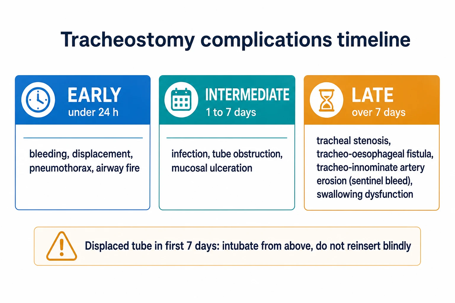
Early vs late complications of tracheostomy
| Timing | Complication | Cause | Recognition | Management |
|---|---|---|---|---|
| Intra-procedural | Paratracheal tube placement | Needle/dilator not midline, guidewire dislodged | No capnography, no CO2, failure to ventilate, bronchoscopy | Oral intubation; re-site after stabilisation |
| Intra-procedural | Posterior tracheal wall injury | Dilator/forceps advanced too deep → perforation | Subcutaneous emphysema, hypoxaemia, bronchoscopic view of oesophagus | Surgical repair if large; conservative if small; high TIF/TOF risk |
| Intra-procedural | Pneumothorax | Apical pleural injury (needle too low) | Post-procedure CXR; hypoxaemia, ↑plateau pressure | Chest tube if symptomatic |
| Early (<7 days) | Bleeding (minor) | Skin edge, anterior jugular vein, thyroid isthmus | Oozing at stoma | Pressure, diathermy, suture ligation; assess coagulation |
| Early | Bleeding (major) | Innominate artery erosion (rare <7 days, usually 1-3 weeks) | Massive bright red blood | Hyperinflate cuff, Foley tamponade, digital pressure, urgent surgery |
| Early | Tube displacement/blockage | Coughing, mucus plug, loose ties | Hypoxaemia, hypercapnia, distress; loss of capnography | If <7 days → oral intubation (do not blind re-insert); if >7 days → re-insert via mature tract |
| Early | Surgical emphysema | Air tracking into tissues (small tracheal tear or cuff leak) | Crackling on palpation, CXR | Usually self-limiting; exclude pneumothorax/pneumomediastinum |
| Early | Infection (stomal) | Skin flora colonisation | Erythema, pus, cellulitis | Local care; swab; topical/systemic antibiotics if cellulitis |
| Late (>7 days) | Tracheal stenosis | Cuff-site ischaemic necrosis → fibrotic scarring | Stridor, dyspnoea, wheeze weeks-months post-decannulation | Bronchoscopy/CT; balloon dilatation, laser, stent, surgical resection |
| Late | Tracheo-oesophageal fistula (TOF) | Cuff pressure necrosis +/- NGT co-pressure | Aspiration, ↑secretions, gastric distension during ventilation | Surgical repair; oesophageal/tracheal stent; reduce cuff pressure |
| Late | Tracheo-innominate artery fistula (TIF) | Erosion into innominate artery (anterior trachea at tube tip) | Sentinel bleed → massive haemorrhage (day 3-50) | Hyperinflate cuff, Foley tamponade, digital pressure, urgent surgery — mortality 50-80% |
| Late | Tracheocutaneous fistula | Stoma fails to close after decannulation (epithelialised tract) | Persistent stoma 3-6 months post-decannulation | Surgical closure |
| Late | Granuloma formation | Foreign-body reaction at tube tip/cuff | Bleeding, obstruction, difficulty weaning/capping | Bronchoscopic excision/laser |
| Late | Swallowing dysfunction/aspiration | Tethered larynx, reduced glottic closure, desensitisation | Cough on secretions/food, recurrent pneumonia | SLP assessment, cuff deflation, speaking valve, thickened fluids |
ICU approach to tracheostomy
- Identify candidate — patient on mechanical ventilation, expected to need >7-14 more days (neurological injury, respiratory failure, weaning difficulty). NOT for: patients likely to extubate within 7 days
- Timing decision — TracMan: early (<10 days) no mortality benefit. Most centres: consider at day 7-14 if prolonged ventilation expected. Discuss with family
- Pre-procedure assessment — (a) Coagulation (INR <1.5, platelets >50). (b) Ventilation (FiO2 <0.6, PEEP <10 — safe for brief disconnection). (c) Anatomy (neck extension possible, no goitre/mass). (d) Consent (or assent from family)
- Technique selection — percutaneous (bedside — most ICU patients) vs surgical (coagulopathy, difficult anatomy, previous neck surgery). Bronchoscopy guidance (percutaneous)
- Procedure — percutaneous: (a) Position (neck extension, shoulders rolled). (b) Bronchoscopy (visualise trachea, confirm needle entry). (c) Needle cricothyroid membrane or tracheal rings 2-3. (d) Seldinger (guidewire). (e) Serial dilation. (f) Insert tracheostomy tube. (g) Confirm position (bronchoscopy, capnography). (h) Secure (sutures + ties)
- Post-procedure care — (a) First trach change: 7-10 days (tract matured — safer). (b) Humidification (heat-moisture exchanger or heated humidifier). (c) Suction (as needed — monitor for blockage). (d) Stoma care (clean, dressing). (e) Cuff pressure 20-25 cmH2O (check daily)
- Weaning and decannulation — (a) Reduce ventilator support (progressive). (b) Deflate cuff (tolerate secretions, use speaking valve). (c) Downsize tube. (d) Capping trials (patient breathes around tube). (e) Decannulate when tolerating spontaneous breathing with effective cough
Percutaneous dilatational tracheostomy (Ciaglia Blue Rhino) — bedside technique
- PREPARATION & TEAM — (a) Two operators: one performs bronchoscopy (via ETT); one performs tracheostomy. (b) Stop enteral feed 4-6 h pre-procedure; aspirate NGT. (c) Sedation + neuromuscular blocker (to prevent cough and bucking — reduces paratracheal injury). (d) Increase FiO2 to 1.0; pre-oxygenate. (e) Continuous monitoring: ECG, SpO2, BP, exhaled CO2 (capnography). (f) Equipment: PDT kit (Blue Rhino single dilator with hydrophilic coating, guidewire, introducer needle, 14G/17G needle, scalpel, trach tube loaded on loading dilator), bronchoscope with stack, cuff pressure manometer, suction, emergency airway kit (oral ETT, laryngoscope, LMA, surgical airway).
- POSITION & ULTRASOUND — (a) SUPINE, neck EXTENDED (shoulder roll transverse, head on doughnut — exposes anterior neck). (b) Reverse Trendelenburg slight. (c) PRE-PROCEDURE ULTRASOUND (linear probe): identify tracheal midline, exclude aberrant vessels (anterior jugular veins, high-riding innominate artery — typically crosses trachea at T3-T4 in children/young, lower in adults), assess tracheal ring depth, mark insertion site (between tracheal rings 2-3, or 1-2 in some). (d) Palpate cricoid cartilage (reference point — insertion is 1-2 rings below cricoid).
- BRONCHOSOPY SETUP — (a) Withdraw ETT to just below vocal cords (under bronchoscopic vision) — exposes the puncture site (rings 2-3) without losing the airway. (b) Keep bronchoscope in ETT, tip just above carina — visualise anterior tracheal wall + transilluminate (light at puncture site confirms midline). (c) Ensure adequate ventilation around bronchoscope (may need to transiently increase tidal volume or pause for short periods). (d) Alternative: exchange ETT for laryngeal mask airway (LMA) or supraglottic airway to give more room — used in some centres (Bickenbach 2014).
- NEEDLE PUNCTURE & SEIDINGER — (a) Local anaesthetic (lidocaine 1% with adrenaline) infiltrated at puncture site (skin + subcutaneous). (b) Small vertical skin incision (1-1.5 cm) at marked site. (c) Blunt dissection with haemostat (separate pretracheal muscles to expose tracheal rings — confirm midline). (d) 14G introducer needle attached to saline-filled syringe, advanced in midline at 45° caudally; aspirate continuously — free air aspirated + bronchoscopic view of needle tip in trachea confirms position. (e) Remove syringe, advance J-tipped guidewire through needle into trachea (bronchoscopic confirmation — wire visible in anterior trachea). (f) Remove needle leaving guidewire.
- BLUE RHINO SINGLE-DILATOR DILATION — (a) Hydrophilic-coated Blue Rhino dilator loaded with loading catheter; activate coating (sterile saline). (b) Advance dilator over guidewire into trachea — SINGLE pass, gentle steady pressure, advancing to the mark (predetermined depth). (c) Bronchoscopic vision throughout: dilator enters trachea anteriorly midline — confirm guidewire and dilator not in posterior wall (TOF risk). (d) Remove dilator, leaving guidewire + 14 Fr loading catheter in place.
- TRACHEOSTOMY TUBE INSERTION — (a) Pre-loaded tracheostomy tube (typically size 8 for adult female, 8-9 for male; cuffed; appropriate length for patient) mounted on loading dilator (the dilator guides the tube through the tract). (b) Advance tube + dilator assembly over guidewire into trachea (rotation may help). (c) Remove guidewire + loading dilator together — leaving trach tube in trachea. (d) Inflate cuff (to seal — verify with cuff pressure manometer to 20-25 cmH2O). (e) Connect to ventilator circuit.
- CONFIRMATION — (a) BRONCHOSCOPY through trach tube: confirm tube position in trachea, above carina, not against posterior wall or main bronchus. (b) CAPNOGRAPHY: exhaled CO2 through trach confirms tracheal position (vs paratracheal). (c) Bilateral chest rise + breath sounds. (d) Chest X-ray post-procedure: tube position, exclude pneumothorax (apical), exclude pneumomediastinum/subcutaneous emphysema. (e) Withdraw oral ETT only after trach position confirmed.
- SECURE & DOCUMENT — (a) SUTURE trach flange to skin at 4 points (or trach ties around neck — firm but allowing one finger underneath). (b) Document procedure note: indication, technique (Blue Rhino), operator, assistants, bronchoscopy findings, complications (none/intra-procedural), tube size + cuff pressure, post-procedure CXR. (c) Plan first trach change at day 7-10 (tract matured). (d) ICU orders: cuff pressure daily, humidification (HME or heated humidifier), stoma care, suction PRN.
Tracheostomy decannulation protocol (stepwise weaning to removal)
- CRITERIA FOR READINESS — (a) Underlying condition resolved/improving (weaning from ventilation successful — patient tolerating spontaneous breathing or low PSV/CPAP). (b) Effective cough and secretion clearance (can manage own secretions). (c) Neurologically intact (awake, follows commands, can protect airway — GCS adequate, no bulbar dysfunction). (d) Haemodynamically stable, no active infection. (e) Adequate oxygenation (SpO2 ≥ 92% on FiO2 ≤ 0.4 or baseline). (f) Cuff leak present (air flows around tube when cuff deflated — confirms patent upper airway and adequate space around tube). (g) Speech-language pathology swallow assessment if aspiration concern.
- STAGE 1 — CUFF DEFLATION + SPEAKING VALVE TRIAL — (a) Deflate cuff (suction above cuff first to clear pooling secretions — reduces post-deflation aspiration). (b) Cap is NOT yet applied. (c) Insert speaking valve (Passy-Muir) on outer trach hub — one-way valve; air IN via trach, OUT via upper airway → speech. (d) Monitor tolerance: respiratory rate, SpO2, comfort, ability to phonate. (e) Start with short trials (30-60 min, tds) and extend as tolerated. (f) COMMON REASIONS FOR FAILURE TO TOLERATE: excessive secretions, inadequate upper airway patency (laryngeal oedema/stenosis), large tube obstructing airflow around tube (consider downsize), anxiety — desensitise.
- STAGE 2 — DOWNSIZE TUBE — (a) Once tolerating speaking valve trials, downsize trach tube (e.g., 8 → 6 → 4 over days-weeks) — smaller tube leaves more space around it for airflow through native upper airway. (b) May switch to uncuffed or fenestrated tube at this stage. (c) Confirm cuff leak is adequate (patient can breathe around tube with cuff deflated). (d) Smaller tube = less airway resistance = easier capping. (e) First tube change MUST occur after tract maturation (day 7-10) — downsize by experienced operator.
- STAGE 3 — CAPPING TRIALS — (a) Place CAP on outer trach hub — patient breathes ENTIRELY around tube via upper airway (cuff must be deflated — else asphyxia). (b) Start short (1-2 h) and extend to overnight (24-48 h continuous). (c) Monitor for: distress, dyspnoea, desaturation, stridor (suggests upper airway obstruction — uncuff, remove cap, assess larynx). (d) Success criteria: tolerates capping 24-48 h without distress, SpO2 maintained, effective cough, no significant stridor. (e) Fenestrated tube — cap directs airflow through fenestration + around tube → improved airflow → easier tolerance; if non-fenestrated, downsize to smaller tube before capping.
- STAGE 4 — DECANNULATION — (a) Once capping tolerated 24-48 h + effective cough + no aspiration → DECANNULATE. (b) Technique: remove ties/sutures → deflate cuff (if still cuffed) → withdraw tube briskly in expiration → apply occlusive dressing (sterile) over stoma. (c) Patient breathes normally through upper airway. (d) Stoma closes by secondary intention over days-weeks (clean dressing changed as needed). (e) If tracheocutaneous fistula persists >3-6 months → surgical closure. (f) Document decannulation note.
- POST-DECANNULATION MONITORING — (a) Monitor SpO2, work of breathing, ability to clear secretions for 12-24 h. (b) Watch for STRIDOR (laryngeal oedema — may need racemic adrenaline/nebulised adrenaline, dexamethasone, rarely re-intubation). (c) Swallow assessment before full oral diet (silent aspiration common even post-decannulation). (d) SLP review if voice change, swallow difficulty. (e) Follow-up at 4-8 weeks for assessment of late complications (stridor → consider tracheal stenosis — bronchoscopy/CT).
Tracheostomy emergency response — the 'BOLD' algorithm (Blocked, Obstructed, Lost, Displaced) tube
- RECOGNISE THE EMERGENCY — Tracheostomy RED FLAGS requiring IMMEDIATE intervention: (a) Sudden deterioration (hypoxaemia, hypercapnia, distress, agitation). (b) Loss of capnography / abnormal waveform. (c) Air leak from stoma (cuff rupture or tube out). (d) Failure to pass suction catheter (blockage or tube displacement). (e) Distress in speaking-valve patient (valve stuck/cuff up). ACTIONS: oxygenate via face mask to stoma AND face mask to mouth simultaneously (cover both routes) — call for help (ENT, anaesthetics, senior).
- 'A' — ASSESS AIRWAY — (a) Is the trach tube IN the trachea (proper position)? (b) Is it patent (suction catheter passes)? (c) Is the cuff intact? (d) Is the upper airway usable (cuff deflated, glottis patent)? Two routes to oxygenate: through trach tube AND through upper airway.
- 'B' — BLOCKED TUBE — (a) SUCTION via trach (deep suction — may dislodge mucus plug). (b) Inner cannula (if double-cannula tube): REMOVE inner cannula (often where plug is). (c) Humidification review (was HME clogged?). (d) If suction cannot pass → tube is BLOCKED or DISPLACED → proceed to removal. (e) Bronchodilator/mucolytic if thick secretions.
- 'C' — CUFF / OUTER LEAK — (a) If air leak from stoma + unable to ventilate: cuff may be ruptured → try to re-inflate (check pilot balloon — if pilot fills but cuff doesn't, cuff is ruptured). (b) If cuff ruptured → tube CHANGE required (mature tract only — day 7-10).
- 'D' — DISPLACED OR REMOVED TUBE — (a) TIME SINCE INSERTION CRITICAL: (i) <7 days post-insertion (tract IMMATURE): DO NOT attempt blind re-insertion → tract collapses → tube may go into false passage (mediastinum). EMERGENCY = OROTRACHEAL INTUBATION (most reliable — secure airway). (ii) >7-10 days (tract MATURE): re-insert trach tube over SUCTION CATHETER GUIDE (pass suction catheter into stoma → advance gently into trachea → railroad new tube over catheter); if any resistance → orotracheal intubation instead. (b) BRONCHOSCOPY if available (visualise tract). (c) Have senior airway operator present.
- 'E' — ESCALATION — (a) If orotracheal intubation fails → SURGICAL AIRWAY (cricothyroidotomy if tube tract lost, or formal tracheostomy in theatre). (b) LMA as bridge if ventilation impossible. (c) Maximal FiO2 throughout. (d) POST-EVENT: root-cause review (why displaced? secure properly? sedation? cuff pressure? tract maturity?). (e) Document thoroughly — medico-legal implications.
Tracheo-innominate artery fistula (TIF) — the life-threatening bleed
- RECOGNISE — (a) Classic: SENTINEL bleed (small haemoptysis or blood at trach) 1-2 days before MASSIVE bleed. ANY bleed day 3 to <50 days post-tracheostomy — suspect TIF until proven otherwise. (b) Massive bright red pulsatile blood from trach site. (c) Hypoxaemia, hypotension, haemorrhagic shock. (d) Often tube is over-inflated or low-lying (tip eroding into innominate artery — artery crosses anterior trachea ~9th tracheal ring).
- IMMEDIATE — TAMponADE — (a) HYPERINFLATE CUFF (over-inflate the trach cuff with >10 mL additional air or to ~50 mL — balloon-tamponade the bleeding point at tube tip against the anterior tracheal wall). (b) If cuff inflation inadequate: pull trach tube OUTWARD slightly and re-inflate (may shift cuff to compress artery). (c) If still bleeding: insert UNTAPPERED Foley catheter through stoma alongside or instead of tube → inflate balloon with 10-30 mL saline → apply outward traction → balloon compresses bleeding point against sternum — MAINTAIN traction. (d) DIGITAL PRESSURE: operator's index finger into stoma, compressing anteriorly against sternum (compresses innominate artery against manubrium) — bridge to theatre.
- DEFINITIVE — SURGICAL — (a) IMMEDIATE thoracic surgery / ENT / vascular surgery activation — sternotomy for proximal control of innominate artery. (b) Ligation or bypass of innominate artery (segmental resection + interposition graft). (c) Some patients tolerate innominate ligation (collateral from left subclavian and circle of Willis); others develop right arm/cerebral ischaemia — assess perfusion postoperatively. (d) MORTALITY 50-80% even with treatment; best outcomes with early recognition (sentinel bleed → intervention).
- PREVENTION — (a) Insert trach tube between rings 2-3 (NOT low — low placement at ring 7-9 risks TIF — tip against innominate artery). (b) Appropriate tube LENGTH (avoid tube tip pressing on anterior tracheal wall — over-long tube erodes). (c) Maintain cuff pressure 20-25 cmH2O (avoid high cuff → mucosal ischaemia → fistula). (d) minimise cuff movement (well-secured tube). (e) Bronchoscopic re-check of tube position. (f) Consider tube change if persistent coughing/bucking.
SAQ — Tracheostomy timing in prolonged ventilation
10 minutes · 10 marks
A 64-year-old man is on day 9 of mechanical ventilation for severe ARDS secondary to pneumococcal pneumonia. He remains on FiO2 0.65, PEEP 12, with a driving pressure of 16 cmH2O, on low-dose propofol and tolerating the endotracheal tube well. The team asks whether a tracheostomy should be performed now or deferred.
SAQ — Tracheostomy decannulation pathway
10 minutes · 10 marks
A 58-year-old woman with Guillain-Barre syndrome required mechanical ventilation and underwent percutaneous tracheostomy on day 12. She is now on day 35, has recovered substantial limb and respiratory strength, is off the ventilator on a T-piece with FiO2 0.3, has an effective cough, and is neurologically intact. The team wishes to decannulate.
Clinical pearls
Red flags
Prognosis
TracMan trial (Young 2013, NEJM) — early vs late tracheostomy
RCT: 909 ICU patients expected to need ventilation >7 days. Early (within 4 days) vs late (after 10 days, if still ventilated).
- Primary outcome (30-day mortality): early 30% vs late 31% (NO difference)
- ICU stay: early SHORTER (by ~2 days)
- Tracheostomy performed: early 92% vs late 45% (many late-group patients extubated before needing trach)
- CONCLUSION: Early tracheostomy does NOT improve survival. Many patients extubate before day 10 (don't need trach). Individualise: consider at day 7-14 if prolonged ventilation still expected. [1]
Meta-analyses: pooled RCTs confirm — no mortality benefit of early trach. Slightly shorter ICU stay, less sedation with early trach. Percutaneous vs surgical: multiple RCTs — similar outcomes (bleeding, infection, stenosis, mortality). Percutaneous preferred (bedside, cheaper).
SETT2 / Scales 2008 — tracheostomy timing & long-term survival
Retrospective cohort (Canada, >10,000 mechanically ventilated ICU patients) — examined timing of tracheostomy and long-term survival beyond index admission.
- Finding: early tracheostomy associated with shorter ICU + hospital stay, shorter mechanical ventilation — but NO long-term mortality benefit (consistent with TracMan).
- Signal: increased long-term survival signal in patients with successful trach decannulation (selection bias — survivors more likely to be decannulated).
- TAKE-HOME: timing is individualised; trach provides comfort and weaning benefits but does not change long-term survival in unselected ICU populations.
Siempos 2015 meta-analysis — early vs late/percutaneous vs surgical (Lancet)
Systematic review & meta-analysis of RCTs (early vs late trach; percutaneous vs surgical trach).
- EARLY vs LATE (timing): pooled analysis — NO significant mortality difference. Early trach: trend to reduced ICU stay, reduced mechanical ventilation duration, less sedation.
- PERCUTANEOUS vs SURGICAL: similar overall complication rates; percutaneous had fewer wound infections; surgical had slightly fewer intra-operative bleeds; long-term stenosis similar.
- CONCLUSION: choose percutaneous as default (bedside, cheaper); individualise timing based on predicted ventilation duration.
Byhahn 2000 — Ciaglia Blue Rhino vs standard Ciaglia multiple-dilator
Prospective randomised comparison of modified single-dilator (Blue Rhino) technique vs original Ciaglia serial-dilator technique in ICU patients.
- Result: Blue Rhino — FASTER procedure (fewer instrument passes), SIMILAR bleeding and complications, equivalent stomal maturation.
- Adoption: Blue Rhino became the dominant percutaneous technique worldwide (single pass, hydrophilic coating reduces friction/trauma).
- Complication profile: posterior tracheal wall injury remains the principal risk of all PDT techniques — bronchoscopic guidance mandatory.
Dulguerov 1999 meta-analysis — percutaneous vs surgical tracheostomy
Pooled analysis of trials comparing PDT vs surgical tracheostomy.
- Findings: (a) OPERATIVE complications (intra-procedural): slightly HIGHER with PDT — paratracheal placement, posterior wall injury, transient hypotension/hypoxaemia. (b) POST-OPERATIVE: similar — bleeding, infection, stenosis. (c) STOMAL infection: LOWER with PDT. (d) Long-term STENOSIS: similar (~5% radiological, <1% symptomatic).
- Caveat: most trials in experienced centres — operator expertise is key. With bronchoscopic guidance + experienced operators, PDT complication rate drops to comparable with surgical.
- Conclusion: PDT is acceptable and preferred for most ICU patients; surgical reserved for selected (coagulopathy, difficult anatomy, paediatric, prior neck surgery).
Engels 2009 systematic review — predictors of weaning from tracheostomy
Systematic review identifying factors associated with successful decannulation in ICU/rehab patients.
- Positive predictors: (a) younger age, (b) intact cognition (GCS, RASS), (c) effective cough (peak cough flow >160 L/min), (d) tolerance of cuff deflation/capping, (e) absence of excessive secretions, (f) successful swallow assessment, (g) adequate oxygenation on minimal support.
- Negative predictors: (a) neurological disease (stroke, TBI, neurodegenerative), (b) recurrent pneumonia/aspiration, (c) high secretion burden, (d) need for re-intubation/ventilation, (e) critical illness polyneuropathy/myopathy.
- TAKE-HOME: standardised weaning protocols (cuff deflation → speaking valve → downsize → capping → decannulate) improve decannulation success and reduce time to decannulation.
French ICU guidelines (Trouillet 2019) — tracheostomy in ICU
Society-level consensus on indications, timing, technique, and decannulation in critically ill adults.
- INDICATION: predicted mechanical ventilation >7-10 days (defer if extubation likely before day 7).
- TIMING: no routine 'early' (day 1-4) trach; consider day 7-10 in patients with ongoing ventilation, neurological failure, or weaning difficulty.
- TECHNIQUE: percutaneous (with bronchoscopy + ultrasound) preferred over surgical in most patients; surgical for specific indications.
- DECANNULATION: structured protocol — cuff leak → cuff deflation + speaking valve → downsize → capping trial (24-48 h) → decannulate.
- FOLLOW-UP: assessment for late complications (stenosis) at 4-8 weeks post-decannulation in patients with stridor/dyspnoea.
Summary of evidence & 2025 practice
- Timing: NO mortality benefit of early trach (TracMan, Siempos meta-analysis, SETT2). Benefits of early trach: shorter ICU stay, less sedation, possibly less VAP. CONSENSUS: individualise — day 7-14 if prolonged course expected.
- Technique: percutaneous (Ciaglia Blue Rhino, bronchoscopy-guided, ultrasound-assessed) = standard for most ICU patients. Surgical reserved for coagulopathy, difficult anatomy, paediatric, prior neck surgery, emergencies.
- Complications: prevention focus — cuff pressure 20-25 cmH2O daily, appropriate tube size + length, humidification, secure fixation.
- Decannulation: standardised protocol (cuff leak → cuff deflation + speaking valve → downsize → capping 24-48 h → decannulate).
- TIF (tracheo-innominate artery fistula): sentinel bleed = emergency — hyperinflate cuff, Foley tamponade, digital pressure, urgent surgery (mortality 50-80%).
- Tracheal stenosis: late — weeks-months post-decannulation; think in any post-trach patient with new stridor/dyspnoea.
- Speaking valve (Passy-Muir): always deflate cuff first; benefits speech, swallow, olfaction, decannulation readiness.
- Tract maturation: 7-10 days — displacement before this = oral intubation (don't blind re-insert).
References
- [1]Young D, et al. Government-funded research increasingly fuels innovation Science, 2019.PMID 31221848
- [2]Trouillet JL, et al. Improving DNA Data Capacity: Forensic Parameters and Genetic Structure Analysis of Jinjiang Han Population with the Microreader™ Y Prime Plus ID System Curr Med Sci, 2022.PMID 35403953
- [3]Siempos II, et al. Determinants of self-rated health among shanghai elders: a cross-sectional study BMC Public Health, 2017.PMID 29029627
- [4]Freeman BD, et al. Can sand nourishment material affect dune vegetation through nutrient addition? Sci Total Environ, 2020.PMID 32278174
- [5]Brenner MJ, et al. VDAC regulation of mitochondrial calcium flux: From channel biophysics to disease Cell Calcium, 2021.PMID 33529977
- [6]Abrams DC, et al. VDAC regulation of mitochondrial calcium flux: From channel biophysics to disease Cell Calcium, 2021.PMID 33529977
- [7]Scales DC, Thiruchelevam D, Kiss A, Redelmeier DA. A behavioral intervention to improve obstetrical care N Engl J Med, 2008.PMID 18450604
- [8]Byhahn C, Wilke HJ, Halbig S, Lischke V, Westphal K. Protective role of medroxyprogesterone acetate on N-methyl-N-nitrosourea-induced lymphomas in BALB/c female mice Leuk Res, 2001.PMID 11166832
- [9]Dulguerov P, Gysin C, Perneger TV, Chevrolet JC. Carotenoid absorption from salad and salsa by humans is enhanced by the addition of avocado or avocado oil J Nutr, 2005.PMID 15735074
- [10]Bickenbach J, Lipinski S, Schälte G, Beckers S, Marx G, Rahmel J. Traffic-related air pollution and noise and children's blood pressure: results from the PIAMA birth cohort study Eur J Prev Cardiol, 2015.PMID 24047569
- [11]Engels PT, Bagshaw SM, Meier M, Brindley PG. [Identification of three novel mutations of IRF6 in Chinese families with Van der Woude syndrome] Zhonghua Yi Xue Yi Chuan Xue Za Zhi, 2006.PMID 16456794
- [12]Sue RD, Susanto I. Computerization in medicine Am J Ther, 2015.PMID 25734589