ICU · Rehabilitation
Acute severe community-acquired pneumonia: recovery and follow-up
Also known as CAP recovery · Post-pneumonia follow-up · Pulmonary rehabilitation after ICU · Vaccination after pneumonia · Non-resolving pneumonia · Post-intensive care syndrome after CAP
Recovery from severe CAP requiring ICU admission is prolonged and multifaceted: physical (ICU-acquired weakness, reduced exercise tolerance, deconditioning), cognitive (impaired memory, executive function — from delirium/hypoxia), psychological (PTSD, depression, anxiety), respiratory (reduced lung function, pulmonary fibrosis in some). Follow-up: ICU follow-up clinic at 2-3 months, CXR at 6 weeks to ensure resolution (non-resolving: investigate malignancy, TB, immunodeficiency), pulmonary rehabilitation, vaccination (pneumococcal, influenza), smoking cessation. Complete recovery may take 6-12 months. 30-50% never return to baseline function.
On this page & tools
Your progress
Saved locally on this device.
Target exams
Red flags
Severe community-acquired pneumonia (CAP) is not "over" when the patient leaves the ICU. The acute infection resolves over days, but the functional, cognitive, psychological and radiological legacy persists for months — and in up to half of survivors, permanently. Recovery is best understood as a trajectory with four interlocking domains — the post-intensive care syndrome (PICS) — superimposed on the specific pulmonary consequences of pneumonia itself (impaired gas transfer, restrictive defect after ARDS, bronchiectasis, and the ever-present question of whether the infiltrate has actually resolved). This topic follows the patient from the ICU bed to the 12-month review: what recovers and when, what predicts poor recovery, how to structure follow-up (clinical review at 6 weeks, repeat chest X-ray at 6–12 weeks), when and how to investigate the non-resolving infiltrate (bronchoscopy, malignancy, TB), and how pulmonary rehabilitation, vaccination and smoking cessation convert a passive convalescence into an active recovery. [1]
Recovery timeline
CAP recovery phases
Acute phase (ICU stay)
Focus: treat infection, support organs, prevent complications. Early rehabilitation: passive ROM day 1, sit out of bed when stable, stand/walk when possible. Prevent ICU-acquired weakness: minimise sedation, early mobilisation, glycaemic control, minimise NMBAs. Prevent delirium: ABCDEF bundle. Begin discharge planning early.
Early recovery (hospital ward)
Continue antibiotics (IV-to-oral switch when improving). Physiotherapy (progressive exercise, gait retraining). Nutritional support (rebuild muscle mass). Assess: swallowing (speech therapy — post-extubation dysphagia common), cognitive function (MoCA — screen for impairment), mood (PHQ-9 — depression screen). Plan: home vs rehabilitation facility, home oxygen if needed, outpatient follow-up.
Medium-term recovery (2-3 months)
ICU follow-up clinic: comprehensive assessment. Physical: 6-minute walk test, MRC score (muscle strength), grip strength. Cognitive: MoCA or MMSE. Psychological: PTSD checklist (PCL-5), depression (PHQ-9), anxiety (GAD-7). Respiratory: spirometry, CXR (ensure resolution — non-resolving: investigate). Address: smoking cessation, vaccination, pulmonary rehabilitation referral, occupational therapy (return to work).
Long-term recovery (6-12 months)
Most recovery occurs in first 6 months. 30-50% never return to baseline physical or cognitive function. Continued: pulmonary rehabilitation, psychological support (PTSD/depression may persist), occupational therapy (workplace modifications). Reassess: lung function (spirometry — some develop restrictive pattern from ARDS/pulmonary fibrosis), exercise tolerance (6-minute walk test trend), quality of life (SF-36). Annual influenza vaccination. Pneumococcal booster as per schedule.
The recovery trajectory is non-linear and differs markedly between domains: physical strength recovers first (weeks to 3 months), respiratory physiology lags (gas transfer normalises over 3–12 months), cognitive function plateaus or slowly improves over a year, and psychological morbidity can first appear months after discharge. Radiographic resolution trails clinical improvement by weeks. Understanding these different time-courses prevents premature reassurance (a patient who feels well at 4 weeks may still have a non-resolving infiltrate) and premature pessimism (gas transfer can still be improving at 12 months). [1]
Weeks 0-4
Acute / early ward
- Clinical recovery: fever, tachypnoea, hypoxaemia resolve; antibiotics completed (5-7 d typical, longer for Legionella/S.aureus)
- Functional: profound weakness, breathlessness on minimal exertion, needs assistance with ADLs
- Radiology: CXR may still WORSEN in first week despite clinical improvement — do not over-interpret
- Cognitive: active delirium may persist into early ward phase; screen with CAM
- Milestone: independent mobilisation around bed; safe swallow (SLT-cleared)
Weeks 4-8
Early convalescence
- Clinical review at 6 weeks: assess symptoms, functional recovery, smoking cessation
- Repeat CXR at 6 weeks (or 6-12 weeks): document radiographic resolution
- Functional: walking independently, but reduced exercise tolerance; breathlessness common
- Psychological: depression and early PTSD symptoms may emerge
- Milestone: return to light ADLs; pulmonary rehabilitation referral if breathless/deconditioned
Months 2-6
Medium-term
- ICU follow-up clinic at 2-3 months: comprehensive PICS assessment
- Spirometry + DLCO: most show mild restrictive/diffusion defect; severe ARDS recovery slower
- 6-minute walk test: objective functional capacity; set baseline and track trend
- Most physical recovery occurs here; cognitive recovery is slower
- Milestone: ~50-70% return to work (often modified/phased)
Months 6-12
Long-term / plateau
- Further CXR only if not previously resolved or new symptoms
- Lung function may still be improving (DLCO recovers latest)
- Cognitive function plateaus; persistent impairment in ~30%
- Psychological morbidity (PTSD, depression) may persist or newly present
- Milestone: final functional status; 30-50% never reach pre-ICU baseline
Recovery milestones by the numbers
Factors affecting recovery
Recovery is not uniform. A young adult with unilateral pneumococcal pneumonia ventilated for 48 h will recover fully within weeks; a 75-year-old with bacteraemic ARDS, multi-organ failure and a month of ventilation may never walk home again. Recognising the predictors of poor recovery early allows targeted rehabilitation and sets realistic expectations for patient and family. [1]
Non-modifiable
Patient factors
- Age >65 years — slower physical recovery, higher persistent cognitive impairment
- Pre-existing comorbidity (COPD, heart failure, CKD, diabetes, frailty)
- Baseline cognitive impairment / low cognitive reserve — magnifies delirium-related injury
- Lower socioeconomic status / limited social support — slower functional reintegration
- Genetic susceptibility to critical-illness myopathy (variable expression)
Illness severity
Drive of PICS
- Duration of mechanical ventilation (each day increases ICU-acquired weakness risk ~3%)
- Severity of ARDS and duration of hypoxaemia (cerebral + tissue hypoxic injury)
- Septic shock duration and peak lactate (organ crosstalk, AKI, mitochondrial dysfunction)
- Delirium: longer coma-free delirium days → worse long-term cognition (BRAIN-ICU)
- Multi-organ failure (SOFA score) and prolonged ICU stay (>1 week)
- Bacteraemia, metastatic infection, necrotising/multilobar pneumonia
Modifiable (ICU)
What we control
- Minimise sedation (daily awakening, light sedation targets)
- Minimise neuromuscular blockade (steroid-associated myopathy risk)
- Early mobilisation (Schweickert trial: PT/OT from day 1-3 improved function)
- Glycaemic control (avoid hyperglycaemia → critical illness polyneuropathy)
- Delirium prevention: ABCDEF bundle (assess pain, both spontaneous awakening/breathing trials, choice of sedation, delirium monitoring, early mobility, family engagement)
- Conservative fluid strategy, lung-protective ventilation (less barotrauma/VILI)
- Glycaemic control 6-10 mmol/L, adequate nutrition (avoid overfeeding and underfeeding)
Modifiable (post-ICU)
Recovery levers
- Structured pulmonary rehabilitation — improves exercise tolerance and QoL
- Smoking cessation — single biggest modifiable CAP recurrence risk factor
- Vaccination: pneumococcal + annual influenza (CAPITA: vaccine prevents first episode)
- Optimise comorbidities (COPD inhalers, heart failure, diabetes)
- Psychological support and ICU diaries (reduce PTSD)
- Nutritional rehabilitation and progressive exercise
Post-intensive care syndrome (PICS)
PICS is the umbrella term for the new or worsening impairment in physical, cognitive or mental health that persists after critical illness and affects discharge outcomes. It is the dominant framework for understanding the post-ICU CAP patient and is near-examined in CICM/FFICM. The three domains are independent: a patient may recover physically but remain cognitively impaired, or vice versa. About a third of survivors have impairment in all three domains at 1 year. [1]
Physical domain
ICUAW + deconditioning
- ICU-acquired weakness (ICUAW): critical illness polyneuropathy (CIP), myopathy (CIM), or both — MRC sum <48, symmetric
- Deconditioning, reduced exercise tolerance, sarcopenia from bed rest
- Joint contractures, pressure injuries, heterotopic ossification
- Screen: MRC sum score, grip strength, 6-minute walk test
- Recovery: physical therapy, gradual; ICUAW may take 6-12 months, some permanent
Cognitive domain
Impaired memory/executive
- Deficits in memory, attention, executive function, visuospatial ability
- Driven by delirium duration, hypoxaemia, sedation, septic encephalopathy (BRAIN-ICU)
- ~30-40% have impairment resembling mild traumatic brain injury at 1 year
- Screen: MoCA, MMSE; formal neuropsychology if impaired
- Recovery: slow; cognitive rehabilitation; some impairment is permanent
Psychological domain
PTSD / depression / anxiety
- Depression (~30%), anxiety (~30%), PTSD (~20%) — common, often coexist
- Risk factors: recall of delirium/psychotic experiences, long sedation, lack of family diaries
- Screen: PHQ-9 (depression), GAD-7 (anxiety), PCL-5/IES-R (PTSD)
- Recovery: psychological therapy (CBT), PTSD-focused treatment, ICU diaries (reduce PTSD)
- Often under-recognised — proactive screening at ICU follow-up clinic essential
BRAIN-ICU — long-term cognitive impairment after critical illness
Prospective cohort (n=821), medical and surgical ICU
Population: Patients with respiratory failure, shock, or septic shock (non-elective)
Key finding
40% had global cognition scores <1.5 SD below mean (TBI-equivalent) at 3 months; 34% at 12 months; 26% had executive scores similar to traumatic brain injury at 12 months. Longer duration of delirium independently predicted worse cognition.
Practice change
Cognitive impairment is the rule, not the exception, after critical illness. Delirium duration is a modifiable driver — prevent and minimise delirium with the ABCDEF bundle.
Schweickert — early physical and occupational therapy during mechanical ventilation
Single-centre RCT (n=104)
Population: Mechanically ventilated patients, ICU stay expected >72h
Key finding
More patients returned to independent function (59% vs 35%, p=0.02), more days without delirium (2.0 vs 0.0, p=0.02), more days out of bed. Trend to shorter ventilation/ICU stay.
Practice change
Early, structured mobilisation during mechanical ventilation improves functional outcomes and reduces delirium. Begin rehabilitation in the ICU — do not wait for the ward.
Post-CAP complications
The pulmonary consequences of pneumonia persist long after the organism is eradicated. Alveolar and airway injury, pleural inflammation, and (in ARDS) diffuse alveolar damage can resolve completely, resolve with scarring (fibrosis), or organise into structural disease (bronchiectasis). Surveillance for these sequelae — and for recurrence — is the respiratory half of post-CAP follow-up. [1]
Pulmonary complications
Lung-limited sequelae
- Pulmonary fibrosis / restrictive defect — especially after severe ARDS; DLCO recovers slowest
- Bronchiectasis — post-infective, particularly after necrotising pneumonia (S. aureus, Klebsiella)
- Organising pneumonia — patchy infiltrates, migratory, steroid-responsive; suspect if non-resolving
- Lung abscess / cavity persistence — may need prolonged antibiotics or surgical resection
- Pleural disease — residual pleural thickening (rind), trapped lung, chronic empyema
- Post-infectious bronchial hyperreactivity — persistent cough/wheeze for weeks-months
Systemic complications
Beyond the lung
- Cardiac: new arrhythmia, heart failure, myocardial infarction — pneumococcal CAP has high cardiac event rate
- Renal: recovery from sepsis-AKI may be incomplete; some progress to CKD
- Neurological: cognitive impairment (PICS), critical illness polyneuropathy/myopathy
- Haematological: recovery of marrow; VTE risk persists for weeks post-discharge
- Nutritional: sarcopenia, weight loss, refeeding issues
- Psychological: depression, anxiety, PTSD
Recurrence risk
Re-admission
- Recurrence risk highest in first 6 months; ~10-15% re-admitted with pneumonia within 30 days
- Drivers: untreated aspiration (post-extubation dysphagia), untreated immunodeficiency, ongoing smoking
- Recurrent lobar pneumonia in the SAME lobe → suspect endobronchial obstruction (cancer, foreign body)
- Prevention: vaccination, smoking cessation, treat dysphagia, optimise comorbidities
Active surveillance for post-CAP complications at follow-up
At 6-week clinical review
Symptom assessment (persistent cough, sputum, breathlessness, haemoptysis, weight loss). Functional status (ADLs, exercise tolerance). Examine: chest (persistent crackles, wheeze, signs of effusion), oxygen saturation, look for clubbing (suggests bronchiectasis or malignancy). Check weight and nutritional recovery.
Repeat CXR at 6-12 weeks
Document radiographic resolution. If fully resolved and asymptomatic → no further imaging. If incomplete resolution but improving → repeat at 3 months. If static/worsening or new cavitation/effusion → investigate (see non-resolving pneumonia section).
Spirometry + DLCO at 3 months
Assess for restrictive defect (ARDS/pulmonary fibrosis) or obstructive defect (underlying COPD revealed, post-infective bronchiectasis). DLCO reduced is commonest abnormality after severe CAP/ARDS. Compare to pre-morbid if available.
Screen for dysphagia if intubated >48 h
Post-extubation dysphagia in up to 50% of long-intubated patients → silent aspiration → recurrent pneumonia. Refer to speech and language therapy; water-swallow test, VFSS/FEES if positive.
Cardiovascular review
Pneumococcal CAP markedly raises cardiac events (MI, AF, heart failure) in the following months. Check ECG, BP, optimise cardiac medications. Consider echocardiogram if new murmur or heart failure signs.
PICS screen at ICU follow-up clinic (2-3 months)
Physical (MRC, grip, 6MWT), cognitive (MoCA), psychological (PHQ-9, GAD-7, PCL-5). Refer impaired domains to pulmonary rehab, neuropsychology, and clinical psychology respectively.
Follow-up schedule
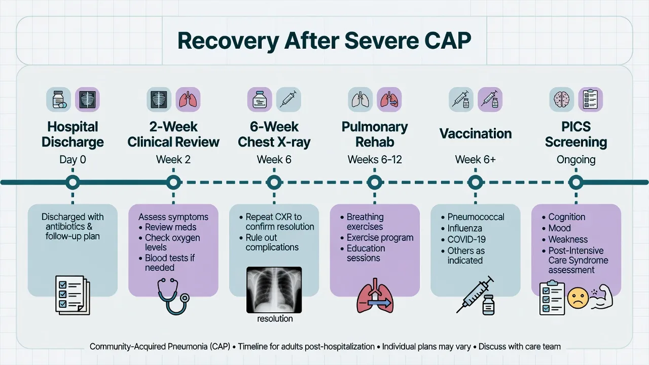
Structured follow-up converts an opportunistic convalescence into a protocolised recovery. The non-negotiable pillars are a clinical review at ~6 weeks, a repeat chest X-ray at 6–12 weeks (to confirm radiographic resolution and catch an underlying malignancy or TB), and a comprehensive ICU follow-up clinic at 2–3 months to detect and triage PICS. [1]
Structured CAP follow-up schedule
2 weeks (post-discharge)
Clinical review: symptoms improving? Tolerating oral intake, medications, home oxygen if discharged on it? Smoking cessation counselling initiated. Functional status. No routine CXR at this point (radiology lags — may still look abnormal). Reinforce pneumonia-recurrence prevention messages.
6 weeks (clinical review)
Symptom assessment: persistent cough, sputum, breathlessness, haemoptysis, weight loss (red flags for malignancy/TB). Functional recovery: exercise tolerance, return to work planning. Smoking cessation follow-through. Vaccination status (pneumococcal, influenza) — give if not yet given. Spirometry if underlying COPD suspected.
6-12 weeks (repeat CXR)
Routine repeat chest X-ray to document radiographic resolution. ~50% clear by 6 weeks, ~90% by 12 weeks. Fully resolved + asymptomatic → no further imaging. Incomplete resolution → repeat at 3 months or investigate (see non-resolving). MANDATORY in smokers and older patients to exclude underlying lung cancer presenting as pneumonia.
3 months (ICU follow-up clinic)
Multidisciplinary review: physical (MRC, grip strength, 6MWT), cognitive (MoCA/MMSE), psychological (PHQ-9, GAD-7, PCL-5). Spirometry + DLCO. Pulmonary rehabilitation referral if breathless/deconditioned. Review ICU diary with patient. Vocational/occupational therapy. Address persistent symptoms and comorbidities.
6 months
Functional and quality-of-life assessment (SF-36). 6MWT trend. Reassess psychological morbidity — PTSD may first present here. Pulmonary rehabilitation progress. Return to work status. Repeat imaging only if not previously resolved or new symptoms.
12 months
Final review: most recovery has plateaued by 12 months. Spirometry + DLCO (may still be improving). Establish final functional status and residual impairment. Confirm annual influenza vaccination and pneumococcal booster schedule. Discharge to primary care / respiratory physician for ongoing prevention.
Follow-up timeline
Repeat chest X-ray and radiographic resolution
Radiographic resolution lags behind clinical recovery by weeks. The alveolar infiltrate of pneumonia represents protein-rich exudate and inflammatory cells; even after the organism is eradicated, this debris must be cleared by macrophages and reabsorbed, and any necrotic tissue must organise. A patient who looks and feels well at 4 weeks may still have a dense infiltrate on CXR — this is expected, not pathological. Conversely, an infiltrate that is static or worsening at 6 weeks despite clinical recovery is a red flag. [1]
Normal resolution
Expected
- ~20-30% of infiltrate clears by 2 weeks; ~50% by 6 weeks; ~70% by 8 weeks; ~90% by 12 weeks
- Faster in young, previously well patients; slower in elderly, bacteraemic, multilobar, ARDS
- Bacteraemic pneumococcal pneumonia may clear faster than expected (often dramatic response)
- Legionella, staphylococcal and gram-negative pneumonia clear SLOWER (necrotising tendency)
- Clinical improvement precedes radiographic improvement — do not be alarmed
Delayed resolution
Watch closely
- Defined as incomplete clearing at 4 weeks in an otherwise improving patient
- Common in age >50, multilobar, alcoholism, COPD, diabetes, immunocompromise
- Action: continue observation, repeat CXR at 6-8 weeks, ensure no underlying cause
- Usually resolves by 12 weeks without intervention if patient is clinically well
Non-resolution
Investigate
- Defined as failure to resolve by 6 weeks, or progression/worsening at any time
- Recurrence in the SAME lobe (even months later) — suspect endobronchial obstruction
- Differential: underlying malignancy, TB, atypical infection, abscess, organising pneumonia
- Action: CT chest with contrast, sputum (incl. AFB), consider bronchoscopy ± biopsy
- Bronchoscopy indicated if no improvement by 6 weeks or earlier if obstruction suspected
Non-resolving pneumonia
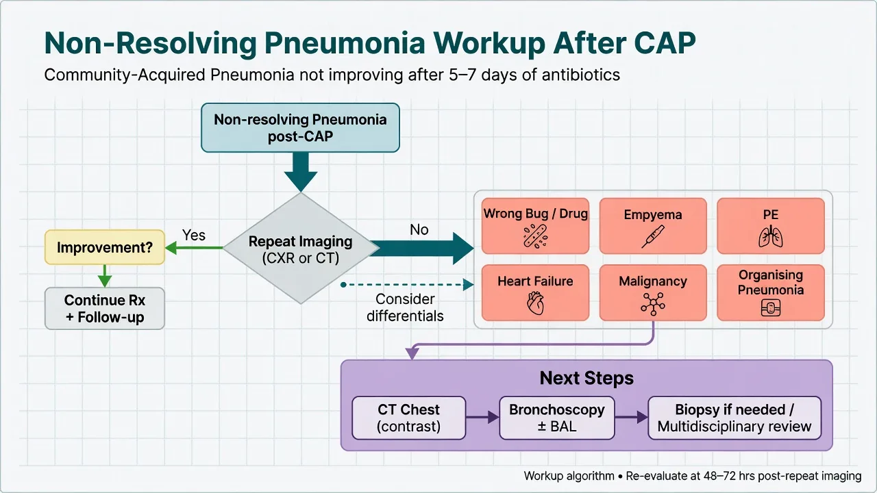
Non-resolving (or "slowly resolving") pneumonia is the failure of clinical and/or radiographic improvement despite what should be adequate antibiotic therapy. It is the single most important diagnosis to actively exclude at the 6-week review, because the commonest underlying cause in adults is bronchogenic carcinoma — a curable (if early) disease that will be missed if the infiltrate is simply dismissed as "slow pneumonia." The other big misses are tuberculosis, atypical/opportunistic infection, lung abscess, and organising pneumonia. [1]
Infectious causes
Wrong bug / resistant
- Tuberculosis — always send sputum for AFB in non-resolving pneumonia (especially upper lobe)
- Atypical organisms: Mycoplasma, Legionella (urine antigen), Chlamydia, Coxiella
- Opportunistic: Pneumocystis (HIV/immunosuppression), fungal (endemic or opportunistic)
- Viral: influenza, COVID-19 ( organise / fibrosis )
- Resistant bacteria: MRSA, Pseudomonas, ESBL — especially in healthcare exposure
- Anaerobic / mixed aspiration with abscess formation
Non-infectious causes
Mimics / obstruction
- Bronchogenic carcinoma — the diagnosis you must not miss; recurrent lobar pneumonia = obstruction
- Foreign body aspiration (especially right lower lobe; adults may not recall the event)
- Bronchiectasis — recurrent infection in structurally damaged airway
- Organising pneumonia (cryptogenic, COP) — patchy, migratory, steroid-responsive
- Interstitial lung disease, eosinophilic pneumonia, vasculitis (ANCA), radiation pneumonitis
- Pulmonary embolism with infarction, atelectasis, heart failure (mimic)
Host / therapy factors
Why it persists
- Immunodeficiency: HIV, malignancy, immunosuppressants, hyposplenism, hypogammaglobulinaemia
- Inadequate antibiotic (wrong drug, wrong dose, poor penetration, non-adherence)
- Un-drained source: empyema, lung abscess, infected pleural collection
- Impaired clearance: severe COPD, bronchiectasis, ciliary dysfunction
- Re-aspiration: untreated dysphagia, recurrent micro-aspiration
Workup of non-resolving pneumonia
Re-confirm the diagnosis and reassess
Is this really pneumonia? Re-take history (symptoms, travel, exposures, HIV risk, aspiration), examine (clubbing, lymphadenopathy, weight loss), review all imaging and cultures from the acute episode. Check immune status (HIV, immunosuppressants). Review antibiotic choice, dose, adherence and duration.
CT chest with contrast
Characterise the non-resolving opacity: mass/cavity/nodule/consolidation/interstitial pattern/lymphadenopathy/pleural disease. CT distinguishes abscess (in lung, air-fluid level) from empyema (in pleura). Cavitation, mass, or hilar/mediastinal lymphadenopathy raise suspicion for malignancy or TB.
Targeted microbiology
Sputum: Gram stain, culture, AND acid-fast bacilli (AFB) x3 + TB PCR/GeneXpert. Urine: pneumococcal and Legionella antigen. Blood cultures. Consider atypical serology (Mycoplasma, Coxiella, Chlamydia). If immunocompromised: Pneumocystis (induced sputum/BAL), fungal studies (galactomannan, beta-D-glucan).
Bronchoscopy (no improvement by 6 weeks, or earlier if obstruction suspected)
Indications: failure to improve by 6 weeks, recurrent lobar pneumonia, suspected endobronchial lesion (mass, foreign body), immunocompromise with no diagnosis, cavitating lesion needing microbiology. Obtains: BAL for microbiology + cytology, transbronchial biopsy, and direct visualisation to exclude/exclude an obstructing tumour or foreign body.
Consider non-infectious mimics
If infective workup negative: organising pneumonia (steroid trial, biopsy), pulmonary vasculitis (ANCA, anti-GBM, urinalysis), eosinophilic pneumonia (differential blood eosinophil count, BAL eosinophilia), malignancy (biopsy, PET-CT, staging). Pulmonary embolism with infarction (D-dimer, CTPA).
Address source and host factors
Drain any undrained collection (empyema, abscess). Treat dysphagia (SLT, VFSS). Optimise immunosuppression reversal if possible. Treat underlying malignancy/structural disease once diagnosed. Smoking cessation.
Bronchoscopy: what it adds in non-resolving pneumonia
Visualisation of the airway
Directly excludes endobronchial obstruction — the single most important miss. A bronchogenic carcinoma, carcinoid, foreign body (e.g. inhaled food/teeth), or bronchial stenosis may present as recurrent/post-obstructive pneumonia and is invisible on plain CXR. Foreign body inhalation is under-diagnosed in adults (large series: many present weeks-months after the event).
Bronchoalveolar lavage (BAL)
Targets the affected segment for microbiology: quantitative bacterial culture, AFB and TB PCR, fungal stains/culture, Pneumocystis (immunofluorescence/PCR), viral PCR, Nocardia, and cytology (malignant cells, eosinophilia of organising pneumonia/eosinophilic pneumonia).
Biopsy
Transbronchial biopsy for organising pneumonia, interstitial lung disease, vasculitis, infection (Pneumocystis, fungal). Endobronchial/mucosal biopsy for visible lesions. EBUS-TBNA for mediastinal/hilar lymphadenopathy (stages malignancy, diagnoses TB/sarcoidosis).
Therapeutic intervention
Foreign body removal (baskets/forceps), debulking of obstructing tumour (laser/cryotherapy/stent), clearance of mucus plugs/bronchial casts in atelectasis. Converts a purely diagnostic procedure into definitive treatment in selected cases.
Pulmonary rehabilitation
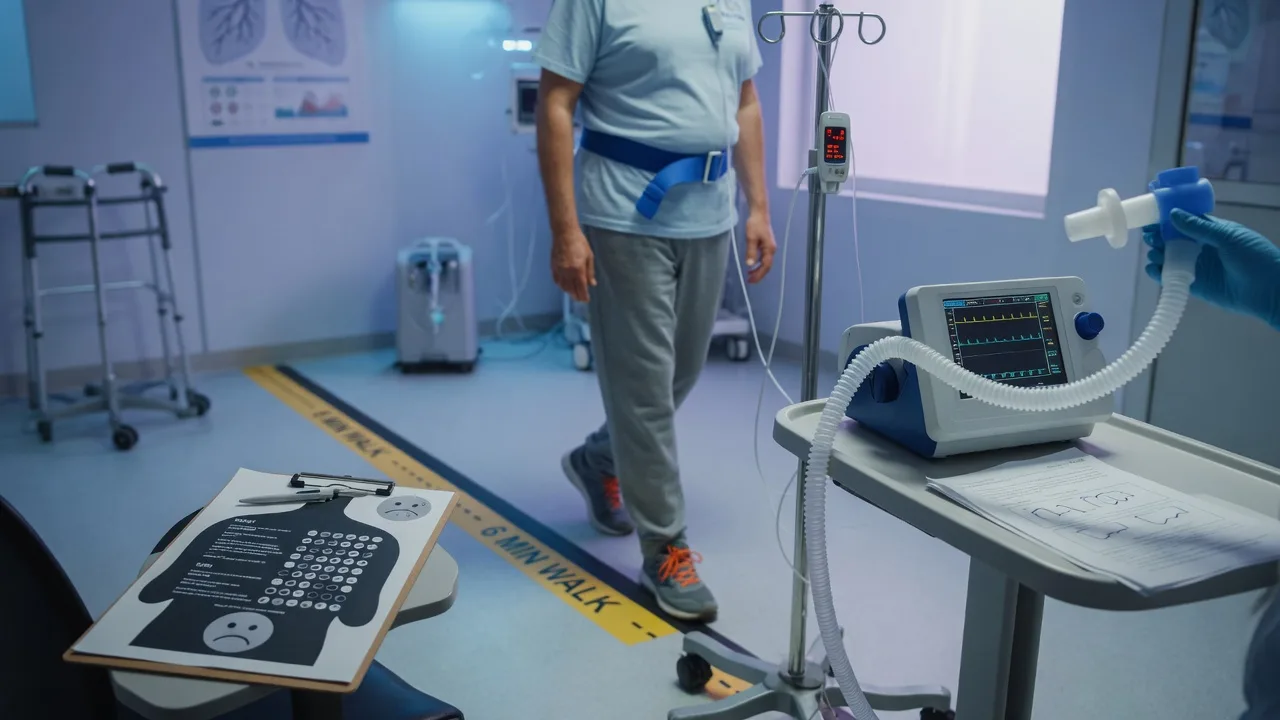
Pulmonary rehabilitation is a structured, multidisciplinary programme of exercise training, education, and behaviour change designed to improve the physical and psychological condition of people with chronic lung disease — and increasingly, to support recovery after an acute severe respiratory illness like CAP. After ICU CAP, it addresses the deconditioning, breathlessness, anxiety and exercise intolerance that dominate the patient's experience. It is one of the most cost-effective interventions in respiratory medicine and is endorsed by ATS/ERS policy. [1]
Pulmonary rehabilitation programme structure
Patient selection and baseline assessment
Refer ICU CAP survivors with persistent breathlessness, exercise intolerance, or functional limitation (typically 4-8 weeks post-discharge, once clinically stable). Baseline: 6-minute walk test (or incremental shuttle walk), MRC dyspnoea scale, grip strength, spirometry + DLCO, quality of life (SF-36, St George Respiratory Questionnaire), mood screen (PHQ-9, HADS).
Exercise training (the core)
6-12 week programme, 2-3 sessions/week, supervised. Aerobic training (cycle/walking) at ~60-80% peak workload, progressive. Resistance training for quadriceps and upper limb (combats sarcopenia/ICUAW). Inspiratory muscle training if respiratory muscle weakness. Education breaks interspersed. Tailored to comorbidities (cardiac, musculoskeletal).
Education and self-management
Topics: understanding pneumonia and recovery, breathing control and paced activity, energy conservation, airway clearance techniques, smoking cessation, medication and inhaler technique, nutrition, recognising and acting on deterioration (respiratory exacerbation action plan), vaccination.
Psychological and nutritional support
Embedded psychologist/CBT for anxiety, depression, PTSD. Dietitian for nutritional rehabilitation (high-protein to rebuild muscle). Social worker for return to work and social reintegration. Peer support from other survivors.
Maintenance and relapse prevention
After programme completion: home exercise programme, community exercise group, annual review. Reinforce exacerbation action plan. Repeat 6MWT at completion to document improvement and motivate. Long-term adherence is the main challenge.
Who benefits
Refer these patients
- Persistent breathlessness or exercise intolerance after ICU CAP (4-8 weeks post-discharge)
- ICU-acquired weakness / deconditioning limiting function
- Underlying chronic lung disease revealed or worsened (COPD, bronchiectasis, ILD)
- Anxiety, depression, or fear-avoidance behaviour limiting activity
- Pre-lung-surgery optimisation or post-resection recovery
Benefits
Evidence-based
- Improved exercise tolerance (6MWT distance increases ~40-50 m — clinically meaningful)
- Reduced breathlessness (MRC dyspnoea score improves)
- Improved quality of life (SF-36, disease-specific questionnaires)
- Reduced anxiety and depression
- Fewer hospital readmissions and exacerbations (in COPD/bronchiectasis)
- Reduced healthcare utilisation — highly cost-effective
Barriers and caveats
Be aware
- Adherence: dropout rates 20-40%; non-attendance worse in low socioeconomic groups
- Access: programme availability varies; rural/remote patients underserved
- Timing: too early (still acutely unwell) reduces benefit; too late loses the recovery window
- Telerehabilitation is an evidence-based alternative for access-limited patients
- Not a substitute for treating the underlying cause of deconditioning (hypothyroidism, anaemia, recurrent infection)
Pulmonary rehabilitation outcomes
Vaccination after CAP
Vaccination after an episode of CAP is one of the most effective, evidence-based prevention interventions available — yet it is frequently omitted at discharge. The objectives are to prevent a recurrent episode (which carries a higher mortality than the first) and to reduce complications of seasonal respiratory viruses that commonly precipitate CAP. [1]
Pneumococcal vaccine
Anti-pneumococcus
- Two formulations: PCV13/15/20 (conjugate) and PPSV23 (polysaccharide)
- CAPITA trial (NEJM 2015): PCV13 reduced vaccine-type pneumococcal pneumonia, invasive disease, and NP pneumonia in adults >65
- Immunocompromised, asplenic, CKD, chronic heart/lung disease: both PCV and PPSV23 (8 weeks apart)
- Schedule: PCV first, then PPSV23 after >=8 weeks; repeat PPSV23 at 5 years for high-risk
- ANZ/CDC guidance: give to all adults recovering from severe CAP with risk factors, and routinely >65
Influenza vaccine
Annual, universal
- Annual inactivated influenza vaccine for ALL CAP survivors (and household contacts)
- Influenza is a major CAP precipitant — vaccination reduces influenza and post-influenza bacterial pneumonia
- Give at any CAP follow-up visit during influenza season; safe post-recovery (usually 2+ weeks after acute illness)
- Influenza vaccination also reduces cardiovascular events (a bonus in this high-risk group)
COVID-19 and others
Don't forget
- COVID-19 vaccination per current guidance — COVID CAP has high recurrence/complication risk
- RSV vaccination for eligible older adults
- Herpes zoster vaccine (reduces zoster, a cause of chest pain/dyspnoea mimicry in elderly)
- Ensure tetanus and other routine vaccines are up to date
CAPITA — 13-valent pneumococcal conjugate vaccine in adults
Randomised, double-blind, placebo-controlled trial (n=~85,000)
Population: Adults >=65 years without prior pneumococcal vaccination
Key finding
Reduced vaccine-type pneumococcal CAP (OR 0.55), invasive pneumococcal disease (OR 0.40), and vaccine-type non-bacteraemic/non-invasive pneumonia. Efficacy against all-cause CAP was modest but significant.
Practice change
Pneumococcal conjugate vaccination prevents a first episode of vaccine-type pneumococcal pneumonia in older adults — a powerful prevention tool that should be offered to all eligible CAP survivors.
Vaccination schedule after CAP
Assess at discharge / first follow-up
Review vaccination history, risk factors (age >65, chronic heart/lung/liver/renal disease, diabetes, immunocompromise, asplenia, CSF leak, cochlear implant, smoking, alcoholism). Determine what is due.
Pneumococcal
If not previously vaccinated and eligible: PCV13/15/20 first, then PPSV23 after >=8 weeks. If previously had PPSV23 only: PCV after >=1 year. Repeat PPSV23 at 5 years for immunocompromised/asplenic. Document and record in immunisation register.
Influenza (annual)
Offer annual inactivated influenza vaccine to all CAP survivors and household contacts. Best given before influenza season but can be given at any CAP follow-up visit. Do NOT give live attenuated intranasal vaccine to immunocompromised patients.
COVID-19 / RSV / others
COVID-19 vaccine per current guidance. RSV vaccine if eligible (older adults). Complete any other due routine vaccines (herpes zoster, tetanus).
Reinforce at every encounter
Vaccination is often missed — re-check at the 6-week, 3-month, and 12-month visits. Combine with smoking cessation advice (the other key prevention lever).
Smoking cessation
Smoking is the single biggest modifiable risk factor for CAP and for recurrence. It impairs mucociliary clearance, reduces alveolar macrophage and neutrophil function, damages the respiratory epithelium, and is the leading cause of underlying COPD and lung cancer — both of which complicate CAP recovery. Every post-CAP encounter is an opportunity (a "teachable moment") for cessation intervention. [1]
Smoking cessation intervention (5 As)
Ask
Document smoking status at every visit, including amount, duration, and prior quit attempts. Use the teachable moment of a life-threatening ICU admission — motivation is often maximal here.
Advise
Clear, personalised, non-judgemental advice to quit, linking it to their recent CAP admission and risk of recurrence/complications (lung cancer, COPD, cardiac disease). Every encounter.
Assess
Assess readiness to quit (pre-contemplation, contemplation, preparation, action). Assess nicotine dependence (Fagerström test) and suitability of pharmacotherapy.
Assist
Pharmacotherapy: nicotine replacement therapy (NRT — patch + short-acting), varenicline (first-line, most effective single agent), bupropion. Behavioural support: quitline referral, counselling. Combination NRT + varenicline most effective. Set a quit date.
Arrange
Follow-up within 1-2 weeks of quit date, then at 1 month and 3 months. Address relapse (common, expected — reframe as a learning step, not failure). Combine with pulmonary rehabilitation.
ICU follow-up clinic and psychological recovery
The ICU follow-up clinic (typically at 2–3 months post-discharge) is the structured setting in which PICS is detected, triaged, and managed. It is multidisciplinary (intensivist, ICU nurse, physiotherapist, clinical psychologist, dietitian, social worker) and serves both clinical and rehabilitative purposes — and, where family diaries are used, reduces post-ICU PTSD. [1]
What the clinic does
Core functions
- Comprehensive PICS assessment: physical (MRC, grip, 6MWT), cognitive (MoCA), psychological (PHQ-9, GAD-7, PCL-5)
- Reviews the ICU stay with the patient (reconstructs the narrative, corrects delirium-based false memories)
- Hands over and reviews the ICU diary (reduces PTSD)
- Triage to pulmonary rehab, neuropsychology, clinical psychology, dietetics, social work
- Coordinates care with primary care, respiratory and other specialist teams
Psychological interventions
Treat the mind
- Screen all survivors — depression, anxiety and PTSD are common and under-recognised
- ICU diaries (written by staff/family during admission) reduce PTSD when reviewed at follow-up (DRIP study)
- CBT and trauma-focused therapy for PTSD
- Pharmacotherapy (SSRIs) for moderate-severe depression/anxiety
- Peer support groups and ICU survivor networks
Return to work
Reintegration
- ~50-70% of ICU survivors return to work within 1 year; many need modified duties or phased return
- Predictors of non-return: older age, pre-existing illness, PICS, manual occupation
- Occupational therapy and employer liaison for workplace modifications
- Driving assessment (cognitive/mobility/seizure considerations)
- Financial and social support for patients and families
DRIP-study — family-authored ICU diaries and PTSD
Multicentre randomised controlled trial
Population: ICU patients (>=48 h) and their relatives
Key finding
Family-authored diaries reduced PTSD symptoms in patients (and did not increase distress). Diaries help patients reconstruct coherent memories of their ICU stay, reducing the intrusive memories that drive PTSD.
Practice change
Offer and facilitate ICU diaries (staff- and family-written) during admission; review them with the patient at the ICU follow-up clinic. A low-cost, high-impact intervention to reduce psychological morbidity.
Clinical pearls
Exam-exhaustive pearls: recovery, non-resolution, and prevention
Red flags
Prognosis
Post-CAP recovery outcomes by the numbers
Exam practice
SAQ — Post-CAP recovery and non-resolving pneumonia
10 minutes · 10 marks
A 68-year-old man, current smoker (40 pack-years), was admitted to ICU 7 weeks ago with severe community-acquired pneumococcal pneumonia requiring 5 days of mechanical ventilation for type 1 respiratory failure. He recovered and was discharged after 12 days. He attends your clinic for his 6-week review. He still has a productive cough, has lost 4 kg since admission, and gets breathless walking 100 m on the flat. His CXR today shows persistent right lower lobe consolidation, little changed from discharge. SpO2 94% on room air.
References
- [1]Martin-Loeches I, Torres A. Severe community-acquired pneumonia Eur Respir Rev, 2022.PMID 36517046
- [2]Needham DM, et al. Notum palmitoleoyl-protein carboxylesterase regulates Fas cell surface death receptor-mediated apoptosis via the Wnt signaling pathway in colon adenocarcinoma Bioengineered, 2021.PMID 34402722
- [3]Pandharipande PP, Girard TD, Jackson JC, et al. Long-term cognitive impairment after critical illness N Engl J Med, 2013.PMID 24088092
- [4]Schweickert WD, Pohlman MC, Pohlman AS, et al. Early physical and occupational therapy in mechanically ventilated, critically ill patients: a randomised controlled trial Lancet, 2009.PMID 19446324
- [5]Marra A, Ely EW, Pandharipande PP, Patel MB. The ABCDEF Bundle in Critical Care Crit Care Clin, 2017.PMID 28284292
- [6]Nielsen AH, Egerod I, Jensen JF, et al. The effect of family-authored diaries on posttraumatic stress disorder in intensive care unit patients and their relatives: A randomised controlled trial (DRIP-study) Aust Crit Care, 2020.PMID 30795978
- [7]Fein AM, Feinsilver SH. The approach to nonresolving pneumonia in the elderly Semin Respir Infect, 1993.PMID 8372276
- [8]Feinsilver SH, Fein AM, Niederman MS, et al. A practical approach to nonresolving pneumonia Semin Respir Infect, 1992.PMID 1307132
- [9]Bonten MJM, Huijts SM, Bolkenbaas M, et al. Vaccine against Pneumococcal Pneumonia in Adults N Engl J Med, 2015.PMID 26132952
- [10]Sehgal IS, Dhooria S, Ram B, et al. Foreign Body Inhalation in the Adult Population: Experience of 25,998 Bronchoscopies and Systematic Review of the Literature Respir Care, 2015.PMID 25969517
- [11]Rochester CL, Vogiatzis I, Holland AE, et al. An Official American Thoracic Society/European Respiratory Society Policy Statement: Enhancing Implementation, Use, and Delivery of Pulmonary Rehabilitation Am J Respir Crit Care Med, 2015.PMID 26623686
- [12]Vogiatzis I, Rochester CL, Spruit MA, et al. Increasing implementation and delivery of pulmonary rehabilitation: key messages from the new ATS/ERS policy statement Eur Respir J, 2016.PMID 27132269