ICU · Renal/Metabolic
Acid-base disorders in the ICU
Also known as Metabolic acidosis · Metabolic alkalosis · Anion gap · Delta gap · Stewart approach · MUDPILES
Acid-base interpretation is fundamental to ICU practice. Use a disciplined 9-step algorithm: (1) pH (acidaemia/alkalaemia); (2) primary process (PaCO2 vs HCO3); (3) expected compensation (Winter, etc.); (4) A-a gradient; (5) anion gap (albumin-corrected); (6) delta-delta gap; (7) osmolar gap; (8) lactate; (9) clinical correlation. High-AG acidosis = GOLDMARK (Glycols, Oxoproline, L-lactate, D-lactate, Methanol, Aspirin, Renal, Ketoacidosis). Normal-AG = hyperchloraemic (diarrhoea, RTA, saline). Metabolic alkalosis: urine chloride separates responsive (<20) from resistant (20). Respiratory acidosis compensation: HCO3 rises 0.1/mmHg acute, 0.35 chronic. Treat the cause, not the number — bicarbonate only for pH <7.1 with shock, hyperkalaemia, TCA, or renal failure.
On this page & tools
Your progress
Saved locally on this device.
Target exams
Red flags

Systematic approach — the 9-step algorithm
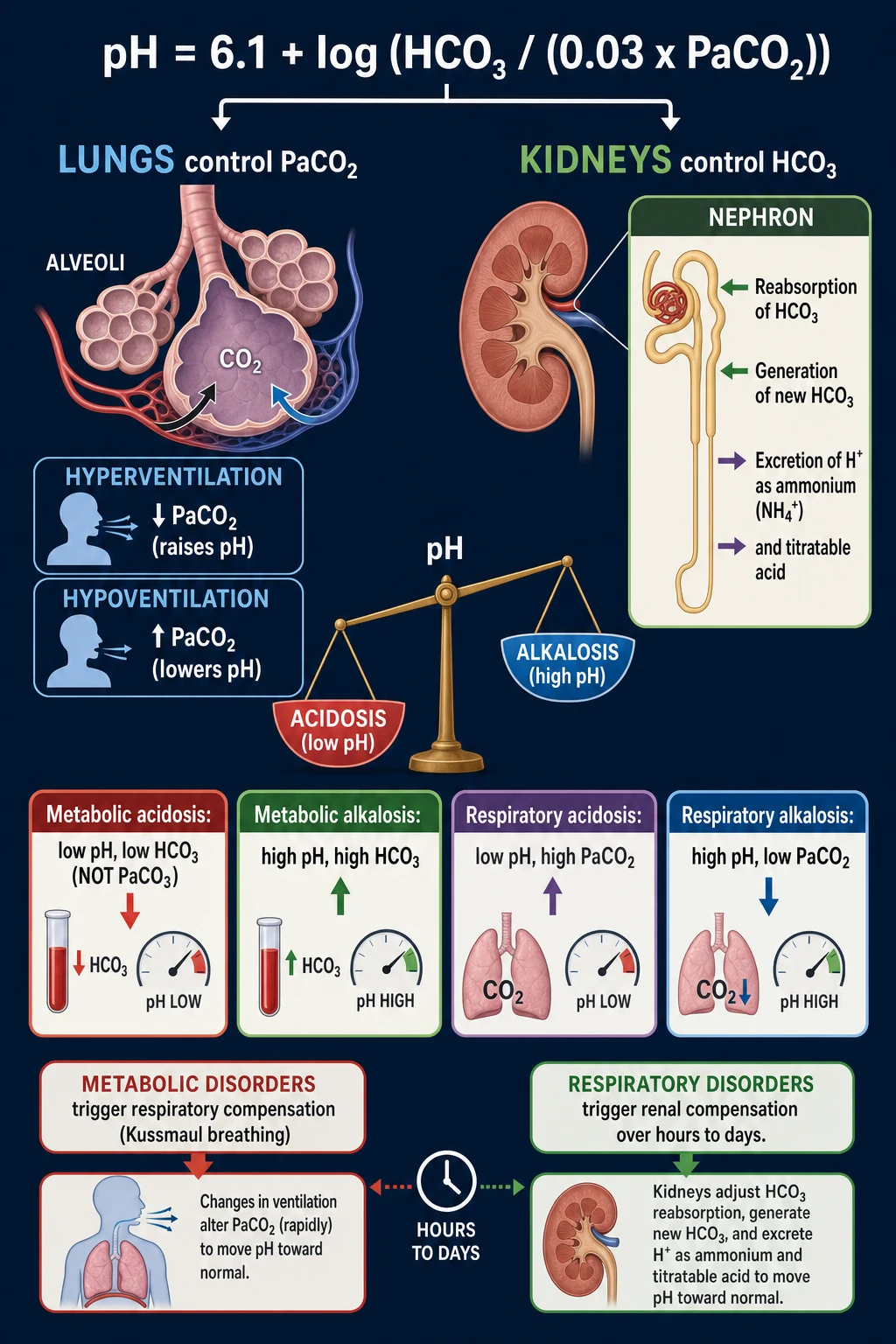
A disciplined, reproducible method is what examiners expect and what catches mixed disorders. Process every blood gas in the same order. [1]
Acid-base interpretation — 9 steps
Step 1 — pH (acidaemia or alkalaemia?)
pH <7.35 = acidaemia; pH >7.45 = alkalaemia; 7.35-7.45 = normal, but a mixed disorder can hide here (e.g., metabolic acidosis + alkalosis cancelling). The suffix "-aemia" refers to the pH; "-osis" refers to the process.
Step 2 — Primary process (PaCO2 vs HCO3)
If PaCO2 moves OPPOSITE to pH, the primary disorder is respiratory. If HCO3 moves in the SAME direction as pH, the primary disorder is metabolic. Low pH + low HCO3 = metabolic acidosis; low pH + high PaCO2 = respiratory acidosis; high pH + high HCO3 = metabolic alkalosis; high pH + low PaCO2 = respiratory alkalosis.
Step 3 — Expected compensation
Apply the formula for the suspected primary disorder (see compensation table). If the measured value falls outside the expected range, a second (mixed) disorder is present. Compensation never overshoots to fully normalise pH — except chronic respiratory alkalosis.
Step 4 — A-a gradient (is the hypoxaemia pulmonary?)
A-a = PAO2 - PaO2. PAO2 = FiO2(Patm - PH2O) - PaCO2/0.8. Normal <15 mmHg (<25 in the elderly; rises ~4 per decade). A high A-a gradient confirms a pulmonary cause (V/Q mismatch, shunt, diffusion); a normal A-a gradient points to hypoventilation or low inspired oxygen.
Step 5 — Anion gap (high or normal?)
AG = Na - (Cl + HCO3). Normal 8-12 mmol/L. Correct for albumin: AG falls ~2.5 mmol/L for every 10 g/L albumin below 40 g/L. High AG = unmeasured anions (organic acids, toxins). Normal AG = hyperchloraemic (bicarbonate lost or chloride gained).
Step 6 — Delta-delta gap (a hidden second disorder?)
Delta ratio = (AG - 12) / (24 - HCO3). ~1 = pure high-AG acidosis. >2 = concurrent metabolic alkalosis (HCO3 higher than expected). <0.4 = concurrent normal-AG acidosis (HCO3 lower than expected from the AG alone).
Step 7 — Osmolar gap (toxins?)
OG = measured osmolality - calculated osmolality. Calculated = 2Na + glucose + urea (+ ethanol if known). Normal <10 mOsm/kg. A high OG + high AG = toxic alcohol (ethylene glycol, methanol, propylene glycol, diethylene glycol).
Step 8 — Lactate (perfusion vs non-perfusion?)
L-lactate (standard assay): Type A (hypoperfusion — sepsis, shock, mesenteric ischaemia) vs Type B (metformin, malignancy, toxins, mitochondrial disease). D-lactate (separate assay): short-gut syndrome, bacterial overgrowth.
Step 9 — Correlate with the clinical picture
No ABG is interpreted in isolation. Integrate with history, examination, electrolytes (K, Cl, albumin), renal function, glucose, ketones, drug levels, and the trajectory of previous gases. Treat the cause, not the number.
Reference ranges (arterial)
Arterial blood gas
Adult normal values
- pH 7.35-7.45
- PaCO2 35-45 mmHg (4.7-6.0 kPa)
- PaO2 80-100 mmHg (10.7-13.3 kPa) on room air
- HCO3 22-26 mmol/L
- Base excess -2 to +2 mmol/L
- Anion gap 8-12 mmol/L
- A-a gradient <15 mmHg (young adult)
- Osmolar gap <10 mOsm/kg
- Lactate <2.0 mmol/L
Kilopascals
ANZ convention
- PaCO2 4.7-6.0 kPa
- PaO2 10.7-13.3 kPa
- 1 kPa = 7.5 mmHg
- Multiply mmHg x 0.133 for kPa
- Most CICM/FFICM exams accept either unit if stated
Compensation formulas — master table
Metabolic acidosis
Respiratory compensation
- Winter formula: PaCO2 = (1.5 x HCO3) + 8 ± 2
- Alternative: PaCO2 ≈ last two digits of pH
- Measured PaCO2 > expected → concurrent respiratory acidosis
- Measured PaCO2 < expected → concurrent respiratory alkalosis
Metabolic alkalosis
Respiratory compensation
- PaCO2 = (0.7 x HCO3) + 20 ± 5
- Or PaCO2 rises ~0.6 mmHg per 1 mmol/L rise in HCO3
- Compensation is limited by hypoxaemia — rarely exceeds 55 mmHg
Respiratory acidosis
Renal compensation (slow, 2-5 days)
- ACUTE: HCO3 rises 1 mmol/L per 10 mmHg rise in PaCO2 (≈ 0.1 per mmHg)
- CHRONIC: HCO3 rises 3.5-4 mmol/L per 10 mmHg rise in PaCO2 (≈ 0.35 per mmHg)
- pH falls 0.08 (acute) or 0.03 (chronic) per 10 mmHg rise in PaCO2
- Exam rule: ΔHCO3/ΔPaCO2 = 0.1 acute, 0.35 chronic (per mmHg)
Respiratory alkalosis
Renal compensation (slow)
- ACUTE: HCO3 falls 2 mmol/L per 10 mmHg fall in PaCO2 (≈ 0.2 per mmHg)
- CHRONIC: HCO3 falls 4-5 mmol/L per 10 mmHg fall in PaCO2 (≈ 0.5 per mmHg)
- pH rises 0.08 (acute) or 0.03 (chronic) per 10 mmHg fall in PaCO2
- Exam rule: ΔHCO3/ΔPaCO2 = 0.2 acute, 0.5 chronic (per mmHg)
Stepwise interpretation
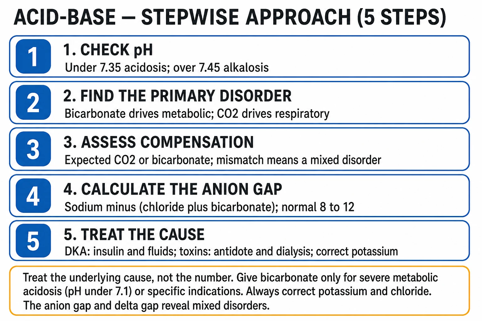
Acid-base interpretation — 6 steps
Step 1: Is there acidosis or alkalosis?
pH <7.35 = acidosis. pH >7.45 = alkalosis. pH 7.35-7.45 = normal (or mixed disorder). Normal pH with abnormal PaCO2/HCO3 = compensated disorder or mixed.
Step 2: Is it respiratory or metabolic?
Look at PaCO2 and HCO3: PaCO2 changes in the SAME direction as pH = metabolic (e.g., low pH + low PaCO2 = metabolic acidosis with respiratory compensation). PaCO2 changes in OPPOSITE direction to pH = respiratory (e.g., low pH + high PaCO2 = respiratory acidosis).
Step 3: Is compensation appropriate?
Metabolic acidosis: PaCO2 = 1.5 x HCO3 + 8 (±2). Winter formula. Metabolic alkalosis: PaCO2 = 0.7 x HCO3 + 20 (±5). Respiratory acidosis (acute): HCO3 rises 1 mmol/L per 10 mmHg rise in PaCO2. Respiratory alkalosis (acute): HCO3 falls 2 mmol/L per 10 mmHg fall in PaCO2.
Step 4: If metabolic acidosis — calculate anion gap
AG = Na - (Cl + HCO3). Normal: 8-12 mmol/L (correct for albumin: subtract 2.5 for every 10 g/L below 40). HIGH AG (>12): MUDPILES. NORMAL AG (8-12): hyperchloraemic acidosis (diarrhoea, RTA, saline).
Step 5: Delta-delta gap (assess mixed disorders)
Delta ratio = (AG - 12) / (24 - HCO3). If =1: pure high AG acidosis. If >2: concurrent metabolic alkalosis (HCO3 is higher than expected). If <0.4: concurrent normal AG acidosis (HCO3 is lower than expected from AG alone).
Step 6: Treat the cause
Do NOT treat the number — treat the underlying disorder. Bicarbonate is RARELY indicated. Treat: hypoperfusion (fluids, inotropes), sepsis, DKA (insulin), renal failure (dialysis), toxic alcohols (fomepizole, dialysis), salicylates (alkalinise urine, dialysis).
The four gaps — quantifying hidden disorders
A-a (alveolar-arterial) gradient
Anion gap (albumin-corrected)
Anion gap
AG = Na − (Cl + HCO3)
- Normal 8-12 mmol/L
- Albumin correction: subtract 2.5 per 10 g/L albumin below 40 g/L
- In hypoalbuminaemia the AG may look normal despite unmeasured anions
- Potassium: each 1 mmol/L K change shifts AG ~ — consider AG with K if borderline
Expected anion gap (albumin-adjusted)
Corrected AG = AG + 2.5 x (40 - albumin)/10
- Albumin 20 g/L → add 5 to measured AG
- Albumin 30 g/L → add 2.5 to measured AG
- Useful in cirrhosis, nephrotic syndrome, critical illness
- A "normal" AG of 12 with albumin 20 is actually a high AG of 17
Delta-delta (gap-gap) — finding the co-existing disorder
Delta ratio — interpretation ladder
Calculate
Delta ratio = (AG − 12) / (24 − HCO3). The numerator is the anion-gap rise; the denominator is the bicarbonate fall. In a pure high-AG acidosis they should match (ratio ≈ 1).
Ratio 0.7-1.4
Consistent with a SINGLE high-AG metabolic acidosis (no additional disorder).
Ratio <0.4 (or <0.7)
HCO3 has fallen MORE than the AG rose → a concurrent NORMAL-AG (hyperchloraemic) acidosis is also present. Classic: DKA with osmotic diuresis volume depletion + saline (ketoacidosis + hyperchloraemic acidosis).
Ratio >2
HCO3 has fallen LESS than expected (or not at all) → a concurrent METABOLIC ALKALOSIS is masking it. Classic: vomiting + lactic acidosis.
Delta gap (alternative form)
Delta gap = (AG − 12) + HCO3. Expected ≈ 24. <24 → extra normal-AG acidosis; >24 → extra metabolic alkalosis.
Osmolar gap
Lactate
L-lactate
Standard assay
- Type A (tissue hypoxia): sepsis, shock, mesenteric ischaemia, burns, trauma, seizures, severe hypoxaemia
- Type B1 (underlying disease): malignancy (lymphoma, leukaemia), diabetes, liver failure, AIDS
- Type B2 (drugs/toxins): metformin, NRTIs, salicylates, propylene glycol, cyanide, β2-agonists, cocaine
- Type B3 (inborn errors): mitochondrial myopathies, GSD, pyruvate dehydrogenase deficiency
D-lactate
Separate assay
- Produced by gut bacteria (not human metabolism)
- Short-bowel syndrome, jejunoileal bypass, bacterial overgrowth
- Carbohydrate malabsorption → bacterial fermentation
- Encephalopathy with high-AG acidosis after a large carbohydrate meal
Davenport diagram — visualising the disorders
[1]Stewart approach (strong ion difference) — brief
[1]Anion gap mnemonic — MUDPILES
High anion gap metabolic acidosis
MUDPILES
- M: Methanol (toxic alcohol — formic acid)
- U: Uraemia (renal failure)
- D: Diabetic ketoacidosis (and other ketoacidoses — alcoholic, starvation)
- P: Propylene glycol (infusion solvent — lorazepam, diazepam)
- I: Iron, Isoniazid (INH)
- L: Lactic acidosis (sepsis, shock, mesenteric ischaemia, metformin)
- E: Ethylene glycol (toxic alcohol — oxalic acid)
- S: Salicylates (aspirin overdose)
Normal anion gap (hyperchloraemic)
HARDUP
- H: Hyperalimentation (TPN)
- A: Acetazolamide (carbonic anhydrase inhibitor)
- R: Renal tubular acidosis
- D: Diarrhoea (GI bicarbonate loss)
- U: Ureteroenteric fistula
- P: Pancreatic fistula (bicarbonate-rich fluid loss)
- Plus: Excess normal saline (hyperchloraemic acidosis)
GOLDMARK — the modern high-AG mnemonic
GOLDMARK
Preferred modern mnemonic
- G — Glycols (ethylene glycol, propylene glycol, diethylene glycol)
- O — Oxoproline (5-oxoproline / pyroglutamic acid — chronic paracetamol, flucloxacillin, malnutrition)
- L — L-lactate (sepsis, shock, mesenteric ischaemia, metformin)
- D — D-lactate (short-gut, bacterial overgrowth)
- M — Methanol (formic acid)
- A — Aspirin (salicylates)
- R — Renal failure (uraemia — sulphates, phosphates)
- K — Ketoacidosis (diabetic, alcoholic, starvation)
Why GOLDMARK over MUDPILES?
Exam nuance
- Replaces outdated/overlapping entries (Iron, INH less common in ICU)
- Explicitly includes oxoproline (commonly missed) and D-lactate
- Recognises propylene glycol as an ICU iatrogenic cause
- Both mnemonics acceptable — be able to list causes WITHOUT a mnemonic
Metabolic acidosis — categorised by anion gap
High anion gap
Unmeasured anions
- Ketoacidosis: DKA (hyperglycaemia, ketones), alcoholic (history, low/normal glucose), starvation
- Lactic acidosis: Type A (hypoperfusion) and Type B (drugs, malignancy) — see lactate section
- Renal failure: sulphates, phosphates, organic anions (AG rises when GFR <20-25)
- Toxins: methanol, ethylene glycol, propylene glycol, salicylates, toluene, paraldehyde
- Oxoproline: chronic paracetamol, sepsis, malnutrition, female sex, renal failure
Normal anion gap (hyperchloraemic)
HARDUP / used-up bicarbonate
- GI bicarbonate loss: diarrhoea, fistulae (pancreatic, biliary), ureteroenteric, villous adenoma
- Renal: RTA (distal/type I, proximal/type II, type IV hypoaldosteronism)
- Drugs: acetazolamide, topiramate, sevelamer, cholestyramine, calcium chloride
- Iatrogenic: rapid normal saline expansion ("saline acidosis"), TPN
- Post-hypocapnia: recovery from chronic respiratory alkalosis
Metabolic alkalosis — chloride-responsive vs resistant
Metabolic alkalosis workup — urine chloride is the key
Step 1 — Recognise
pH >7.45, HCO3 >26, with compensatory PaCO2 rise. Two mechanisms: (a) GENERATION (gain of bicarbonate or loss of acid) + (b) MAINTENANCE (failure to excrete the excess bicarbonate, usually from chloride depletion, hypokalaemia, or hyperaldosteronism).
Step 2 — Measure urine chloride
Urine Cl <20 mmol/L = chloride-RESPONSIVE (extracellular volume depletion / chloride loss). Urine Cl >20 mmol/L = chloride-RESISTANT (mineralocorticoid excess or retained bicarbonate).
Step 3 — If urine Cl <20 (responsive)
Causes: vomiting / NG suction (loss of HCl), diuretics (loop/thiazide — late, after diuresis), post-hypercapnia, cystic fibrosis (sweat Cl loss), villous adenoma, laxative abuse. Treat with chloride: normal saline + KCl correction.
Step 4 — If urine Cl >20 (resistant)
Check BP and renin/aldosterone. Hypertensive + low renin + low aldosterone = Liddle. Low renin + high aldosterone = Conn (primary hyperaldosteronism). High renin + high aldosterone = renal artery stenosis, Cushing. Normotensive = Bartter (loop-like), Gitelman (thiazide-like), severe K/Mg depletion.
Step 5 — Acetazolamide
If saline is contraindicated (heart failure, pulmonary oedema), acetazolamide 250-500 mg promotes bicarbonate diuresis. Watch for worsening hypokalaemia.
Chloride-responsive
Urine Cl <20
- Vomiting / NG suction (most common in ICU)
- Diuretics (loop, thiazide) — post-diuresis
- Post-hypercapnia (after correcting chronic CO2 retention)
- Villous adenoma, laxative/cystic fibrosis Cl loss
- Corrected by saline + KCl (chloride repletion stops the alkalosis)
Chloride-resistant
Urine Cl >20
- Hypertensive + low renin + low aldosterone: Liddle syndrome, exogenous mineralocorticoid
- Hypertensive + low renin + high aldosterone: primary hyperaldosteronism (Conn)
- Hypertensive + high renin + high aldosterone: renal artery stenosis, reninoma, Cushing
- Normotensive: Bartter, Gitelman, severe K+/Mg2+ depletion, ongoing diuretic
Respiratory acidosis — hypoventilation
[1]Causes
Anywhere in the ventilatory pathway
- CNS: opioids, sedatives, stroke, brainstem lesion, sleep apnoea
- Neuromuscular: myasthenia, GBS, ALS, electrolyte (low K/Mg/P), muscle relaxants
- Airway: obstruction, laryngospasm, asthma (late/fatiguing)
- Lung/chest wall: COPD (most common), severe pneumonia, ARDS (late), flail chest, kyphoscoliosis, obesity hypoventilation
Management
Reverse the cause + ventilate
- Reverse sedatives/opioids (naloxone, flumazenil)
- Treat the underlying cause (bronchodilators, NM weakness)
- NIV (BiPAP) for COPD exacerbation — first-line
- Invasive ventilation if failing NIV, GCS low, or aspiration risk
- Do NOT abruptly correct chronic hypercapnia — risks post-hypercapnic alkalosis and seizures
Respiratory alkalosis — hyperventilation
[1]Causes
Hyperventilation
- Hypoxaemia / hypoxia (any cause — drives respiratory centre)
- Pulmonary: PE, pneumonia, oedema, early asthma (tachypnoea before fatigue)
- Stimulation: pain, anxiety, fever, sepsis, salicylates (early), progesterone, hepatic failure
- CNS: stroke, tumour, trauma; physiological: pregnancy, high altitude
- Iatrogenic: over-ventilation on a ventilator (permissive or unintentional)
Management
Treat the cause
- Correct hypoxaemia (O2, treat PE/pneumonia)
- Treat pain, anxiety, fever
- Reduce minute volume if ventilated
- Watch for post-hypocapnic metabolic acidosis if a chronic CO2 retainer is over-ventilated
Mixed acid-base disorders
[1]Classic mixed patterns
High-yield exam scenarios
- Salicylate toxicity: respiratory alkalosis (early, direct stimulation) + high-AG metabolic acidosis
- Vomiting + keto/lactic: metabolic alkalosis + high-AG metabolic acidosis (delta ratio >2)
- COPD + vomiting: chronic respiratory acidosis + metabolic alkalosis
- Sepsis + salicylate: high-AG metabolic acidosis + respiratory alkalosis
- DKA + saline: high-AG + normal-AG (hyperchloraemic) acidosis (delta ratio <1)
- Renal failure + COPD: metabolic acidosis + chronic respiratory acidosis
- Post-arrest: mixed respiratory + lactic metabolic acidosis
Triple disorders
Example: COPD + vomiting + sepsis
- Chronic respiratory acidosis (baseline raised PaCO2 and HCO3)
- + Metabolic alkalosis (vomiting — pushes HCO3 up further)
- + High-AG metabolic acidosis (sepsis — pushes HCO3 down)
- Diagnosed by calculating compensation AND the delta-delta in a patient with one obvious disorder
Renal tubular acidosis (RTA)
Type I (distal)
Failure to excrete H+
- Severe metabolic acidosis (HCO3 often <10-20)
- Urine pH >5.5 (cannot acidify urine)
- Normal AG (hyperchloraemic)
- Hypokalaemia
- Nephrocalcinosis / nephrolithiasis
- Causes: autoimmune (Sjögren, SLE), amphotericin, toluene, genetic
- Treatment: bicarbonate (replace losses) + K
Type II (proximal)
Failure to reabsorb HCO3
- Mild-moderate acidosis (HCO3 usually 12-20)
- Urine pH <5.5 once serum HCO3 below threshold
- Normal AG (hyperchloraemic), hypokalaemia
- Fanconi syndrome (glycosuria, aminoaciduria, phosphaturia)
- Causes: myeloma, ifosfamide, tenofovir, carbonic anhydrase inhibitors
- Treatment: high-dose bicarbonate (matches losses) + K + thiazide
Type IV (hypoaldosteronism)
Most common RTA in adults
- Mild acidosis (HCO3 usually >17)
- Urine pH <5.5
- Normal AG (hyperchloraemic), HYPERkalaemia (distinguishes it)
- Hyporeninaemic hypoaldosteronism: diabetic nephropathy, CKD
- Adrenal insufficiency: Addisons
- Drugs: ACEi/ARB, K-sparing diuretics, NSAIDs, heparin, calcineurin inhibitors
- Treatment: fludrocortisone (if adrenal), correct K, treat cause
Toxic alcohol poisoning
Ethylene glycol
Antifreeze
- Metabolised by alcohol dehydrogenase → glycolate (acid) → oxalate
- High AG + HIGH osmolar gap
- Crystalluria (calcium oxalate) → AKI, oxalate crystals in urine
- Calcium oxalate stones, hypocalcaemia, QT prolongation
- Three stages: CNS depression (1-12 h), cardiopulmonary (12-24 h), renal failure (>24 h)
Methanol
Windscreen washer, illicit spirits
- Metabolised → formaldehyde → formic acid (acid, retinal toxin)
- High AG + HIGH osmolar gap
- Visual disturbance: blurred vision, "snowfield" vision, optic disc oedema, blindness
- Basal ganglia infarction on imaging
- Latent period before acidosis develops (while metabolising)
Salicylate (aspirin) toxicity
[1]When to give bicarbonate
[2]Evidence base — landmark trials
BICARB-ICU — bicarbonate for severe metabolic acidosis
Lancet
Population: Critically ill adults with severe metabolic acidosis (pH ≤7.20)
Key finding
No difference in the primary composite (28-day mortality or day-7 organ failure). Pre-specified AKI subgroup showed lower mortality (46% vs 63%) and less need for renal replacement therapy.
Practice change
Bicarbonate does not improve overall outcomes in severe metabolic acidosis, but may benefit the AKI subgroup — hence selective, not routine, use.
SPLIT — buffered crystalloid vs saline (ICU)
JAMA
Population: 2278 ICU patients (New Zealand)
Key finding
No difference in AKI incidence, RRT use, or hospital mortality. Did NOT support the saline-AKI hypothesis in this ICU population.
Practice change
First large blinded RCT on balanced vs saline was negative; saline-induced hyperchloraemic acidosis was not enough to change renal outcomes here.
SMART — balanced crystalloids vs saline
NEJM
Population: 15,802 critically ill adults (single-centre, pragmatic)
Key finding
Lower MAKE30 (death, new RRT, or persistent renal dysfunction) with balanced crystalloids (14.3% vs 15.4%, p=0.04). Sepsis subgroup: lower 30-day mortality (26.3% vs 31.2%).
Practice change
Balanced crystalloids reduce major adverse kidney events vs saline — preferentially avoid large-volume saline to prevent iatrogenic hyperchloraemic acidosis.
Worked ABG examples — exam drills
Worked example 1 — the classic mixed disorder
ABG
pH 7.40, PaCO2 20 mmHg, HCO3 12, PaO2 95. Na 138, Cl 102.
Read pH
7.40 is normal — but PaCO2 and HCO3 are BOTH abnormal → a mixed disorder is hiding.
Anion gap
AG = 138 − (102 + 12) = 24. High (corrected normal ~12). High-AG metabolic acidosis present.
Winter formula
Expected PaCO2 = 1.5 × 12 + 8 = 26 ± 2. Measured is 20 — LOWER → concurrent respiratory alkalosis.
Diagnosis
Mixed high-AG metabolic acidosis + respiratory alkalosis. Classic for SEPSIS (lactic acidosis + hyperventilation) or SALICYLATE toxicity.
Worked example 2 — delta ratio in DKA + vomiting
ABG
pH 7.25, PaCO2 24, HCO3 10. Na 135, Cl 95.
Anion gap
AG = 135 − (95 + 10) = 30. High.
Delta ratio
(30 − 12) / (24 − 10) = 18/14 = 1.3 → within 0.7-1.4 → essentially a single high-AG acidosis.
Now add vomiting
Same patient but Cl 88: AG = 135 − (88 + 10) = 37. Delta ratio = (37 − 12)/(24 − 10) = 25/14 = 1.8 → trending >2 → concurrent metabolic alkalosis from vomiting is unmasked.
Diagnosis
DKA (high-AG) + metabolic alkalosis (vomiting). Always check the delta ratio when an AG acidosis "looks too mild" for the pH.
Worked example 3 — chronic COPD compensation
ABG
pH 7.34, PaCO2 60, HCO3 32.
Primary disorder
Low pH + high PaCO2 = respiratory acidosis.
Acute or chronic?
Expected HCO3 if ACUTE = 24 + 1×(60−40)/10 = 26. If CHRONIC = 24 + 3.5×2 = 31. Measured 32 → CHRONIC respiratory acidosis (compensated COPD).
Is there a second disorder?
HCO3 of 32 matches chronic expectation — single disorder. If HCO3 were 38, a metabolic alkalosis (diuretics, vomiting) would be added.
Exam practice — SAQs
SAQ — A near-normal pH with three acid-base disorders (COPD, vomiting and sepsis)
10 minutes · 10 marks
A 68-year-old man with severe COPD on long-term oxygen and home BiPAP is admitted with three days of intractable vomiting from a small-bowel obstruction and a 24-hour history of fever and a productive cough. He is drowsy, RR 28, SpO2 88% on his usual 2 L/min, BP 96/60, lactate 3.2. Arterial gas on 2 L/min: pH 7.34, PaCO2 64 mmHg, PaO2 58 mmHg, HCO3 34 mmol/L. Bloods: Na 140, Cl 92, K 4.0, albumin 30 g/L.
SAQ — The delta ratio unmasks a second acidosis in treated diabetic ketoacidosis
10 minutes · 10 marks
A 23-year-old woman presents in new-onset diabetic ketoacidosis (glucose 28 mmol/L, ketones 5.2, pH 6.98). After 3 L of 0.9% saline and a fixed-rate insulin infusion in the emergency department she is reassessed. Arterial gas: pH 7.20, PaCO2 27 mmHg, HCO3 12 mmol/L, lactate 1.4. Venous biochemistry: Na 138, Cl 112, K 4.2, albumin 38 g/L. The registrar states the anion gap is now only 14 and plans to stop the insulin infusion.
Clinical pearls
Red flags
[1]Summary — the bedside algorithm in one breath
[1]References
- [1]Seifter JL Integration of acid-base and electrolyte disorders N Engl J Med, 2014.PMID 25372090
- [2]Kraut JA, Madias NE. Metabolic acidosis: pathophysiology, diagnosis and management Nat Rev Nephrol, 2010.PMID 20308999
- [3]Emmett M, Narins RG Clinical use of the anion gap Medicine (Baltimore), 1977.PMID 401925
- [4]Jaber S, Paquette F, Girard M, et al. Sodium bicarbonate therapy for patients with severe metabolic acidaemia in the intensive care unit (BICAR-ICU): a multicentre, open-label, randomised controlled, phase 3 trial Lancet, 2018.PMID 29910040
- [5]Self WH, Semler MW, Wanderer JP, et al. Balanced Crystalloids versus Saline in Critically Ill Adults N Engl J Med, 2018.PMID 29485925
- [6]Young P, Bailey M, Beasley R, et al. Effect of a Buffered Crystalloid Solution vs Saline on Acute Kidney Injury Among Patients in the Intensive Care Unit: The SPLIT Randomized Clinical Trial JAMA, 2015.PMID 26444692
- [7]Kraut JA, Madias NE. Lactic acidosis N Engl J Med, 2014.PMID 25494270