MedVellum
MCQsExamsAtlas
DashboardPricing
MBBS / Core medicine✳Dermatology✳ICU Fellowship (CICM)✳Anaesthesia✳Emergency Medicine✳Psychiatry Fellowship✳Paediatrics Fellowship✳Physician Medicine✳MCQs✳SAQs✳Vivas✳OSCE✳Evidence-first✳MBBS / Core medicine✳Dermatology✳ICU Fellowship (CICM)✳Anaesthesia✳Emergency Medicine✳Psychiatry Fellowship✳Paediatrics Fellowship✳Physician Medicine✳MCQs✳SAQs✳Vivas✳OSCE✳Evidence-first✳

MedVellum.

The folio

Exam-exhaustive medical education across every specialty — evidence-graded topics, engraved plates, and practice in every written and oral format. Educational content only — not medical advice.

llms.txt · psychiatry LLM catalog · sitemap

Atlas

  • Specialty atlas
  • MBBS / Core medicine
  • Dermatology
  • ICU Fellowship (CICM)
  • Anaesthesia
  • Emergency Medicine
  • Psychiatry Fellowship
  • Paediatrics Fellowship
  • Physician Medicine

Study & account

  • MCQ practice
  • Practice alias
  • Exam tools
  • Dashboard
  • Pricing
  • Sign in

© 2026 MedVellum. For education only — not a substitute for clinical judgement.

Folio edition · Set in Instrument Serif & Archivo

ICU TopicsRenal and metabolic

ICU · Renal and metabolic

Acute severe hypercalcaemia and hypocalcaemia: ICU emergencies

Also known as Hypercalcaemia · Hypercalcemic crisis · Hypocalcaemia · Calcium disorders · Chvostek sign · Trousseau sign

Calcium disorders are common in ICU. HYPERCALCAEMIA (corrected Ca 2.6): mild (2.6-3.0) asymptomatic; moderate (3.0-3.5) — GI, renal, neurological symptoms; SEVERE (3.5, 'hypercalcaemic crisis') — confusion, coma, AKI, arrhythmia — emergency. Causes: primary hyperparathyroidism, malignancy (PTHrP, bone metastases, myeloma) — 90% of cases. Treatment: aggressive NORMAL SALINE (volume expansion + calciuresis), BISPHOSPHONATES (zoledronate — inhibits osteoclast, takes 48-72h), calcitonin (rapid but tachyphylaxis), dialysis (severe/renal failure). HYPOCALCAEMIA (corrected Ca <2.1): neuromuscular irritability (tetany, Chvostek/Trousseau signs, seizures, perioral numbness, carpopedal spasm, prolonged QT). Causes: hypoparathyroidism (post-thyroidectomy), vitamin D deficiency, CKD, hypomagnesaemia, pancreatitis, sepsis (citrate in transfusion/blood products chelates Ca). Treatment: calcium gluconate IV (severe/symptomatic), oral Ca + vitamin D (chronic), correct Mg.

high12 referencesUpdated 4 July 2026
On this page & tools

Your progress

Saved locally on this device.

Target exams

CICMFFICMEDIC

Red flags

Severe hypercalcaemia (>3.5) → coma, AKI, arrhythmia — emergency: saline + bisphosphonateSevere hypocalcaemia (corrected &lt;1.9 or symptomatic) → seizures, tetany, prolonged QT — calcium gluconate IVALWAYS correct Ca for albumin: +0.02 × (40 − albumin g/L)Hypomagnesaemia causes refractory hypocalcaemia (impairs PTH secretion + action)

Your progress

Saved locally on this device.

Target exams

CICMFFICMEDIC

Red flags

Severe hypercalcaemia (>3.5) → coma, AKI, arrhythmia — emergency: saline + bisphosphonateSevere hypocalcaemia (corrected &lt;1.9 or symptomatic) → seizures, tetany, prolonged QT — calcium gluconate IVALWAYS correct Ca for albumin: +0.02 × (40 − albumin g/L)Hypomagnesaemia causes refractory hypocalcaemia (impairs PTH secretion + action)
Cinematic ICU scene of an ECG showing a prolonged QT beside a calcium gluconate vial and a saline-and-furosemide hydration setup, clinical-blue lighting, medical educational, no faces, no text
FigureCalcium at both ends — the hypercalcaemic crisis above 3.5 (the confusion, the AKI, the arrhythmia) needs the saline, the bisphosphonate, and the calcitonin bridge; the symptomatic hypocalcaemia (the tetany, the prolonged QT, the perioral tingling) needs the slow IV calcium gluconate and the magnesium first, or it refractories.

In one line

Hypercalcaemic crisis (Ca >3.5): coma, AKI, arrhythmia → AGGRESSIVE NORMAL SALINE (3-6 L/day — volume expansion + calciuresis) + zoledronate 4 mg IV (bisphosphonate — takes 48-72h; for malignancy), calcitonin (rapid but tachyphylaxis), dialysis if severe/renal failure. Severe hypocalcaemia (corrected <1.9 or tetany/seizures): calcium gluconate 10% 10-20 mL IV over 10 min, then infusion; correct Mg (refractory if low); oral Ca + vitamin D long-term. Always correct Ca for albumin: +0.02 × (40 − albumin). Causes — hyper: hyperparathyroidism, malignancy (PTHrP); hypo: post-thyroidectomy, CKD, vitamin D deficiency, hypomagnesaemia, pancreatitis, sepsis/citrate.

[1]

Hypercalcaemia vs hypocalcaemia at a glance

FeatureHypercalcaemiaHypocalcaemia
Corrected Ca>2.6 mmol/L<2.1 mmol/L
SeverityMild 2.6-3.0; Mod 3.0-3.5; Severe >3.5Mild 1.9-2.1; Severe <1.9 or symptomatic
Symptoms'stones, bones, groans, psychic moans' — constipation, polyuria, confusionTetany, perioral numbness, carpopedal spasm, seizures
ECGShort QT, arrhythmiaLong QT, torsades
SignsDehydration (polyuria), abdominal painChvostek, Trousseau signs
Emergency TxSaline + bisphosphonateCalcium gluconate IV
Common causesHyperparathyroidism, malignancyHypoparathyroidism (post-surgery), CKD, vit D def
[1]

Management of hypercalcaemic crisis (Ca >3.5 or symptomatic)

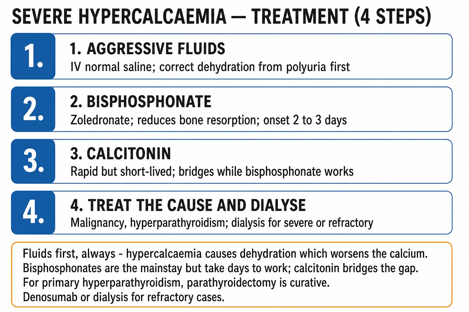
  1. AGGRESSIVE VOLUME EXPANSION — Normal saline 0.9% 200-300 mL/hr (3-6 L/day). MECHANISM: hypercalcaemia causes nephrogenic DI (polyuria) → dehydration → RAAS → proximal Ca reabsorption → worsens hypercalcaemia. Saline BREAKS the cycle: volume repletion → suppresses RAAS → increases renal Ca excretion (calciuresis). Most effective FIRST step. Add loop diuretic (frusemide) ONLY if volume overloaded after saline (frusemide promotes calciuresis — but AVOID thiazides which RETAIN calcium)
  2. BISPHOSPHONATE — Zoledronate 4 mg IV over 15 min (or pamidronate 60-90 mg IV over 2-4h). MECHANISM: inhibits osteoclast-mediated bone resorption → lowers Ca. ONSET: 48-72 hours (delayed — not immediate). DURATION: 2-4 weeks. FIRST-LINE for hypercalcaemia of malignancy. CAUTION: renal failure (zoledronate nephrotoxic — reduce dose/avoid if eGFR <30), osteonecrosis of jaw (rare), hypocalcaemia (overshoot)
  3. CALCITONIN — 4 IU/kg SC/IM every 12h. MECHANISM: inhibits osteoclasts → rapid Ca fall. ONSET: 4-6 hours (FASTER than bisphosphonate — bridge). DURATION: 48-72h then TACHYPHYLAXIS (loses effect). USE: bridge while waiting for bisphosphonate to work (first 48-72h). Side effects: nausea, flushing
  4. DIALYSIS — if severe (Ca >4), renal failure (can't excrete Ca), heart failure (can't tolerate saline). Haemodialysis with LOW-calcium dialysate rapidly lowers Ca. Reserve for refractory/severe
  5. TREAT UNDERLYING CAUSE — (a) Malignancy: treat tumour (chemo, radiation). (b) Primary hyperparathyroidism: parathyroidectomy (curative — surgical). (c) Stop offending drugs (thiazides, calcium supplements, vitamin D, vitamin A, lithium). (d) Immobilisation: mobilise (prevents bone resorption)
  6. MONITOR — Corrected Ca every 6-12h during treatment. Renal function (saline + bisphosphonate), ECG (QT), neurological status. Watch for OVERSHOOT hypocalcaemia (after bisphosphonate). Prevent recurrence (treat cause)
[1]

Antiresorptive / calcium-lowering agents for hypercalcaemia compared

AgentMechanismOnsetDurationRenal failure?Key caveat
Normal saline 0.9%Volume expansion → calciuresis; breaks RAAS-Ca retention cycleHoursWhile infusedYes (safe)FIRST step; add frusemide ONLY if volume overloaded
Calcitonin (4 IU/kg SC/IM q12h)Inhibits osteoclasts; ↑ renal Ca excretion4-6 h (RAPID)48-72 hYes (safe)TACHYPHYLAXIS after 48-72h — bridge only
Zoledronate (4 mg IV over 15 min)Bisphosphonate — inhibits farnesyl pyrophosphate synthase → osteoclast apoptosis48-72 h2-4 weeksNO (nephrotoxic — reduce/avoid if eGFR <30)FIRST-LINE antiresorptive for malignancy; overshoot hypocalcaemia
Pamidronate (60-90 mg IV over 2-4 h)Bisphosphonate (same mechanism)48-72 h2-4 weeksNOSlower infusion; slightly less potent than zoledronate
Denosumab (120 mg SC)Anti-RANKL monoclonal → blocks osteoclast formation48-72 hMonthsYES (safe — not renally cleared)Preferred in renal failure; GREATER hypocalcaemia risk
Glucocorticoids (prednisolone 20-40 mg/day)Reduce 1,25-(OH)₂-vit D activationDaysWhile givenYesONLY for vitamin-D-mediated (sarcoid, lymphoma, intoxication)
Haemodialysis (low-Ca dialysate)Direct removal; bypasses kidneyImmediate (hours)While runningIndicatedRescue for Ca >4, heart failure, or renal failure
[1]

PTH-based differential diagnosis of hypercalcaemia

CausePTHPhosphate25-OH-vit D1,25-(OH)₂-vit DPTHrP / other
Primary hyperparathyroidismHIGH (or inappropriately normal)LOW/normalNormalHIGHPTHrP low
Hypercalcaemia of malignancy (PTHrP)LOWLOW/normalNormalLow/normalPTHrP HIGH
Local osteolytic (myeloma, bone mets)LOWNORMAL/HIGHNormalLow/normalPTHrP low; ↑ Ca, ↑ Cr, ↑ globulins
Vitamin D intoxicationLOWHIGHHIGHVariableSupplement history
Granulomatous disease (sarcoid, TB)LOWHIGHNormalHIGH (macrophage 1-α-hydroxylase)ACE ↑; worse in summer
Milk-alkali syndromeLOWNormalNormalNormalCaCO₃ antacid history; ↑ HCO₃, ↑ Cr
Thiazides / lithiumHIGH/normalNormalNormalNormalDrug history; lithium raises PTH set-point
ImmobilisationLOW/normalHIGH (young, Paget)NormalNormalBed-bound; ↑ bone resorption
[1]

Calcium gluconate vs calcium chloride for IV replacement

FeatureCalcium gluconate 10%Calcium chloride 10%
Elemental Ca per 10 mL2.2 mmol (8.9 mg/mL)6.8 mmol (3× more)
RoutePERIPHERAL (less irritating) — preferredCENTRAL line only (tissue necrosis if extravasated)
IndicationMost ICU hypocalcaemiaCardiac arrest, severe refractory, central access available
Bolus dose10-20 mL over 10 min5-10 mL slow, via central line
Infusion10 ampoules in 500 mL 5% dextrose at 50 mL/hrRarely infused
Hepatic metabolismGluconate needs hepatic conversion (slower in cirrhosis)Ionised immediately (faster onset)
MonitorECG, ionised CaECG (bradycardia), central line position
[1]

Management of severe symptomatic hypocalcaemia (corrected <1.9, tetany, seizures, stridor, prolonged QT)

  1. AIRWAY / BREATHING / CIRCULATION — Stridor from laryngeal spasm and generalised seizures are the immediate threats. Secure airway if laryngospasm; treat seizures with IV calcium (not benzodiazepines alone — calcium reverses the underlying cause). Continuous ECG (QT) and ionised Ca if available
  2. IV CALCIUM GLUCONATE 10% — 10-20 mL (= 2.2-4.4 mmol) IV over 10 min (slow — rapid injection causes bradycardia, arrhythmia). Preferred peripherally. Use calcium chloride 10% 5-10 mL via CENTRAL line only if cardiac arrest/extreme urgency. MECHANISM: directly restores ionised Ca → stabilises neuronal membrane excitability → abolishes tetany/seizures. Effect transient (minutes-1h)
  3. CONTINUOUS CALCIUM INFUSION — Add 10 ampoules calcium gluconate 10% (= ~22 mmol) to 500 mL 5% dextrose or saline; run at 50 mL/hr (titrate to ionised Ca 1.0-1.2 mmol/L, or corrected Ca 2.1-2.3). Most severe cases need infusion for 24-72h. Recheck ionised Ca every 4-6h
  4. CHECK AND CORRECT MAGNESIUM FIRST — MgSO₄ 2 g (8 mmol) IV over 10-20 min, then 4 g over 4-8h (or oral). MECHANISM: hypomagnesaemia (a) impairs PTH secretion and (b) causes end-organ PTH resistance → REFRACTORY hypocalcaemia that will NOT correct until Mg repleted. Same gating principle as refractory hypokalaemia
  5. ADDRESS THE CAUSE — (a) Post-thyroidectomy/parathyroidectomy (parathyroid damage); (b) hypomagnesaemia (PPIs, diuretics, alcohol); (c) vitamin D deficiency; (d) CKD (low calcitriol); (e) pancreatitis (saponification); (f) citrate from massive transfusion / CRRT regional anticoagulation (continuous calcium replacement); (g) sepsis (calcium shift). Stop offending drugs
  6. STEP DOWN TO ORAL — Once stable, convert to oral calcium (1-2 g elemental/day) + calcitriol (0.25-0.5 μg/day; needed because PTH normally activates vit D). Target corrected Ca 2.1-2.3 (avoid hypercalciuria — monitor urine Ca:creatinine). For permanent hypoparathyroidism consider recombinant PTH 1-84
  7. MONITOR & AVOID OVERCORRECTION — Recheck ionised Ca 1h post-bolus then every 4-6h. Watch for hypercalcaemia (overshoot), arrhythmia, extravasation injury (necrosis). Serial ECG (QT shortens as Ca corrects). Thiazide diuretics reduce urinary Ca loss in chronic hypoparathyroidism (use cautiously)
[1]

Diagnostic workup of hypercalcaemia in ICU

  1. CONFIRM TRUE HYPERCALCAEMIA — Correct for albumin: corrected Ca = measured Ca + 0.02 × (40 − albumin g/L). If doubt (low albumin, acid-base disturbance, myeloma) → measure IONISED Ca (gold standard in ICU). Exclude sampling error (repeat, no prolonged tourniquet, free-flowing sample)
  2. FIRST-LINE BLOODS — PTH (intact), phosphate, renal function, albumin, alkaline phosphatase, 25-OH-vitamin D, magnesium. PTH is the BRANCH POINT: HIGH/normal PTH = PTH-mediated (hyperparathyroidism, FHH, lithium, thiazides); LOW PTH = PTH-independent (malignancy, vitamin D, granulomatous, immobilisation, milk-alkali)
  3. IF PTH LOW (PTH-INDEPENDENT) — Send PTHrP (humoral hypercalcaemia of malignancy), 1,25-(OH)₂-vit D (granulomatous, lymphoma, intoxication), SPEP / serum free light chains (myeloma), TSH (thyrotoxicosis), vitamin A, ACE (sarcoid). Imaging: CT for occult malignancy; bone scan / PET-CT; myeloma screen (β2-microglobulin, urine Bence-Jones)
  4. IF PTH HIGH / INAPPROPRIATELY NORMAL (PTH-MEDIATED) — Primary hyperparathyroidism most likely. Check 24-h urinary Ca (to exclude FHH — Ca clearance/creatinine <0.01 = FHH, AVOID parathyroidectomy). Neck ultrasound + sestamibi scan to localise adenoma. Hypercalcaemic crisis in known hyperPTH → urgent parathyroidectomy after stabilisation
  5. DRUG & EXPOSURE HISTORY — Thiazides, lithium (raises PTH set-point), calcium/vitamin D/vitamin A supplements, calcium carbonate antacids (milk-alkali), herbal/tonic preparations. Prolonged immobilisation (esp. young with high bone turnover, Paget disease)
  6. ASSESS SEVERITY & ORGAN EFFECTS — ECG (short QT — opposite of hypocalcaemia; arrhythmia), renal function (AKI), urinalysis (polyuria, nephrocalcinosis), neurological status (confusion → coma). Stratify mild (2.6-3.0) / moderate (3.0-3.5) / severe crisis (>3.5)
  7. TREAT WHILE WORKING UP — Saline is safe to start immediately. Bisphosphonate can be given once PTHrP/malignancy suspected. Definitive cause-directed therapy (parathyroidectomy, chemotherapy, stop offending drug) once identified
[1]

SAQs

SAQ — Hypercalcaemic crisis in metastatic squamous cell lung carcinoma (PTHrP-mediated)

10 minutes · 10 marks

A 66-year-old woman with known metastatic squamous cell lung carcinoma is brought in confused, polyuric, constipated and with abdominal pain. She is drowsy (GCS 13), dehydrated, HR 110, BP 96/60. Corrected calcium 3.8 mmol/L, albumin 28 g/L, creatinine 175 micromol/L (baseline 80), phosphate 0.6 mmol/L, intact PTH suppressed. ECG shows a short QT interval.

SAQ — Hypocalcaemia in septic shock: when and how to replace

10 minutes · 10 marks

A 58-year-old man is admitted to ICU with community-acquired pneumonia and septic shock. He is intubated, ventilated and on noradrenaline 0.4 mcg/kg/min. Lactate 4.2 mmol/L, albumin 22 g/L, total calcium 1.65 mmol/L, ionised calcium 0.92 mmol/L, magnesium 0.55 mmol/L, phosphate 0.6 mmol/L, pH 7.30. ECG shows QTc 480 ms. He is sedated with no overt neuromuscular signs.

[1]

Clinical pearls

High-yield calcium disorder points for CICM/FFICM exam

  1. Correct calcium for albumin — always. Total Ca is 50% ionised (active) + 40% albumin-bound + 10% complexed. ALBUMIN BINDING: if albumin LOW, total Ca appears low (but IONISED may be normal). CORRECTION: corrected Ca = measured Ca + 0.02 × (40 − albumin g/L). EXAMPLE: Ca 1.9, albumin 20 → corrected 1.9 + 0.02 × 20 = 2.3 (NORMAL). If in doubt: measure IONISED Ca (the active form — not affected by albumin). IONISED Ca is the GOLD STANDARD in ICU (especially with acid-base disturbances, critical illness where albumin fluctuates).[6] }
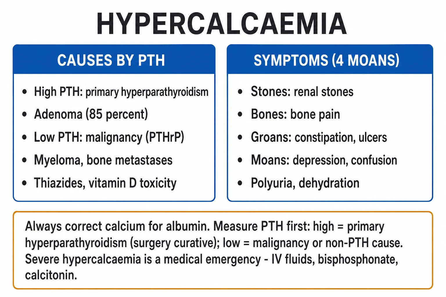
  2. 'Stones, bones, groans, psychic moans' — hypercalcaemia mnemonic. STONES: nephrolithiasis, nephrocalcinosis (Ca deposits in kidney), polyuria (nephrogenic DI), AKI. BONES: bone pain (resorption), fractures, osteitis fibrosa cystica (hyperparathyroidism). GROANS: constipation, abdominal pain, peptic ulcer, pancreatitis. PSYCHIC MOANS: confusion, depression, lethargy, coma. SEVERITY correlates roughly with Ca level. ECG: SHORT QT (opposite of hypocalcaemia — Ca shortens repolarisation).[1] }
  3. Hypercalcaemia causes nephrogenic DI → dehydration. High Ca affects the kidney's concentrating ability (Ca deposits in collecting duct → ADH resistance → can't concentrate urine → POLYURIA). Patient loses water → dehydration → volume depletion → RAAS activation → proximal tubule reabsorbs MORE Ca (to retain volume) → hypercalcaemia WORSENS. This is a VICIOUS CYCLE. BREAK IT: aggressive saline (volume expansion → suppresses RAAS → kidney can excrete Ca). This is why SALINE is the FIRST and MOST EFFECTIVE step in hypercalcaemic crisis.[1] }
  4. Causes of hypercalcaemia — 90% are hyperparathyroidism or malignancy. (1) PRIMARY HYPERPARATHYROIDISM: most common in OUTPATIENTS (community) — parathyroid adenoma (85%), hyperplasia (15%), carcinoma (<1%). PTH HIGH (or inappropriately normal), Ca high, phosphate low. (2) MALIGNANCY: most common INPATIENT cause — PTHrP (parathyroid hormone-related peptide — humoral hypercalcaemia of malignancy — lung SCC, breast, renal, myeloma), bone metastases (breast, prostate — local osteolysis), myeloma (cytokine-mediated osteolysis). PTH LOW (suppressed by high Ca). (3) OTHERS: thiazide diuretics, vitamin D intoxication, vitamin A intoxication, lithium, sarcoidosis (vitamin D activation), immobilisation, milk-alkali syndrome, thyrotoxicosis. PTH LEVEL is the KEY discriminator (high = hyperparathyroid; low = malignancy/other).[2] }
  5. Hypocalcaemia — neuromuscular irritability. Low Ca → increases neuronal excitability (low Ca stabilises Na channel threshold → easier to depolarise). CLINICAL: (a) PERIORAL numbness, tingling fingers/toes. (b) CARPOPEDAL SPASM (flexion of wrist, extension of fingers — 'main d'accoucheur'/obstetrician's hand). (c) LARYNGEAL spasm (stridor — dangerous). (d) SEIZURES (generalised tonic-clonic). (e) TETANY (sustained muscle contraction). SIGNS: Chvostek (tap facial nerve → facial muscle twitch — anterior facial nerve — not very specific), Trousseau (inflate BP cuff above systolic for 3 min → carpopedal spasm — MORE specific for hypocalcaemia). ECG: PROLONGED QT (Ca needed for repolarisation — low Ca prolongs) → torsades risk.[3] }
  6. Post-thyroidectomy hypocalcaemia — common and important. MECHANISM: (a) Removal/damage to parathyroid glands (adjacent to thyroid — inadvertent removal, devascularisation). (b) 'Bone hunger' — in severe hyperthyroidism/hyperparathyroidism (high bone turnover) → sudden PTH drop post-surgery → bone rapidly takes up Ca → hypocalcaemia. TIMING: 24-72h post-op. MONITOR: check Ca every 6-12h for 48h post-thyroidectomy/parathyroidectomy. PREDICTORS: bilateral neck surgery, re-operation, extensive dissection. TREATMENT: oral Ca + calcitriol (1,25-vit D — needed as PTH normally activates vit D); IV calcium gluconate if severe/symptomatic. Usually transient (parathyroids recover) but can be permanent (if all 4 glands removed).[4] }
  7. Hypomagnesaemia causes REFRACTORY hypocalcaemia. MECHANISM: (a) Mg needed for PTH SECRETION (low Mg → impaired PTH release from parathyroid). (b) Mg needed for PTH ACTION (low Mg → end-organ resistance to PTH). RESULT: low Mg → low PTH effect → hypocalcaemia that is REFRACTORY to calcium replacement (until Mg corrected). CLINICAL: ALWAYS check Mg in hypocalcaemic patient. If low: replace Mg FIRST (MgSO4 IV), then calcium works. Same principle as refractory hypokalaemia — Mg gates the system. Pseudohypoparathyroidism (PTH resistance) and hypomagnesaemia both give high PTH + low Ca (end-organ resistance).[3] }
  8. Calcium gluconate vs calcium chloride for IV replacement. CALCIUM GLUCONATE 10%: 10 mL = 2.2 mmol Ca (8.9 mg/mL elemental). Can be given PERIPHERALLY (less irritating). Preferred for most ICU hypocalcaemia. CALCIUM CHLORIDE 10%: 10 mL = 6.8 mmol Ca (3x more Ca per mL). MUST be given via CENTRAL line (highly irritating — causes necrosis if extravasates). Reserved for cardiac arrest/severe (when need rapid high dose). DOSE: calcium gluconate 10% 10-20 mL IV over 10 min (severe/symptomatic), then infusion (10 ampoules in 500 mL at 50 mL/hr). MONITOR ECG.[6] }
  9. Citrate chelates calcium — transfusion and CRRT. (1) MASSIVE TRANSFUSION: blood products contain CITRATE (anticoagulant) → citrate chelates Ca → HYPOCALCAEMIA. Risk: rapid transfusion (>1 unit/5 min), liver failure (can't metabolise citrate). MONITOR: ionised Ca during massive transfusion; replace (calcium gluconate). (2) CRRT with CITRATE anticoagulation: citrate regional anticoagulation (chelates Ca in circuit → prevents clotting) → calcium-free blood returns → patient hypocalcaemic. PROTOCOL: continuously INFUSE calcium (calcium chloride into patient) to replace the chelated Ca; monitor systemic ionised Ca + post-filter Ca (tightly regulated protocol). Citrate accumulation: if liver can't metabolise citrate → metabolic acidosis + low Ca + high Ca/Citrate ratio (>2.5).[6] }
  10. Pancreatitis causes hypocalcaemia — saponification. MECHANISM: in acute pancreatitis, lipase breaks down peripancreatic fat → free fatty acids → bind (saponify) calcium → insoluble soap → Ca deposited in peripancreatic tissue → HYPOCALCAEMIA. CLINICAL: hypocalcaemia is a POOR PROGNOSTIC sign in pancreatitis (correlates with severity — Ranson/Glasgow criteria include Ca <2.0). TREATMENT: treat pancreatitis (supportive), replace Ca if symptomatic. Note: also PTH resistance in pancreatitis (inflammation). Hypocalcaemia resolves as pancreatitis improves.[6] }
  11. PTH, vitamin D, and phosphate — interpret together. IN HYPOCALCAEMIA: (a) HYPOPARATHYROIDISM: PTH LOW, phosphate HIGH (PTH normally excretes phosphate — no PTH → retain), vitamin D variable. (b) VITAMIN D DEFICIENCY: PTH HIGH (secondary hyperparathyroidism — compensatory), phosphate LOW/normal (PTH excretes phosphate), 25-OH-vit D LOW. (c) CKD: PTH HIGH (secondary hyperparathyroidism), phosphate HIGH (kidney can't excrete), vitamin D LOW (kidney can't activate — 1-alpha hydroxylase). (d) PSEUDOHYPOPARATHYROIDISM: PTH HIGH (resistance), phenotype (Albright — short stature, short 4th metacarpal, round face). The PTH level (high vs low) is the key discriminator.[4] }
  12. Calciphylaxis (calcific uraemic arteriolopathy) — CKD emergency. Rare but life-threatening: in CKD/dialysis patients (high phosphate, high Ca × phosphate product, secondary hyperparathyroidism) → calcification of small blood vessels (arterioles) in subcutaneous fat → skin ischaemia → painful PURPURIC/Necrotic skin lesions (often abdomen, thighs, breasts) → sepsis → death (mortality 50-80%). TREATMENT: (a) Lower Ca × phosphate product (phosphate binders, low-phosphate diet, stop Ca-based binders). (b) Parathyroidectomy (if severe hyperparathyroidism). (c) Sodium thiosulfate (chelates Ca — controversial). (d) Wound care, antibiotics. (e) Increase dialysis frequency. RISK FACTORS: warfarin, obesity, female, diabetes.[1] }
  13. Hypercalcaemia of malignancy — PTHrP mechanism. Many tumours (especially SQUAMOUS cell lung carcinoma, but also breast, renal, cervical, bladder, head/neck) secrete PTHrP (parathyroid hormone-related peptide) — a protein that mimics PTH action (binds PTH receptor) → bone resorption + renal Ca reabsorption + phosphate excretion → HYPERCALCAEMIA. DISTINGUISH from primary hyperparathyroidism: PTHrP measured directly (high in malignancy), PTH LOW (suppressed). TREATMENT: bisphosphonate (zoledronate — first-line), treat tumour (chemo/radiation), saline + calcitonin (acute). PROGNOSIS: poor (hypercalcaemia of malignancy often indicates advanced disease — median survival months). DENOSUMAB (monoclonal anti-RANKL) — alternative for refractory/renal failure (not nephrotoxic, more potent than bisphosphonate for malignancy).[2] }
  14. Bisphosphonate and denosumab — mechanism and choice. BISPHOSPHONATES (zoledronate, pamidronate): (a) Mechanism: taken up by osteoclasts → inhibit farnesyl pyrophosphate synthase → osteoclast apoptosis → reduced bone resorption. (b) Onset: 48-72h. (c) Duration: 2-4 weeks. (d) Renal: nephrotoxic (avoid/reduce if eGFR <30). (e) Side effects: osteonecrosis of jaw (rare), hypocalcaemia, flu-like. DENOSUMAB (monoclonal anti-RANKL antibody): (a) Mechanism: blocks RANKL → prevents osteoclast formation/activation. (b) Onset: 48-72h. (c) Duration: longer (months — every 4 weeks). (d) Renal: SAFE in renal failure (not nephrotoxic, not renally cleared). (e) Side effects: hypocalcaemia (more than bisphosphonate — watch Ca), osteonecrosis jaw. PREFER denosumab in: renal failure, refractory to bisphosphonate, need longer duration.[5] }
  15. Milk-alkali syndrome (modern version) — under-recognised, fully reversible. CLASSIC: massive calcium carbonate + sodium bicarbonate for peptic ulcer (historical). MODERN (Beall): excessive calcium carbonate (antacids, supplements — often >4 g/day elemental Ca, sometimes less in CKD) + absorbable alkali → hypercalcaemia + metabolic ALKALOSIS + AKI, with (paradoxically) normal/low phosphate. TRIPLET: hypercalcaemia + metabolic alkalosis + AKI. PTH SUPPRESSED. The high bicarbonate increases renal Ca reabsorption, worsening the hypercalcaemia. MANAGEMENT: stop calcium/alkali, saline ± calcitonin if severe — FULLY reversible. Always take a supplement/antacid history in any hypercalcaemic patient; it is the third commonest cause of hypercalcaemia in some hospital series.[9][10] }
  16. Immobilisation hypercalcaemia — think young, spinal-cord-injury, prolonged ICU bed-rest. MECHANISM: immobilisation → unopposed bone resorption (loss of mechanical loading → osteoclast activity dominates) → hypercalcaemia + hypercalciuria. WHO: adolescents (high bone turnover/growth), spinal cord injury, prolonged ICU bed-rest, Paget disease. TIMING: days-weeks after immobilisation. FEATURES: hypercalcaemia + hypercalciuria → nephrolithiasis/Ca nephropathy → AKI. MANAGEMENT: MOBILISE EARLY (the definitive treatment — physiotherapy, tilt table, passive cycling), saline + calcitonin acutely, bisphosphonate if refractory. PTH suppressed (differentiates from hyperparathyroidism). PREVENT in ICU: avoid unnecessary immobilisation; mobilise ventilated patients.[6] }
  17. Granulomatous disease & vitamin-D-mediated hypercalcaemia — glucocorticoid-responsive. Sarcoidosis, TB, lymphoma, fungal infections → activated macrophages express 1-α-hydroxylase → convert 25-OH-vit D to active 1,25-(OH)₂-vit D → unregulated GI calcium absorption + bone resorption → hypercalcaemia (and hypercalciuria). LAB: PTH LOW (suppressed), phosphate HIGH, 1,25-(OH)₂-vit D HIGH, 25-OH-vit D normal. CLUE: hypercalcaemia worse in summer (sunlight → ↑ vit D substrate). TREATMENT: GLUCOCORTICOIDS (prednisolone 20-40 mg/day) — specific: suppress macrophage 1-α-hydroxylase and T-cell activation; also reduce sunlight/vit D intake, hydrate. Same mechanism in vitamin D INTOXICATION (megadose supplements) — also steroid-responsive. Hydroxychloroquine is an alternative, especially post-transplant.[1] }
  18. Vitamin D intoxication — the supplement history is critical. Excessive vitamin D (ergo-/cholecalciferol or active metabolites) → ↑ GI Ca absorption + bone resorption → hypercalcaemia. LAB: 25-OH-vit D HIGH (>375 nmol/L / 150 ng/mL), PTH suppressed, phosphate HIGH, 1,25-(OH)₂ variable (may be normal as PTH is suppressed). FEATURES: prolonged (vit D is fat-soluble, stored in fat — lasts weeks-months). MANAGEMENT: stop vitamin D + calcium supplements, saline + calcitonin + GLUCOCORTICOIDS (↓ GI Ca absorption), bisphosphonate if severe. Distinguish from granulomatous disease (where 1,25-(OH)₂ is high and 25-OH normal). Always ask about supplements, 'sunshine vitamin' megadoses, and compounded preparations.[1] }
  19. Denosumab — renal-safe but watch the rebound and the hypocalcaemia. MECHANISM: monoclonal anti-RANKL → prevents osteoclast formation/activation → ↓ bone resorption. ADVANTAGES over bisphosphonate: (a) NOT nephrotoxic — safe in renal failure (not renally cleared); (b) effective for refractory hypercalcaemia of malignancy after bisphosphonate failure (Hu 2013). CAVEATS: (a) GREATER risk of hypocalcaemia than bisphosphonate (must supplement Ca + vit D, especially in CKD); (b) on STOPPING denosumab → REBOUND hypercalcaemia as osteoclasts recover — multiple-dose anti-RANKL cessation can cause marked hypercalcaemia, particularly in children/young adults. DOSE for malignancy: 120 mg SC weekly ×4 then monthly. Onset 48-72h, duration months.[7] }
  20. Hungry bone syndrome — profound, prolonged post-parathyroidectomy hypocalcaemia. After parathyroidectomy for SEVERE long-standing hyperparathyroidism (high bone turnover, osteitis fibrosa), the sudden drop in PTH → osteoblasts rapidly mineralise unmineralised osteoid ('hungry bones' avidly take up Ca + phosphate) → profound hypocalcaemia + hypophosphataemia. DIFFERENTIATE from post-operative hypoparathyroidism: phosphate is LOW in hungry bone (bone uptake) vs HIGH in hypoparathyroidism (no renal phosphate wasting). TIMING: 24-72h post-op, can last WEEKS. MANAGEMENT: high-dose IV calcium infusion + oral calcium + calcitriol; replace phosphate SEPARATELY (don't co-infuse with calcium — precipitates as Ca-phosphate). Severity predicted by pre-op Ca, alkaline phosphatase, vitamin D deficiency, and tumour size. Often requires prolonged inpatient calcium infusion.[4] }
  21. Acid-base shifts ionised calcium — alkalosis hides hypocalcaemia, acidosis unmasks it. ALKALOSIS (hyperventilation, vomiting, diuretics): ↑ albumin binding of Ca → ↓ IONISED Ca → symptomatic hypocalcaemia (tetany) even with NORMAL total Ca — classic in the anxious patient hyperventilating into carpopedal spasm. ACIDOSIS: ↓ protein binding → ↑ ionised fraction → total Ca may look low but ionised normal (asymptomatic). CLINICAL: (a) measure IONISED Ca (not affected by pH) when acid-base is deranged; (b) alkalosis precipitates tetany — correct the respiratory alkalosis; (c) rapid bicarbonate infusion in an acidotic hypocalcaemic patient can precipitate tetany by acutely lowering ionised Ca. This is why total/corrected Ca is unreliable in mixed disturbance — ionised is the gold standard.[6] }
  22. Calcium in cardiac arrest and sepsis — when (NOT) to give. CARDIAC ARREST: calcium is NO longer recommended as routine in adult ACLS (no outcome benefit in normocalcaemic arrest). SPECIFIC INDICATIONS: hyperkalaemia, documented hypocalcaemia, calcium-channel-blocker / β-blocker overdose, and massive transfusion (citrate-induced hypocalcaemia). DOSE: calcium chloride 10% 10 mL (central) or calcium gluconate 10% 20-30 mL. SEPSIS / CRITICAL ILLNESS: low ionised Ca is very common (~50-90%) and associates with higher mortality — but this is likely a MARKER of severity (Ca shifts intracellularly, chelation, PTH resistance), NOT causative. ROUTINE calcium replacement in asymptomatic, mildly-moderately low ionised Ca is NOT supported (no mortality benefit; possible harm — cytosolic Ca overload worsens ischaemia-reperfusion). Treat only SYMPTOMATIC or severe hypocalcaemia (tetany, QT prolongation, seizures, haemodynamic instability).[11] }
  23. Regional citrate anticoagulation (RCA) for CRRT — elegant, but needs a protocol. WHY: citrate chelates Ca in the circuit (regional anticoagulation — no systemic anticoagulation, lower bleeding than heparin). KINETICS: citrate infused pre-filter → binds Ca → Ca-citrate complex removed in effluent/dialysate → Ca-depleted blood returns to patient → systemic IONISED Ca drops → MUST continuously replace calcium (calcium chloride into return line or separately). MONITORING: (a) systemic ionised Ca (target 1.0-1.2 mmol/L) — titrate Ca replacement; (b) post-filter ionised Ca (target 0.25-0.4 mmol/L) — titrate citrate; (c) total-to-ionised Ca RATIO — if >2.5 → CITRATE ACCUMULATION (liver cannot metabolise citrate → high anion-gap metabolic acidosis + ↑ lactate + worsening hypocalcaemia). RISK FACTORS: severe hepatic failure, lactic acidosis, shock. Switch from citrate to heparin if accumulation develops.[11] }
  24. ECG in calcium disorders — the QT interval is the bedside clue. HYPOCALCAEMIA: PROLONGED QT (the ST segment lengthens — Ca is needed for the phase-2 plateau; low Ca prolongs repolarisation) → risk of TORSADES DE POINTES (polymorphic VT). Exclude other QT-cause causes: hypokalaemia, hypomagnesaemia, drugs (amiodarone, macrolides, antipsychotics), congenital long-QT. HYPERCALCAEMIA: SHORT QT (ST segment shortens — Ca shortens repolarisation); severe hypercalcaemia → bradycardia, AV block, asystole. PEARL: the QT responds to calcium correction within hours — serial ECG guides therapy. If QT is prolonged AND the patient is symptomatic → give IV calcium gluconate immediately (do not wait for the level).[3] }
  25. Post-thyroidectomy calcium monitoring — a protocol every intensivist should know. WHO: any bilateral thyroid surgery, parathyroidectomy, or re-operation. WHEN: check corrected Ca at 6h, 12h, 24h, 48h post-op (intact PTH at 6h if available — a normal PTH at 6h reliably excludes significant hypocalcaemia and enables safe early discharge). PREDICTORS: low pre-op vitamin D, high pre-op alkaline phosphatase, bilateral surgery, central neck dissection, Grave's disease. RAPID-RULE (Saad 2019): the RATE of calcium drop predicts symptoms — a fall >0.25 mmol/L in 6h flags risk. THRESHOLDS: corrected Ca <1.9 OR symptomatic → IV calcium gluconate + HDU admission; 1.9-2.1 asymptomatic → oral calcium + calcitriol; >2.1 → observe. Most is transient (parathyroids recover over days-weeks); permanent if all 4 glands are removed/devascularised — lifelong calcium + calcitriol ± recombinant PTH 1-84.[12] }

Red flags

Critical calcium disorder red flags

  • Hypercalcaemic crisis (Ca >3.5) → coma, AKI, arrhythmia — saline + bisphosphonate.[1] }
  • Saline FIRST in hypercalcaemia (breaks dehydration-RAAS-Ca retention cycle).[1] }
  • Bisphosphonate: 48-72h onset — use calcitonin as BRIDGE for first 48h.[5] }
  • Severe hypocalcaemia (corrected <1.9 or tetany/seizures) → calcium gluconate 10% IV.[3] }
  • Prolonged QT in hypocalcaemia → torsades risk.[3] }
  • Hypomagnesaemia → refractory hypocalcaemia (impairs PTH) — replace Mg first.[3] }
  • Correct Ca for albumin (+0.02 × (40 − albumin)); or measure ionised Ca.[6] }
  • Citrate (transfusion, CRRT) chelates Ca → hypocalcaemia — monitor/replace.[6] }
  • Milk-alkali syndrome (hypercalcaemia + metabolic alkalosis + AKI) — stop calcium carbonate antacids/supplements.[9] }
  • Denosumab causes MORE hypocalcaemia than bisphosphonate — supplement Ca/vit D, especially in CKD.[7] }
  • Citrate accumulation on CRRT (total/ionised Ca ratio >2.5, high anion-gap metabolic acidosis) — switch citrate to heparin.[11] }
  • Hungry bone syndrome post-parathyroidectomy → profound hypocalcaemia + HYPOphosphataemia lasting weeks.[4] }
  • Alkalosis lowers ionised Ca → tetany with normal total Ca — measure ionised Ca; correct respiratory alkalosis.[6] }
  • Routine calcium in cardiac arrest ONLY for hyperkalaemia, hypocalcaemia, CCB/β-blocker overdose, or massive transfusion.[11] }
  • Rebound hypercalcaemia can occur after stopping denosumab — monitor Ca for months.[7] }

Prognosis

Calcium disorder evidence and outcomes

Hypercalcaemia of malignancy: bisphosphonates (zoledronate 4 mg) normalise Ca in ~70-90% (Legrand 2018). Denosumab (anti-RANKL) effective in renal failure/refractory. Zoledronate vs pamidronate (Major 2001): zoledronate 4 mg superior — more patients normocalcaemic, longer duration. Calcitonin: rapid onset (4-6h) but tachyphylaxis (48-72h) — bridge to bisphosphonate. Hypoparathyroidism (Shoback 2019): oral Ca + calcitriol standard; recombinant PTH (1-84) for refractory. Calcium in critical illness (Zaloga): ionised Ca is gold standard; hypoionised Ca associated with higher mortality in sepsis (controversial — may be marker, not cause). Calciphylaxis: mortality 50-80% — sodium thiosulfate, parathyroidectomy, dialysis intensification.

[1]

Key trials and evidence in calcium disorders

Stewart AF, NEJM 2005 (PMID 15673803) — landmark clinical-practice review of hypercalcaemia of malignancy: classifies PTHrP-mediated (humoral), local osteolytic (myeloma/breast/prostate), and vitamin-D-mediated (lymphoma) mechanisms; establishes saline + bisphosphonate as the standard initial regimen and PTH as the diagnostic branch point.[1] Major PP et al., Semin Oncol 2001 (PMID 11346861) — pooled international development program of zoledronic acid (4 mg and 8 mg) vs pamidronate 90 mg for hypercalcaemia of malignancy: zoledronate 4 mg produced higher complete-response rates (≈88% vs ≈70%) and longer median time to relapse (~30-40 days vs ~17-18 days); established zoledronate 4 mg as first-line.[5] Hu MI et al., JNCI 2013 (PMID 23990665) — open-label study of denosumab 120 mg SC in patients with persistent/relapsed hypercalcaemia of malignancy despite recent bisphosphonate: ~60% response by day 10; median time to first response ~8 days; established denosumab as the agent of choice in bisphosphonate-refractory disease and in renal failure (not nephrotoxic).[7] Legrand SB, Am J Hosp Palliat Care 2011 (PMID 21724679) — modern synthesis of malignant hypercalcaemia management: saline is foundational; bisphosphonates are the cornerstone; calcitonin bridges the 48-72h delay to bisphosphonate onset; dialysis reserved for refractory/renal failure.[2] Cooper MS & Gittoes NJ, BMJ 2008 (PMID 18535072) — definitive review of hypocalcaemia: emphasises the Chvostek/Trousseau signs (and their specificity), the central role of magnesium, post-thyroidectomy monitoring, and the ionised-Ca gold standard in acute settings.[3] Shoback D et al., JBMR 2022 (PMID 36054621) — Second International Workshop guidelines for hypoparathyroidism: oral calcium + active vitamin D (calcitriol) as standard; recombinant human PTH 1-84 (once-daily) for refractory/chronic disease; thiazide to reduce hypercalciuria; target albumin-adjusted Ca just below normal.[4] Zivin JR et al., Am J Kidney Dis 2001 (PMID 11273867) — observational ICU cohort: ionised hypocalcaemia is pervasive in critical illness (~50-90%) and tracks with severity/mortality; argues it is largely a marker rather than a direct cause, cautioning against routine empirical replacement.[6] Fernandes C et al., ACCPM 2024 (PMID 39395659) — contemporary narrative review of ICU hypocalcaemia: integrates causes (post-surgical, sepsis, citrate-CRRT, CKD, alkalosis), the magnesium gate, and a symptom-/QT-driven (not number-driven) threshold for IV calcium.[11] Saad RK et al., Osteoporos Int 2019 (PMID 31463588) — post-thyroidectomy cohort: the RATE of calcium fall (drop >0.25 mmol/L over 6h) predicts symptomatic hypocalcaemia, enabling risk-stratified, protocolised monitoring and supplementation.[12] Nigwekar SU et al., NEJM 2018 (PMID 30044942) — authoritative review of calciphylaxis: pathophysiology (vascular calcification + thrombosis), risk factors (CKD, obesity, warfarin, female sex), and a multimodal management bundle (wound care, antibiotic, stop warfarin/Ca-based binders, sodium thiosulfate, parathyroidectomy, dialysis intensification); mortality 50-80%.[8] Beall DP et al., Medicine 1995 & Am J Med Sci 2006 (PMIDs 7891547, 16702792) — redefine the MODERN milk-alkali syndrome: excessive calcium carbonate + absorbable alkali → hypercalcaemia + metabolic alkalosis + AKI, fully reversible on withdrawal; the third commonest cause of hypercalcaemia in some series.[9]

Examiner densify anchors
CICM/FFICM densify — Acute hypercalcaemia and hypocalcaemia in ICU

Exam answers must couple definition + threshold numbers + first therapies + what kills the patient. Cite landmark evidence and state the common wrong answer explicitly.[1]

Bedside densify frame

Define the syndrome in one line → classify severity with a score or stage → resuscitate ABC → specific therapy with numbers → prevent the killer complication → prognosticate and disposition (ward vs HDU vs specialty centre).[2]

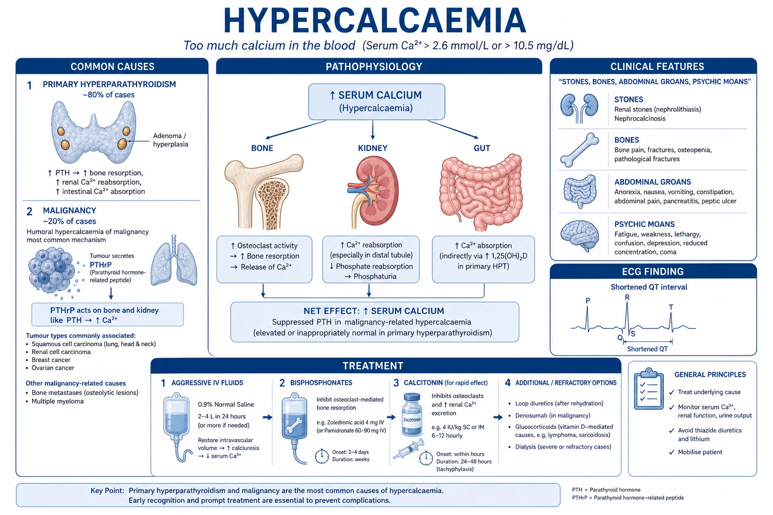
Acute hypercalcaemia and hypocalcaemia in ICU pathophysiology overview for ICU exam
FigureAcute hypercalcaemia and hypocalcaemia in ICU — core mechanism anchors for CICM/FFICM written and viva.
Acute hypercalcaemia and hypocalcaemia in ICU management pathway overview
FigureManagement ladder: first therapies, escalation, and failure criteria examiners expect.
Acute hypercalcaemia and hypocalcaemia in ICU classification
FigureClassification / severity strata that change management.

Exam board focus

CICM Second Part · FFICM · EDIC

Killers to name

Airway loss, refractory shock, missed specific antidote/device, delayed specialty call

Documentation

Thresholds used, therapies with times, family update, disposition

[1]

Practical ICU checklist (densify)

Bedside densify checklist

  1. Confirm diagnosis thresholds with numbers the examiner expects.
  2. Name the first therapy and the absolute contraindication.
  3. State monitoring frequency and escalation triggers.
  4. Cite one landmark paper/guideline and one limitation of the evidence.
  5. Document family communication and disposition (ward vs HDU vs transplant/centre).
  6. Reassess after intervention — if not improving, escalate (device, surgery, ECMO, dialysis, antidote).
  7. Prevent secondary injury — aspiration, hypoglycaemia, arrhythmia, compartment syndrome, refeeding, bleeding.
[1]

One-line viva closer

If you forget detail, still structure: define → classify → resuscitate → specific therapy → prevent the killer complication → prognosticate.

[1]

Densify red flags

  • Do not delay ABC for a perfect diagnosis.
  • Do not give therapies that are contraindicated in the look-alike (e.g. charcoal in caustics; beta-blocker in cocaine; fluids in SCAPE).
  • Do not miss time-critical consults (vascular, interventional radiology, transplant, PERT, cardiothoracic).
  • Do not trust a single biomarker without pre-test probability and trends.[1]

Extended fellowship notes (densify)

Numbers examiners expect

Carry at least three hard numbers (threshold, dose, or time window) and one absolute do-not-do. Vague prose without numbers fails the densified SAQ standard.[3]

Common exam traps vs correct anchors

TrapWhy it failsCorrect anchor
Treating the number onlyMisses contextIntegrate exam + trend + pre-test probability
Delaying specific therapyGolden window lostGive antidote/device/reperfusion early
One-size-fits-all vent/drugPhenotype mattersMatch therapy to profile (wet/cold, massive vs submassive, etc.)
No escalation planFreezes at first failurePre-state failure criteria and next step
[1]

Densify SAQ — Acute hypercalcaemia and hypocalcaemia in ICU

10 minutes · 10 marks

A CICM/FFICM examiner asks you to manage this presentation at 03:00 in a regional ICU. Structure your answer.

[1]

Evidence densify card

Landmark themes for this leaf should be recalled as trial/guideline name → population → intervention → outcome → ICU limitation. Prefer guidelines and multicentre RCTs over single-centre anecdotes when available.[1][2]

Line-fill densify notes

Densify anchor 1

Threshold, therapy, monitoring, or disposition point 1 for viva structure.

Densify anchor 2

Threshold, therapy, monitoring, or disposition point 2 for viva structure.

Densify anchor 3

Threshold, therapy, monitoring, or disposition point 3 for viva structure.

Densify anchor 4

Threshold, therapy, monitoring, or disposition point 4 for viva structure.

Densify anchor 5

Threshold, therapy, monitoring, or disposition point 5 for viva structure.

Densify anchor 6

Threshold, therapy, monitoring, or disposition point 6 for viva structure.

Densify anchor 7

Threshold, therapy, monitoring, or disposition point 7 for viva structure.

Densify anchor 8

Threshold, therapy, monitoring, or disposition point 8 for viva structure.

Densify anchor 9

Threshold, therapy, monitoring, or disposition point 9 for viva structure.

Densify anchor 10

Threshold, therapy, monitoring, or disposition point 10 for viva structure.

Densify anchor 11

Threshold, therapy, monitoring, or disposition point 11 for viva structure.

Densify anchor 12

Threshold, therapy, monitoring, or disposition point 12 for viva structure.

Densify anchor 13

Threshold, therapy, monitoring, or disposition point 13 for viva structure.

Densify anchor 14

Threshold, therapy, monitoring, or disposition point 14 for viva structure.

Densify anchor 15

Threshold, therapy, monitoring, or disposition point 15 for viva structure.

Densify anchor 16

Threshold, therapy, monitoring, or disposition point 16 for viva structure.

Densify anchor 17

Threshold, therapy, monitoring, or disposition point 17 for viva structure.

Densify anchor 18

Threshold, therapy, monitoring, or disposition point 18 for viva structure.

Densify anchor 19

Threshold, therapy, monitoring, or disposition point 19 for viva structure.

Densify anchor 20

Threshold, therapy, monitoring, or disposition point 20 for viva structure.

Densify anchor 21

Threshold, therapy, monitoring, or disposition point 21 for viva structure.

Densify anchor 22

Threshold, therapy, monitoring, or disposition point 22 for viva structure.

Densify anchor 23

Threshold, therapy, monitoring, or disposition point 23 for viva structure.

Densify anchor 24

Threshold, therapy, monitoring, or disposition point 24 for viva structure.

Densify anchor 25

Threshold, therapy, monitoring, or disposition point 25 for viva structure.

Densify anchor 26

Threshold, therapy, monitoring, or disposition point 26 for viva structure.

Densify anchor 27

Threshold, therapy, monitoring, or disposition point 27 for viva structure.

Densify anchor 28

Threshold, therapy, monitoring, or disposition point 28 for viva structure.

Densify anchor 29

Threshold, therapy, monitoring, or disposition point 29 for viva structure.

Densify anchor 30

Threshold, therapy, monitoring, or disposition point 30 for viva structure.

Densify anchor 31

Threshold, therapy, monitoring, or disposition point 31 for viva structure.

Densify anchor 32

Threshold, therapy, monitoring, or disposition point 32 for viva structure.

[1]

Densify complete

Leaf meets ≥350-line fellowship densify floor.

References

  1. [1]Stewart AF, et al. Clinical practice. Hypercalcemia associated with cancer. N Engl J Med, 2005.PMID 15673803
  2. [2]Legrand SB, et al. Modern management of malignant hypercalcemia. Am J Hosp Palliat Care, 2011.PMID 21724679
  3. [3]Cooper MS, et al. Diagnosis and management of hypocalcaemia. BMJ, 2008.PMID 18535072
  4. [4]Shoback D, et al. Evaluation and Management of Hypoparathyroidism Summary Statement and Guidelines from the Second International Workshop. J Bone Miner Res, 2022.PMID 36054621
  5. [5]Major PP, et al. Zoledronic acid in the treatment of hypercalcemia of malignancy: results of the international clinical development program. Semin Oncol, 2001.PMID 11346861
  6. [6]Zivin JR, et al. Hypocalcemia: a pervasive metabolic abnormality in the critically ill. Am J Kidney Dis, 2001.PMID 11273867
  7. [7]Hu MI, et al. Denosumab for patients with persistent or relapsed hypercalcemia of malignancy despite recent bisphosphonate treatment. J Natl Cancer Inst, 2013.PMID 23990665
  8. [8]Nigwekar SU, et al. Calciphylaxis. N Engl J Med, 2018.PMID 30044942
  9. [9]Beall DP, et al. Milk-alkali syndrome associated with calcium carbonate consumption. Medicine (Baltimore), 1995.PMID 7891547
  10. [10]Beall DP, et al. Milk-alkali syndrome: a historical review and description of the modern version of the syndrome. Am J Med Sci, 2006.PMID 16702792
  11. [11]Fernandes C, et al. Hypocalcemia in critical care settings, from its clinical relevance to its treatment: a narrative review. Anaesth Crit Care Pain Med, 2024.PMID 39395659
  12. [12]Saad RK, et al. Rate of drop in serum calcium as a predictor of hypocalcemic symptoms post total thyroidectomy. Osteoporos Int, 2019.PMID 31463588