ICU · Renal and metabolic
Acute severe hyponatraemia: emergency correction, ODS risk, and guidelines
Also known as Hyponatraemia · Acute hyponatraemia · Severe hyponatraemia · Hypotonic hyponatraemia · SIADH · Cerebral salt wasting · Osmotic demyelination syndrome · Central pontine myelinolysis
Hyponatraemia (Na <135 mmol/L) is the commonest electrolyte disorder in hospitalised patients. SEVERE (<120) and ACUTE (<48h) hyponatraemia cause cerebral oedema (seizures, coma, death). CHRONIC (48h) hyponatraemia risks OSMOTIC DEMYELINATION SYNDROME (ODS / central pontine myelinolysis) if corrected too rapidly. CORRECTION RATES (critical): severe symptoms → 3% hypertonic saline bolus (Na rises 4-6 mmol/L), then STOP; max rise ≤8 mmol/L in any 24h (chronic), ≤10-12 mmol/L (acute). NEVER give 100 mL 3% saline as a single rapid bolus without reassessment. Workup: plasma osmolality (hypo = <275), urine osmolality (100 = ADH active), urine Na (SIADH 30, hypovolaemic <20), volume status. The BRAIN adapts to chronic hyponatraemia (extrudes osmolytes) — too-rapid correction → brain shrinks → myelinolysis.
On this page & tools
Your progress
Saved locally on this device.
Target exams
Red flags

Acute vs chronic hyponatraemia — critical distinction
| Feature | ACUTE (<48 hours) | CHRONIC (>48 hours) |
|---|---|---|
| Brain adaptation | NOT yet adapted (still has brain oedema) | ADAPTED (extruded osmolytes — brain water normal) |
| Symptoms | Severe (seizure, coma, cerebral oedema) at higher Na (115-125) | Mild/moderate (often asymptomatic) even at very low Na (<110) |
| Main risk | CEREBRAL OEDEMA (herniation, death) | ODS (if corrected too fast) |
| Correction rate | Can correct faster (≤10-12 mmol/L in 24h) | SLOWER (≤8 mmol/L in 24h) — ODS risk |
| Examples | Psychogenic polydipsia, post-op, MDMA, TURP syndrome | SIADH, heart failure, cirrhosis, thiazides, malnutrition |
| Hypertonic saline | Indicated for severe symptoms | Indicated for severe symptoms (with strict limits) |
Management of severe symptomatic hyponatraemia (European/Australian guideline — bolus approach)
- RECOGNISE SEVERE SYMPTOMS — seizure, coma, severe confusion, vomiting, cardiorespiratory distress. These = cerebral oedema → need IMMEDIATE 3% saline. Don't wait for tests. Estimate duration (acute <48h: post-op, polydipsia, MDMA; chronic >48h: SIADH, heart failure, cirrhosis)
- GIVE 3% HYPERTONIC SALINE BOLUS — 100 mL (or 2 mL/kg) of 3% NaCl IV over 10 MINUTES. This rapidly raises Na by ~1-2 mmol/L and reduces brain oedema. Measure Na after each bolus
- REPEAT BOLUS (up to 3 times) — if severe symptoms persist, repeat 100 mL bolus (up to total 300 mL, or target Na rise 4-6 mmol/L). Once symptoms resolve or Na risen 4-6 mmol/L → STOP boluses. The goal is symptom relief (brain oedema reduction), NOT normalising Na
- SWITCH TO ONGOING MANAGEMENT — once stabilised: (a) identify and treat cause (SIADH fluid restrict; hypovolaemic give normal saline; adrenal insufficiency give steroids). (b) Measure Na every 4-6h. (c) Stay WITHIN correction limits (≤8 mmol/L/24h chronic, ≤10-12 acute)
- MONITOR FOR OVERCORRECTION — if Na rising too fast (>8 in 24h chronic): STOP hypertonic; give DDAVP (1-2 mcg IV) + free water/glucose to RE-LLOWER Na back to target range. Overcorrection is the #1 preventable cause of ODS
- ADDRESS UNDERLYING CAUSE + K+ — hypokalaemia INDEPENDENTLY raises ODS risk and contributes to Na (correcting K shifts Na up) — replace K. Treat SIADH (fluid restrict 800 mL/day, salt tablets, urea, vaptans — rarely in ICU). Stop offending drugs (SSRIs, thiazides, carbamazepine)
SAQ — Post-operative hyponatraemia with seizure
10 minutes · 10 marks
A 26-year-old woman is reviewed on the surgical ward 36 hours after an uneventful laparoscopic appendicectomy. She is on a morphine PCA and has received 3 L of 5% dextrose as maintenance fluid. The nursing staff have called because she has just had a witnessed 3-minute generalised tonic-clonic seizure; she is post-ictal, GCS 12. Serum sodium is 112 mmol/L.
Clinical pearls
Red flags
Prognosis
Hyponatraemia evidence and outcomes
Epidemiology: hyponatraemia in 15-30% of hospitalised patients; severe (<120) in ~1%. Hypertonic saline bolus (European 2014, Australian guidelines): 100 mL 3% over 10 min — predictable ~1-2 mmol/L rise, rapid symptom relief, less overcorrection than infusion. Correction limits: ≤8 mmol/L in 24h (chronic), ≤10-12 (acute) — Sterns, Spasovski. Exceeding → ODS. ODS: mortality 40-50% (Sterns 2009). Risk factors: Na ≤105, hypokalaemia, alcoholism, malnutrition, liver disease. Saline Trial (Hoorn 2020, Lancet): NaCl vs NaHCO3 for hyponatraemia — equivalent (bicarbonate no advantage). Vaptans (SALT-I/II): modest Na rise, no outcome benefit; overcorrection risk (limit ICU use). Adrenal insufficiency: must exclude in every unexplained hyponatraemia — hydrocortisone corrects.
Pathophysiology: brain adaptation and the ODS mechanism
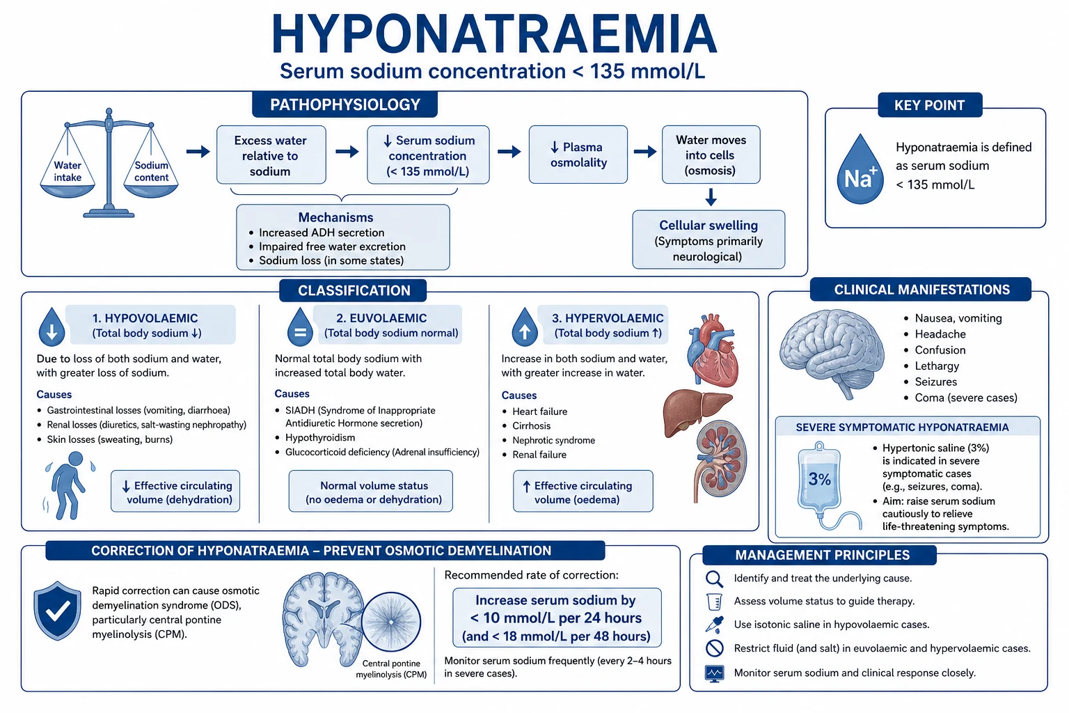
The clinical behaviour of hyponatraemia is dictated almost entirely by brain water handling, and mastery of this single concept explains the symptoms, the correction limits, and ODS.[1] }
- Acute onset (<48 h) — plasma becomes hypotonic → osmotic gradient favours water moving into brain astrocytes (the blood–brain barrier water channel aquaporin-4 sits on astrocytic end-feet). Astrocytes swell → compression of the rigid calvarium → raised intracranial pressure → headache, nausea, vomiting, somnolence, seizures, coma, and ultimately tentorial/tonsillar herniation. Because the brain has not yet adapted, symptoms occur at relatively modest sodium (115–125 mmol/L) and the priority is rapid partial correction to relieve cerebral oedema.
- Chronic onset (>48 h) — over 2–3 days astrocytes actively extrude osmolytes (first potassium and chloride, then organic osmolytes: glutamate, taurine, myo-inositol, glutamine) so that intracellular osmolality falls to match the hypotonic plasma. Brain water normalises and the patient is frequently asymptomatic even at Na 108–112. The now-adapted brain is, however, exquisitely vulnerable to a rising plasma sodium: if Na rises faster than osmolytes can be re-accumulated, water is pulled out of the brain → astrocytic shrinkage → disruption of the blood–brain barrier → oligodendrocyte injury → non-inflammatory demyelination (ODS).[6] }
- Osmotic demyelination syndrome (ODS) — classically in the basis pontis (central pontine myelinolysis) but in ~50 % also extrapontine (basal ganglia, thalamus, cerebellar peduncles, lateral geniculate). Onset is delayed 2–6 days after an over-rapid correction with a characteristic biphasic course: initial improvement followed by dysarthria, dysphagia, quadriparesis, pseudobulbar palsy, "locked-in" syndrome, seizures, coma. Mortality 20–40 %; many survivors have permanent disability. MRI (T2/FLAIR hyperintensity, often with restricted diffusion on DWI) lags clinically by 1–2 weeks, so a normal early MRI does not exclude ODS.[6] }
High-risk ODS phenotypes (apply the slowest correction target, ≤6 mmol/L/24 h, and consider prophylactic DDAVP): sodium ≤105 mmol/L, hypokalaemia, alcoholism, malnutrition/cachexia, advanced liver disease, burns, thiazide-induced, post-operative female, and hypoxia/anoxia preceding the hyponatraemia.[6] }
Acute symptomatic hyponatraemia — what counts as 'severe symptoms' (the trigger for 3% saline)
| Symptom tier | Examples | Action |
|---|---|---|
| Severe (cerebral oedema) | Seizure, coma, obtundation, severe confusion/agitation, vomiting, Cheyne-Stokes/respiratory arrest, decerebrate posturing | 100 mL 3% NaCl over 10 min — repeat up to 3×; do NOT wait for the laboratory |
| Moderate | Headache, nausea, lethargy, disorientation, muscle cramps, falls | Investigate and treat cause; hypertonic saline not automatically indicated — assess duration and risk |
| Mild / incidental | Asymptomatic, found on routine panel | Treat cause; fluid restriction if SIADH; no hypertonic saline |
The severity of symptoms, not the absolute sodium, dictates whether hypertonic saline is given. A patient with chronic Na 110 who is alert and oriented does not need 3% saline, whereas a postoperative patient at Na 122 having a seizure does.[5] }
The 3% saline bolus approach — practical detail

Administering 3% hypertonic saline for severe symptomatic hyponatraemia (bolus algorithm)
- Confirm severe symptoms (seizure/coma/severe obtundation). Do not wait for plasma/urine results — clinical diagnosis. Draw blood for Na, K, osmolality, urine Na, urine osm, glucose, cortisol, TSH on the way.
- Give 100 mL of 3% NaCl IV over 10 minutes (alternatively 2 mL/kg lean body weight). Use a pumped infusion or push; 3% is irritant so prefer a central line if repeated boluses or a peripheral line with a small vein. Each 100 mL bolus raises plasma Na by a predictable ~1–2 mmol/L (Adrogué-Madias ≈ (infusate Na + K − plasma Na)/(TBW + 1); for 100 mL 3% in a 70 kg man ≈ +1.7 mmol/L).[7] }
- Reassess symptoms and recheck Na at 10 min, after each bolus. The therapeutic end-point is resolution of severe symptoms or a rise of 4–6 mmol/L, whichever comes first — not a "normal" sodium.
- Repeat the bolus up to a total of 3 (300 mL) if severe symptoms persist and the Na has risen <4–6 mmol/L. Stop earlier if the patient improves.
- Lock in the gain. Once symptoms resolve, stop boluses, set a written 24-hour correction ceiling (≤8 mmol/L chronic, ≤10–12 acute), and recheck Na every 4–6 h for the first 24 h, then 6–12 h.[2] }
- Treat the cause in parallel — fluid restriction for SIADH, isotonic saline for hypovolaemia, hydrocortisone for adrenal insufficiency, stop thiazides/SSRIs, address the underlying malignancy/infection.
Why bolus over continuous infusion. A continuous 3% infusion (e.g. 0.5–1 mL/kg/h) is unpredictable because the rise depends on ADH tone, renal concentrating ability and total body water; the same infusion can correct slowly in one patient and over-correct dangerously in another (especially when ADH is suppressed after volume resuscitation). Discrete, reassessed boluses give a quantal, reproducible Na rise, lower the risk of over-correction, and free the team from a continuously running pump.[5] }
Predicting the serum Na change — the Adrogué–Madias formula
| Formula / value | |
|---|---|
| Change in Na per 1 L infusate | ΔNa = (Na_infusate + K_infusate − Na_serum) / (TBW + 1) |
| TBW (total body water) | 0.6 × weight (kg) men; 0.5 women; 0.45 elderly men; 0.4 elderly women |
| 3% saline (Na 513 mmol/L) | 100 mL in a 70 kg man → ΔNa ≈ (513 − 110)/(42 + 1) × 0.1 ≈ +1 mmol/L (1 L → ~+10) |
| 0.9% saline (Na 154 mmol/L) | 1 L in same patient → (154 − 110)/43 ≈ +1 mmol/L |
| 0.45% saline (Na 77) | can lower Na if ADH active |
Caveat (Berl). The Adrogué–Madias formula predicts the initial change, but ongoing renal water excretion (which depends on ADH) dominates the final Na. After volume repletion ADH may switch off and a patient who retains free water can correct far faster than the formula predicts — hence the formula is a ceiling estimate, never a substitute for measured Na every 4–6 h.[8] }
Over-correction and the DDAVP rescue
Over-correction is the single most preventable cause of ODS and usually arises from (a) ongoing renal free-water excretion after ADH suppression (e.g. after volume resuscitation or stopping the offending drug), (b) co-administered hypotonic losses, or (c) concurrent potassium repletion that shifts Na upward.[10] }
Rescue protocol when the 24-hour correction ceiling is breached
- Recognise early — if Na has risen >8 mmol/L in 24 h (chronic) or >10–12 mmol/L (acute), or is on trajectory to do so, act immediately. Measure Na every 2–4 h during active correction.
- Stop all sodium-containing and hypertonic fluids. Replace maintenance with 5% dextrose or oral water.
- Give DDAVP (desmopressin) 1–2 mcg IV every 6–8 h to abolish free-water excretion (the dominant driver of late over-correction). This "re-locks" the renal concentrating mechanism so the administered water lowers Na back to target.[9] }
- Re-lower Na to within 8 mmol/L of the pre-treatment value (i.e. undo the excess). Aim for a gradual fall over the next 12–24 h; do not overshoot downward.
- Continue DDAVP until Na is stable in the target band, then taper. Recheck Na every 4 h while active manipulation is under way.
- Review the cause of over-shoot — typically ADH switched off after volume repletion (give DDAVP prophylactically if you anticipate this in a high-risk patient), or potassium repletion.
Prophylactic DDAVP (1–2 mcg IV q8h) plus 3% saline as boluses is an emerging strategy in high-risk, very-low-Na patients: it decouples correction from unpredictable renal water handling and gives the team control of both directions.[9] }
Differentiating the hypotonic hyponatraemias by volume status
Once hypotonicity is confirmed (plasma osm <275 mOsm/kg), the urine osmolality, urine sodium and clinical volume status partition patients into three buckets — each with a different first-line therapy. Giving the wrong fluid (e.g. fluid-restricting a hypovolaemic patient, or saline-loading a dilutional SIADH) worsens the sodium.[2] }
Volume-status classification of hypotonic hyponatraemia
| Bucket | Clinical signs | Urine osm | Urine Na | Typical causes | First-line treatment |
|---|---|---|---|---|---|
| Hypovolaemic (true Na + water loss) | Dry mucosae, low JVP, tachycardia, orthostasis, ↓skin turgor, raised urea:creatinine | >100 (ADH high from hypovolaemia) | <20 = extrarenal (GI, sweat, burns); >20 = renal (diuretics, mineralocorticoid deficit, osmotic diuresis) | Vomiting, diarrhoea, burns, pancreatitis, thiazide, diuretic, cerebral salt wasting, mineralocorticoid deficiency | 0.9% saline / crystalloid to restore volume → switches off ADH → Na rises (watch for over-correction) |
| Euvolaemic — SIADH (water retained, Na normal) | Clinically euvolaemic — no oedema, no dehydration | >100 (inappropriately concentrated) | >30 (often >40) | SIADH (SCLC, CNS disease, pneumonia, SSRIs, carbamazepine, MDMA, cyclophosphamide), glucocorticoid deficiency, hypothyroidism, pain/nausea/post-op, water intoxication | Fluid restriction 800 mL/day + high salt/protein; urea 30 g/day; tolvaptan (cautious); 3% saline if severe symptoms |
| Hypervolaemic (oedema — effective arterial volume low) | Peripheral/pulmonary oedema, raised JVP, ascites | Variable (often >100) | Usually <20 (unless renal failure) | Heart failure, cirrhosis, nephrotic syndrome, advanced CKD | Water + sodium restriction, treat the underlying state; loop diuretic; tolvaptan in select HF; 3% saline only if severe symptoms (then carefully) |
Pitfall: SIADH criteria require exclusion of hypothyroidism, glucocorticoid deficiency and recent diuretic use before the label is applied. A random cortisol and TSH is mandatory in every unexplained hyponatraemia; thiazides reproduce the SIADH biochemical pattern and are the commonest "SIADH mimic" in elderly women.[3] }
Diagnostic workup of confirmed hypotonic hyponatraemia (osm <275)
- Plasma osmolality → confirm hypotonic (<275). If >295 think hypertonic causes (hyperglycaemia — correct Na +2 per 4 mmol/L glucose above 5.5; mannitol; glycine/sorbitol irrigation). If 275–295 consider pseudohyponatraemia (hyperlipidaemia/hyperproteinaemia on older assays).
- Urine osmolality → <100 implies appropriately suppressed ADH (primary polydipsia, low solute "tea-and-toast" or beer potomania); >100 implies ADH is active (most pathologic hyponatraemias).
- Urine sodium → >30 suggests renal sodium loss (SIADH, CSW, diuretics, mineralocorticoid deficiency); <20 suggests extrarenal loss or hypervolaemic states with low effective arterial volume.
- Clinical volume status (mucosae, JVP, perfusion, oedema, postural vitals, weight trend, fluid balance) → allocate to hypo-/eu-/hypervolaemic bucket.
- Endocrine exclude — cortisol (± short Synacthen), TSH, and consider ACTH. Glucocorticoid deficiency mimics SIADH exactly.
- Drug & toxin screen — thiazides, SSRIs, carbamazepine, oxcarbazepine, MDMA, cyclophosphamide, vincristine, desmopressin, NSAIDs, MDMA; ask about beer/low-solute diet (potomania) and excessive water intake (psychogenic polydipsia, endurance sport).
- Imaging / context — chest X-ray/CT for SCLC and pneumonia, CT brain for CNS cause, and review the operative/irrigation history (TURP, hysteroscopy, endoscopic uterine surgery).
Cerebral salt wasting vs SIADH — the neuro-ICU distinction
Both CSW and SIADH produce hypotonic hyponatraemia with inappropriately concentrated urine (osm >100) and high urine sodium (>30) — biochemically near-identical. The distinction rests on volume status, and getting it wrong (fluid-restricting a volume-depleted CSW patient) can precipitate cerebral vasospasm and infarction, particularly after subarachnoid haemorrhage.[14] }
Cerebral salt wasting (CSW) vs SIADH — definitive distinction
| Feature | CSW | SIADH |
|---|---|---|
| Volume status | Hypovolaemic (true renal Na loss) | Euvolaemic |
| Setting | Brain injury — SAH, TBI, neurosurgery, meningitis, encephalitis, post-pituitary surgery | SCLC, CNS disease, pneumonia, drugs, idiopathic |
| Onset | Days 2–10 after brain insult; may be polyuric phase | Variable; often insidious |
| JVP / CVP | Low | Normal |
| Urea / creatinine | Raised (haemoconcentration) | Normal / low |
| Haematocrit / albumin | Raised | Normal |
| Serum urate | Low | Low |
| Fractional excretion of urate (FE_UA) | High — stays high after correction | High — normalises after correction |
| Urine output | Often high (polyuria) | Variable; often low |
| Response to isotonic saline | Improves (volume restored, Na rises) | Worsens (NaCl is excreted, retained water dilutes further; urine Na rises further) |
| Response to fluid restriction | Worsens (exacerbates hypovolaemia → cerebral ischaemia) | Improves |
| Treatment | Volume + salt replacement (normal or hypertonic saline to match urine losses); fludrocortisone 0.1–0.2 mg bd if persistent | Fluid restriction; salt + protein; urea; tolvaptan |
Practical rule. In a neurosurgical/SAH patient with hyponatraemia, assume CSW until proven otherwise, keep the patient euvolaemic-to-slightly hypervolaemic (avoid fluid restriction — hypovolaemia drives vasospasm and delayed cerebral ischaemia, the dominant killer after SAH), and treat with saline ± fludrocortisone. Reserve fluid restriction for biochemically confirmed, clearly euvolaemic SIADH.[14] }
Common pitfalls in ICU hyponatraemia
3% saline — common errors and how to avoid them
| Error | Why it happens | Fix |
|---|---|---|
| Waiting for labs before treating a seizing patient | Reflex caution | Severe symptoms = treat on clinical grounds; draw labs on the way |
| Continuous infusion instead of boluses | Familiarity with infusion pumps | Use discrete 100 mL boluses reassessed at 10 min |
| Treating to a "normal" sodium | Misunderstanding the end-point | End-point = symptom relief / rise of 4–6; the rest is gradual |
| No written correction ceiling | Assumed "everyone knows" | Write the 24 h ceiling on the chart and at handover |
| Infrequent Na checks | "Stable" assumption | q4–6 h while correcting; q2 h if over-correcting |
| Forgetting K⁺ | Focus on Na | Replace K first; it independently raises Na and lowers ODS risk |
| No DDAVP rescue plan | Over-correction seen as unavoidable | Have DDAVP drawn up; give 1–2 mcg IV at first sign of overshoot |
| Restricting fluids in CSW | Biochemistry looks like SIADH | Volume status decides; CSW needs salt + water |
Special situations
Special-situation hyponatraemias — cause, danger, key action
| Situation | Mechanism / danger | Key action |
|---|---|---|
| Post-operative (young women highest risk) | Pain/nausea ADH + hypotonic fluids → acute cerebral oedema | Avoid hypotonic maintenance fluids post-op (use balanced isotonic); 3% saline for symptoms |
| Cirrhosis with ascites | High ADH, total-body-Na-overload, malnutrition → very high ODS risk | Water + Na restrict; tolvaptan cautiously; ceiling ≤6 mmol/L/24 h |
| Heart failure | Low effective arterial volume → ADH; often hypervolaemic | Water restrict; treat HF; tolvaptan select cases; not 3% saline unless severe symptoms |
| Subarachnoid haemorrhage | CSW vs SIADH; hypovolaemia → vasospasm | Assume CSW; keep euvolaemic-hypervolaemic; saline ± fludrocortisone |
| Pregnancy / pre-eclampsia | Reset osmostat, SIADH-like; oxytocin has ADH activity | Obstetric-medicine jointly; correct gently |
| End-stage renal disease | Cannot excrete free water; Na swings with dialysate | Dialyse against a tailored dialysate Na; sequential therapy; slow correction |
Key trials and prognosis
Landmark evidence in severe hyponatraemia
European clinical practice guideline (Spasovski 2014) — established the bolus approach (100 mL 3% over 10 min, repeat up to 3×) and the ≤8–10–12 mmol/L/24 h correction ceilings now adopted worldwide.[2] } American expert panel (Verbalis 2013) — symptom-based, duration-based classification; confirmed hypertonic saline for severe symptoms and fluid restriction for chronic SIADH; defined symptomatic thresholds.[3] } Adrogué–Madias formula (NEJM 2000) and Berl's reappraisal (CJASN 2007) — bedside prediction of ΔNa from any infusate; Berl emphasised ongoing renal water handling dominates the final value, mandating frequent rechecks.[8] } Mohmand (CJASN 2007) — quantified the rate of inadvertent over-correction with hypertonic saline (≈30% of patients), underpinning frequent monitoring and DDAVP rescue.[10] } Perianayagam (CJASN 2008) — DDAVP effectively prevents and reverses inadvertent over-correction, now standard rescue therapy.[9] } SALT-1 and SALT-2 (Schrier NEJM 2006) — tolvaptan raised Na modestly in euvolaemic/hypervolaemic hyponatraemia but no outcome benefit and over-correction risk; basis for narrow ICU use.[4] } Ayus/Arieff/Moritz (NEJM 2005) and Hew-Butler 3rd EAH Consensus (2015) — exercise-associated hyponatraemia is acute cerebral oedema; 100 mL 3% saline boluses are life-saving on the field.[13] } Moritz & Ayus, Front Med 2019 — dismantled barriers to hypertonic saline use; case for bolus over infusion in ICU.[5] } Outcomes: hospital mortality with hyponatraemia roughly doubles vs normonatraemic matched controls; severe (<120) carries 20–60 % mortality depending on setting, largely driven by the underlying illness. ODS mortality 20–40 %; survivors often have residual disability.
ICU densification — examiner checklist
[1]Practical Adrogué–Madias bedside use
The expected change in serum sodium from 1 L of infusate is approximately: [1]
ΔNa ≈ (infusate Na + infusate K − serum Na) / (TBW + 1) [1]
where TBW ≈ 0.6 × weight (men) or 0.5 × weight (women). The formula is a planning tool, not a guarantee — ongoing urinary free-water losses (especially once ADH falls) commonly drive faster rises than predicted. That is why the charted 24-hour ceiling and 2–4-hourly sodium checks matter more than the first-pass arithmetic.[7][8]
Charted correction plan (exam-ready)
- Document starting Na, estimated duration (acute vs chronic), and ODS risk factors.
- Write the maximum permitted 24-hour rise on the drug chart.
- Order 3% saline as boluses with reassessment, not an open-ended infusion, for severe symptoms.
- Replace potassium concurrently if low.
- If the trajectory overshoots, escalate immediately to DDAVP + free water rather than waiting for symptoms of ODS.
- Reassess the diagnosis if Na fails to move — wrong volume status (CSW vs SIADH), ongoing hypotonic intake, or adrenal crisis. [1]
High-yield viva traps
- Treating the number without symptoms using aggressive 3% saline in asymptomatic chronic hyponatraemia.
- Fluid-restricting a volume-depleted neurosurgical patient (CSW).
- Ignoring concurrent hypokalaemia while congratulating yourself on a “perfect” Na rise.
- Labelling SIADH without cortisol and thyroid exclusion.
- Using tolvaptan as first-line ICU rescue for seizures (too slow, unpredictable overshoot). [1]
References
- [1]Sterns RH Disorders of plasma sodium. New England Journal of Medicine, 2015.PMID 25806924
- [2]Spasovski G, Vanholder R, Allolio B, et al. Clinical practice guideline on diagnosis and treatment of hyponatraemia. Nephrology Dialysis Transplantation, 2014.PMID 24569496
- [3]Verbalis JG, Goldsmith SR, Greenberg A, et al. Diagnosis, evaluation, and treatment of hyponatremia: expert panel recommendations. American Journal of Medicine, 2013.PMID 24074529
- [4]Schrier RW, Gross P, Gheorghiade M, et al. Tolvaptan, a selective oral vasopressin V2-receptor antagonist, for hyponatremia. New England Journal of Medicine, 2006.PMID 17105757
- [5]Moritz ML, Ayus JC Misconceptions and Barriers to the Use of Hypertonic Saline to Treat Hyponatremic Encephalopathy. Frontiers in Medicine, 2019.PMID 30931308
- [6]Sterns RH Adverse consequences of overly-rapid correction of hyponatremia. Frontiers of Hormone Research, 2019.PMID 32097948
- [7]Adrogué HJ, Madias NE Hyponatremia. New England Journal of Medicine, 2000.PMID 10824078
- [8]Berl T The Adrogue-Madias formula revisited. Clinical Journal of the American Society of Nephrology, 2007.PMID 17928464
- [9]Perianayagam A, Sterns RH, Silver SM, et al. DDAVP is effective in preventing and reversing inadvertent overcorrection of hyponatremia. Clinical Journal of the American Society of Nephrology, 2008.PMID 18235152
- [10]Mohmand HK, Issa D, Ahmad Z, et al. Hypertonic saline for hyponatremia: risk of inadvertent overcorrection. Clinical Journal of the American Society of Nephrology, 2007.PMID 17913972
- [11]Ayus JC, Arieff AI, Moritz ML Hyponatremia in marathon runners. New England Journal of Medicine, 2005.PMID 16050061
- [12]Moritz ML, Ayus JC The syndrome of inappropriate antidiuresis. New England Journal of Medicine, 2007.PMID 17806140
- [13]Hew-Butler T, Rosner MH, Fowkes-Godek S, et al. Statement of the 3rd International Exercise-Associated Hyponatremia Consensus Development Conference. Clinical Journal of Sport Medicine, 2015.PMID 26227507
- [14]Cole CD, Gottfried ON, Liu JK, Couldwell WT Hyponatremia in the neurosurgical patient: diagnosis and management. Neurosurgical Focus, 2004.PMID 15191338