MedVellum
MCQsExamsAtlas
DashboardPricing
MBBS / Core medicine✳Dermatology✳ICU Fellowship (CICM)✳Anaesthesia✳Emergency Medicine✳Psychiatry Fellowship✳Paediatrics Fellowship✳Physician Medicine✳MCQs✳SAQs✳Vivas✳OSCE✳Evidence-first✳MBBS / Core medicine✳Dermatology✳ICU Fellowship (CICM)✳Anaesthesia✳Emergency Medicine✳Psychiatry Fellowship✳Paediatrics Fellowship✳Physician Medicine✳MCQs✳SAQs✳Vivas✳OSCE✳Evidence-first✳

MedVellum.

The folio

Exam-exhaustive medical education across every specialty — evidence-graded topics, engraved plates, and practice in every written and oral format. Educational content only — not medical advice.

llms.txt · psychiatry LLM catalog · sitemap

Atlas

  • Specialty atlas
  • MBBS / Core medicine
  • Dermatology
  • ICU Fellowship (CICM)
  • Anaesthesia
  • Emergency Medicine
  • Psychiatry Fellowship
  • Paediatrics Fellowship
  • Physician Medicine

Study & account

  • MCQ practice
  • Practice alias
  • Exam tools
  • Dashboard
  • Pricing
  • Sign in

© 2026 MedVellum. For education only — not a substitute for clinical judgement.

Folio edition · Set in Instrument Serif & Archivo

ICU TopicsRenal and metabolic

ICU · Renal and metabolic

Acute severe metabolic acidosis: approach, anion gap, and lactate

Also known as Metabolic acidosis · High anion gap metabolic acidosis · HAGMA · Lactic acidosis · Normal anion gap acidosis · NAGMA · Delta gap · Cohen-Woods lactic acidosis · Toxic alcohol poisoning · Diabetic ketoacidosis

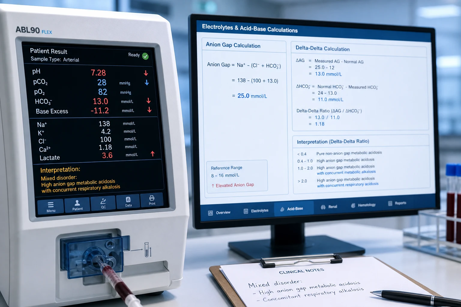
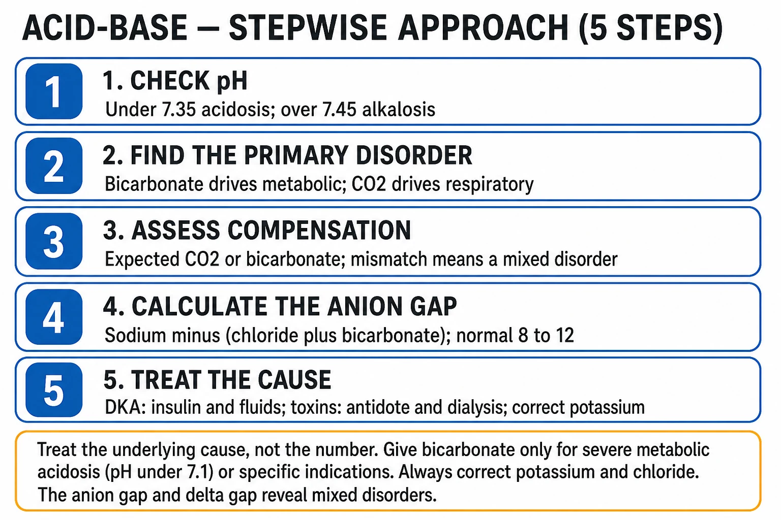
Metabolic acidosis (pH <7.35, HCO3 <22, low/normal PaCO2 compensatory) is ubiquitous in ICU. SYSTEMATIC approach: (1) Is it metabolic acidosis? (2) Calculate ANION GAP: Na − (Cl + HCO3); normal 8-12. (3) HIGH anion gap (HAGMA — 12): lactate (sepsis, shock, metformin), ketones (DKA, alcoholic, starvation), toxins (methanol, ethylene glycol, salicylates, paraldehyde), renal failure (uraemic acids). Mnemonic: MUDPILES / GOLDMARK. (4) NORMAL anion gap (NAGMA): GI bicarbonate loss (diarrhoea), renal tubular acidosis, acetazolamide, ureteroenteric fistula, hyperalimentation. (5) DELTA GAP / delta-delta: assess for concurrent disorders (e.g., metabolic alkalosis from vomiting + HAGMA). (6) Treat CAUSE — bicarbonate generally NOT indicated (unless pH <7.1 with haemodynamic instability, or specific causes like TCA overdose, hyperkalaemia).

high19 referencesUpdated 4 July 2026
On this page & tools

Your progress

Saved locally on this device.

Target exams

CICMFFICMEDIC

Red flags

pH &lt;7.1 → cardiac dysfunction, vasopressor resistance — consider bicarbonateHIGH anion gap (>12) → MUDPILES/GOLDMARK: lactate, ketones, toxins, renal failureLactate >4 + not clearing → sepsis/shock — mortality risesBicarbonate generally NOT indicated except pH &lt;7.1 + unstable, TCA overdose, hyperkalaemiaPropylene glycol toxicity from lorazepam/diazepam/etomidate infusions → HAGMA + high osmolar gapSalicylate toxicity — avoid intubation if possible; hypoventilation worsens CNS toxicityGive thiamine BEFORE dextrose in suspected alcoholic ketoacidosis (Wernicke prevention)

Your progress

Saved locally on this device.

Target exams

CICMFFICMEDIC

Red flags

pH &lt;7.1 → cardiac dysfunction, vasopressor resistance — consider bicarbonateHIGH anion gap (>12) → MUDPILES/GOLDMARK: lactate, ketones, toxins, renal failureLactate >4 + not clearing → sepsis/shock — mortality risesBicarbonate generally NOT indicated except pH &lt;7.1 + unstable, TCA overdose, hyperkalaemiaPropylene glycol toxicity from lorazepam/diazepam/etomidate infusions → HAGMA + high osmolar gapSalicylate toxicity — avoid intubation if possible; hypoventilation worsens CNS toxicityGive thiamine BEFORE dextrose in suspected alcoholic ketoacidosis (Wernicke prevention)
Cinematic ICU scene of a lactate level and an anion-gap calculation on a monitor beside an ECG showing hyperkalaemic changes and a fomepizole vial, clinical-blue lighting, medical educational, no faces, no text
FigureMetabolic acidosis is everywhere in the ICU — sort it by the anion gap. The high gap (the HAGMA) is the lactate, the ketones, the toxins, the renal; the normal gap is the GI or the renal tubular. Treat the cause, not the bicarbonate (reserved for the pH below 7.1); in the toxic alcohol the fomepizole and the dialysis are the antidote.

In one line

Metabolic acidosis approach: (1) Confirm on ABG (pH <7.35, HCO3 <22, low PaCO2). (2) ANION GAP = Na − (Cl + HCO3) (normal 8-12). (3) HIGH AG (HAGMA) >12 → GOLDMARK: Glycols (ethylene/propylene), Oxoproline, L-lactate, D-lactate, Methanol, Aspirin, Renal failure, Ketoacidosis. (4) NORMAL AG (NAGMA): diarrhoea (GI HCO3 loss), renal tubular acidosis, acetazolamide. (5) DELTA GAP (ΔAG/ΔHCO3): 1-2 = pure HAGMA; <1 = HAGMA + NAGMA; >2 = HAGMA + metabolic alkalosis. (6) Treat cause; bicarbonate reserved for pH <7.1 + unstable (BICAR-ICU), TCA overdose, hyperkalaemia.

[1]
acute severe metabolic acidosis clinical overview for ICU fellowship exams
FigureExam overview — key physiology, red flags and first-hour management.
Pathophysiology of acute severe metabolic acidosis
FigureCore mechanism linking insult to organ failure — CICM/FFICM viva scaffold.
Management algorithm for acute severe metabolic acidosis
FigureStepwise ICU management: immediate priorities, disease-specific therapy, escalation.

High vs normal anion gap metabolic acidosis

FeatureHigh AG (HAGMA)Normal AG (NAGMA)
Anion gap>12 mmol/L8-12 mmol/L
MechanismUnmeasured acid added (lactate, ketones, toxins) — anions not measured, so gap widensLoss of bicarbonate (GI/renal) — replaced by Cl (measured) — gap unchanged
ChlorideUsually normal (unmeasured anions fill the gap)HIGH (hyperchloraemic — Cl replaces lost HCO3)
Common causesGOLDMARK/MUDPILES: lactate, ketones, toxins, renal failureDiarrhoea, RTA, acetazolamide, ureteroenteric
Delta gapΔAG/ΔHCO3 = 1-2 (pure HAGMA)Not applicable (AG normal)
Urine anion gapNot helpful (AG already high)Negative (GI loss) or positive (RTA) — localises NAGMA
[1]

Systematic approach to metabolic acidosis

  1. CONFIRM METABOLIC ACIDOSIS — ABG: pH <7.35, HCO3 <22 (primary). Check COMPENSATION: PaCO2 should fall (Winter's formula: expected PaCO2 = 1.5 × HCO3 + 8 ± 2). If PaCO2 higher than expected → concurrent respiratory acidosis; if lower → respiratory alkalosis. Assess A-a gradient if hypoxic
  2. CALCULATE ANION GAP — AG = Na − (Cl + HCO3). Normal 8-12 mmol/L. (Correct for albumin: AG decreases by 2.5 for each 10 g/L albumin below 40 — hypoalbuminaemia narrows the gap; a 'normal' AG in hypoalbuminaemia may actually be elevated.) HIGH (>12) → HAGMA pathway. NORMAL (8-12) → NAGMA pathway
  3. IF HIGH AG — IDENTIFY CAUSE (GOLDMARK/MUDPILES) — (a) LACTATE: sepsis, shock, metformin, malignancy, thiamine deficiency. (b) KETONES (β-hydroxybutyrate): DKA, alcoholic ketoacidosis, starvation. (c) RENAL FAILURE: urea, organic acids. (d) TOXINS: methanol (toxic alcohol — osmolar gap), ethylene glycol (osmolar gap + calcium oxalate crystals in urine), salicylates (respiratory alkalosis + metabolic acidosis), paraldehyde. Measure: lactate, ketones, creatinine, salicylate level, osmolar gap
  4. IF NORMAL AG — GI vs RENAL (urine anion gap) — Urine AG = (Na + K) − Cl. NEGATIVE (<0) = GI loss (diarrhoea — kidney excreting NH4Cl to compensate). POSITIVE (>0) = renal cause (RTA — kidney can't excrete acid). Distinguish RTA types: type 1 (distal — urine pH >5.5, K low), type 2 (proximal — urine pH <5.5, K low, Fanconi), type 4 (hypoaldosteronism — K HIGH)
  5. DELTA GAP (assess concurrent disorders) — ΔAG = AG − 12 (normal). ΔHCO3 = 24 − HCO3. Ratio ΔAG/ΔHCO3: (a) 1-2 = pure HAGMA. (b) <1 = HAGMA + concurrent NAGMA (e.g., sepsis + diarrhoea). (c) >2 = HAGMA + concurrent metabolic alkalosis (e.g., sepsis + vomiting). Also check 'corrected bicarbonate' = HCO3 + ΔAG; if >24 → concurrent metabolic alkalosis
  6. TREAT CAUSE + CONSIDER BICARBONATE — (a) SEPSIS: antibiotics, fluids, source control. (b) DKA: insulin, fluids, K. (c) TOXINS: fomepizole/haemodialysis (methanol/ethylene glycol), urinary alkalinisation (salicylates). (d) RENAL FAILURE: dialysis. (e) BICARBONATE: generally NO — EXCEPT pH <7.1 + haemodynamically unstable (BICAR-ICU: trended to benefit in AKI grade 3/4), TCA overdose (NaHCO3 for sodium load + alkalinisation), hyperkalaemia. Goal: pH >7.1-7.15, NOT normalise
[1]

Clinical pearls

High-yield metabolic acidosis points for CICM/FFICM exam

  1. Anion gap — the foundation. AG = Na − (Cl + HCO3). Normal 8-12. RATIONALE: plasma must be electrically neutral. Na (main cation) = Cl + HCO3 (main anions) + 'unmeasured anions' (proteins, phosphate, sulphate, organic anions). The 'gap' = unmeasured anions. When an ACID is added (lactic acid → lactate + H+; H+ buffered by HCO3 → HCO3 drops; lactate accumulates as unmeasured anion → gap WIDENS). When HCO3 is LOST (diarrhoea), Cl rises to maintain neutrality → gap UNCHANGED (NAGMA). ALWAYS correct for albumin (hypoalbuminaemia falsely lowers AG by ~2.5 per 10 g/L below 40).[5] }
  2. GOLDMARK (modern mnemonic) replaces MUDPILES. GOLDMARK: G — Glycols (ethylene glycol, propylene glycol). O — Oxoproline (5-oxoproline — chronic paracetamol/glutathione depletion). L — L-lactate (sepsis, shock). D — D-lactate (short bowel, bacterial overgrowth). M — Methanol. A — Aspirin (salicylates). R — Renal failure (uraemia). K — Ketoacidosis (DKA, alcoholic, starvation). MUDPILES older: Methanol, Uraemia, DKA, Paraldehyde/phenformin, Iron/INH, Lactic acidosis, Ethylene glycol, Salicylates. Either works — know the modern drugs (metformin replacing phenformin; propylene glycol; 5-oxoproline from chronic paracetamol).[1] }
  3. Lactic acidosis — types A and B. TYPE A (tissue hypoperfusion — most common): sepsis, shock (cardiogenic, hypovolaemic, haemorrhagic), severe hypoxia, mesenteric ischaemia, seizures (transient). TYPE B (no overt hypoperfusion): (a) B1 — drugs/toxins: metformin (biguanide — inhibits complex I), cyanide, cocaine, ethanol, propofol infusion syndrome, NRTIs, linezolid. (b) B2 — systemic: malignancy (leukaemia, lymphoma — Warburg effect), liver failure (impaired lactate clearance), diabetes, thiamine deficiency. (c) B3 — inborn errors of metabolism (mitochondrial, GSD). D-LACTATE: gut bacteria produce D-lactate (short bowel, bacterial overgrowth) — not detected by standard lactate assay (measures L-lactate) — measure D-lactate specifically.[2] …
  4. Lactate clearance predicts mortality — use it. (1) Admission lactate >4 mmol/L = severe (sepsis — SSC recommends). (2) LACTATE CLEARANCE (≥10% over 2-6h): associated with improved survival (Jansen 2010, Arnold 2009). (3) LACTATE NORMALISATION within 24h: best prognosis. (4) RISING or PERSISTENTLY high lactate: ongoing hypoperfusion/injury → reassess (source control in sepsis, mesenteric ischaemia, occult bleed). (5) MECHANISM: lactate is a MARKER of hypoperfusion (anaerobic glycolysis) AND a MEDIATOR (inflammation, myocardial depression). (6) CAVEAT: lactate can be elevated from β-agonists (salbutamol — stimulate glycolysis), malignancy, liver failure (impaired clearance) — not always hypoperfusion.[6] }
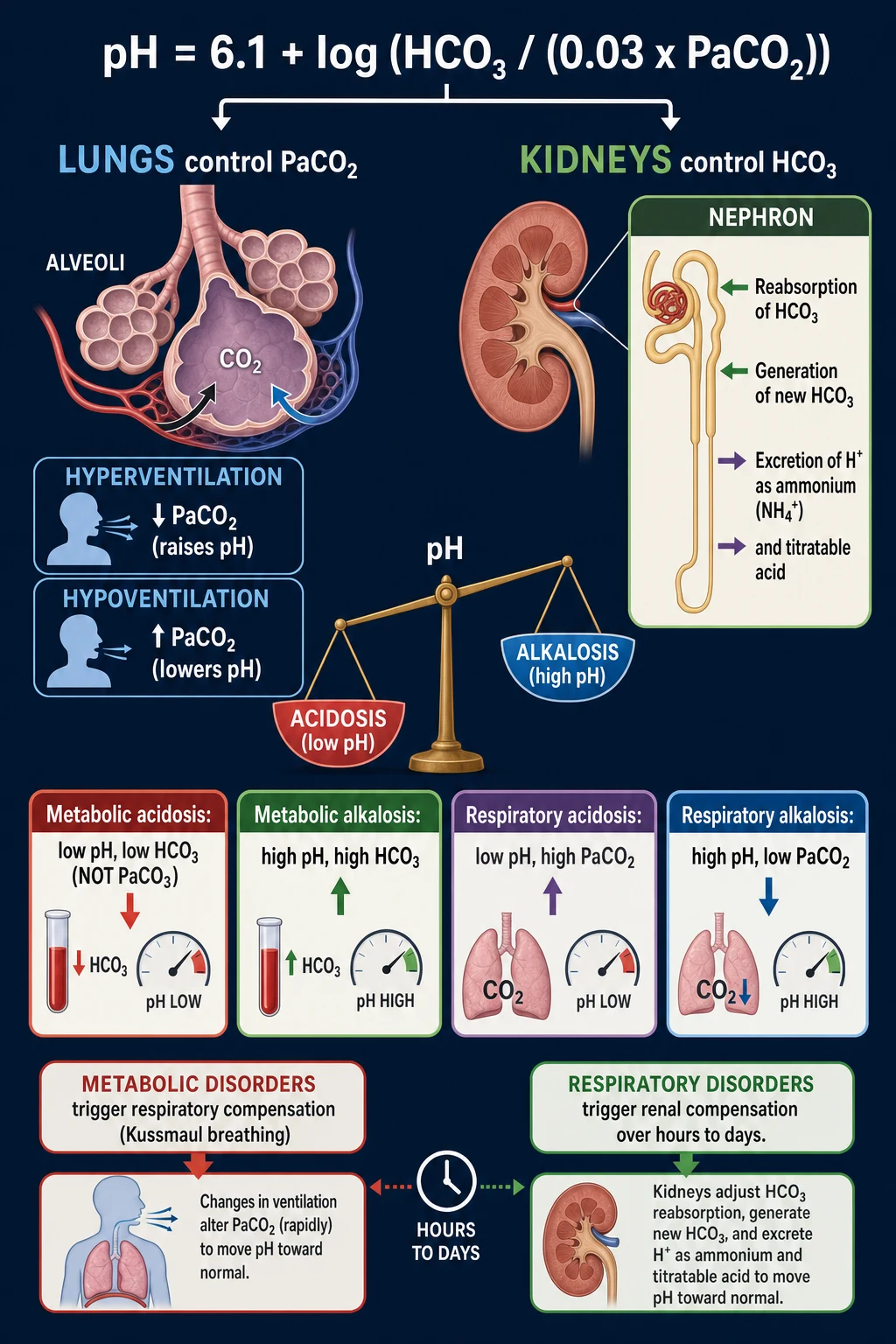
  5. Winter's formula — check compensation. For metabolic acidosis: expected PaCO2 = 1.5 × HCO3 + 8 ± 2. EXAMPLE: HCO3 12 → expected PaCO2 = 1.5 × 12 + 8 = 26 ± 2 (24-28). If measured PaCO2 = 26 → appropriate compensation. If PaCO2 = 35 → respiratory acidosis too (hypoventilating — fatigue, sedation). If PaCO2 = 20 → respiratory alkalosis too (sepsis, anxiety, salicylates early). ALWAYS check — reveals mixed disorders. (For metabolic alkalosis: expected PaCO2 = 0.7 × HCO3 + 20 ± 5.)[4] }
  6. Delta gap — detect mixed disorders. ΔAG (rise in anion gap above normal) = AG − 12. ΔHCO3 (fall in bicarbonate below normal) = 24 − HCO3. In PURE HAGMA: ΔAG ≈ ΔHCO3 (each mmol of acid adds 1 mmol to gap and consumes 1 mmol HCO3) → ratio = 1-2. IF RATIO <1: ΔAG < ΔHCO3 → extra HCO3 lost beyond what acid consumed → concurrent NAGMA (e.g., sepsis lactic acidosis + diarrhoea). IF RATIO >2: ΔAG > ΔHCO3 → less HCO3 fall than expected → concurrent metabolic alkalosis (e.g., sepsis + vomiting). ALTERNATIVE: corrected HCO3 = HCO3 + ΔAG; if >26 → metabolic alkalosis; if <22 → NAGMA.[5] }
  7. Toxic alcohols — methanol and ethylene glycol. Both cause HIGH AG metabolic acidosis + HIGH OSMOLAR GAP. METHANOL → metabolised (alcohol dehydrogenase) → formaldehyde → formic acid (toxic to retina → blindness). Presentation: headache, visual changes ('snowstorm' vision), abdominal pain, severe acidosis. ETHYLENE GLYCOL → metabolised → glycolic/oxalic acid → calcium oxalate crystals in urine (envelope-shaped) → AKI. Presentation: CNS depression, hypocalcaemia (Ca precipitates as oxalate), renal failure. TREATMENT: FOMEPIZOLE (4-methylpyrazole — blocks alcohol dehydrogenase → stops toxic metabolite formation) OR ethanol (competitive substrate — if no fomepizole); HAEMODIALYSIS (removes alcohol + metabolites); folate/thiamine (cofactors for metabolism).[1] }
  8. Salicylate toxicity — classic mixed disorder. EARLY: respiratory ALKALOSIS (salicylates directly stimulate medullary respiratory centre → hyperventilation). THEN: metabolic ACIDOSIS (uncoupling oxidative phosphorylation → lactate + organic acids). So CLASSIC: respiratory alkalosis + high AG metabolic acidosis (the 'mixed' pattern is the clue). OTHER features: tinnitus, deafness, hyperthermia, agitation, seizures, pulmonary oedema (non-cardiogenic). TREATMENT: (a) Activated charcoal (if recent ingestion). (b) URINARY ALKALINISATION (NaHCO3 — trap salicylate ionised in alkali urine → excreted). (c) HAEMODIALYSIS (severe: level >100 mg/dL, severe acidosis, end-organ damage). (d) Avoid intubation if possible (hypoventilation worsens acidosis → salicylate …
  9. BICAR-ICU trial — bicarbonate evidence. Jaber (2018, Lancet): RCT of 4% sodium bicarbonate vs no bicarbonate in ICU patients with severe acidosis (pH ≤7.20). PRIMARY OUTCOME (composite of death + need for RRT by day 28): NO significant difference overall. BUT: SUBGROUP with AKI grade 3-4 (severe AKI): bicarbonate REDUCED primary outcome (55% vs 67%) AND fewer need for RRT. CONCLUSION: bicarbonate may benefit severe acidosis WITH severe AKI. STILL controversial — most guidelines: bicarbonate for pH <7.1 + haemodynamic instability (when pH itself impairs cardiac function/vasopressor response).[3] }
  10. Why bicarbonate is usually NOT given (the harms). (1) GENERATES CO2 (HCO3 + H+ → H2O + CO2) → CO2 diffuses into cells (including CSF/brain) faster than HCO3 → paradoxical INTRACELLULAR/CNS acidosis (briefly worsens). (2) HYPERNATRAEMIA (sodium load). (3) HYPEROSMOLARITY. (4) HYPOKALAEMIA (H+ shifts into cells, K out — but also correction of acidosis shifts K into cells). (5) OVERSHOOT metabolic alkalosis (when cause resolves). (6) Does NOT improve outcomes in most studies (except BICAR-ICU AKI subgroup). INDICATIONS (limited): pH <7.1 + unstable (haemodynamic), TCA overdose (Na load + alkalinisation), hyperkalaemia, specific toxin (methanol/ethylene glycol — as adjunct to fomepizole/dialysis).[3] }
  11. Renal tubular acidosis (RTA) — NAGMA classification. TYPE 1 (DISTAL): impaired H+ secretion (distal tubule) → can't acidify urine (pH >5.5) → metabolic acidosis. K LOW (impaired H+ secretion → more K secreted). CAUSES: autoimmune (Sjögren, SLE), amphotericin, lithium, genetic. Stones (calcium phosphate — alkaline urine). TYPE 2 (PROXIMAL): impaired HCO3 reabsorption → HCO3 lost in urine → acidosis (until new steady state at lower HCO3). K LOW. Fanconi syndrome (generalised proximal dysfunction — glycosuria, aminoaciduria, phosphaturia). CAUSES: myeloma, ifosfamide, acetazolamide, genetic. TYPE 4 (HYPOALDOSTERONISM): low aldosterone → impaired H+ and K secretion → acidosis + HYPERKALAEMIA (distinguishes type 4 — others are hypokalaemic). CAUSES: diabetic nephropathy, ACEi/ARB, heparin, adrenal insufficiency, K-sparing diuretics.[19] }
  12. Metformin-associated lactic acidosis (MALA). Metformin (biguanide) → inhibits mitochondrial complex I → impairs gluconeogenesis from lactate → lactate accumulates → lactic acidosis. RISK FACTORS: CKD (metformin accumulates — eGFR <30 high risk), acute kidney injury (from any cause), sepsis, hypoxia, contrast (hold metformin). PRESENTATION: severe lactic acidosis (lactate often >10), low mortality if recognised. TREATMENT: STOP metformin, supportive, RRT (haemodialysis removes metformin + corrects acidosis), methylene blue (mitochondrial electron carrier — investigational). PREVENTION: hold metformin if eGFR <30, acute illness, contrast, surgery. Most 'metformin-associated' acidosis is actually SEPSIS in a diabetic on metformin (metformin may be …
  13. Diarrhoea vs vomiting — opposite acid-base. DIARRHOEA: loss of HCO3-rich pancreatic/intestinal secretions → metabolic ACIDOSIS (NAGMA — Cl rises to replace HCO3). Low K (stool K). VOMITING: loss of HCl-rich gastric secretions → metabolic ALKALOSIS (Cl falls). Low K (renal loss from aldosterone). Low Cl (hypochloraemic alkalosis). TREATMENT: diarrhoea → replace HCO3 (or let kidney compensate); vomiting → normal saline (volume) + KCl → corrects alkalosis. This distinction is a CLASSIC exam question — know the acid-base consequences.[4] }
  14. Kussmaul breathing — compensatory hyperventilation. Metabolic acidosis → low pH sensed by peripheral (carotid) and central (medullary) chemoreceptors → stimulate respiratory centre → DEEP, RAPID breathing (Kussmaul respirations) → blow off CO2 → raise pH toward normal. This is the COMPENSATORY mechanism (Winter's formula predicts it). CLINICAL: patient with DKA/sepsis breathing fast and deep. NOTE: if patient can't compensate (fatigue, sedation, neuromuscular disease) → pH drops further → respiratory failure → intubation (but ventilation settings must allow high minute ventilation to maintain compensation — don't 'normalise' PaCO2).[4] }

Cohen-Woods classification of lactic acidosis (Type A vs Type B)

ClassMechanismTypical causesKey teaching point
Type ATissue hypoperfusion / impaired O2 delivery (most common)Septic / cardiogenic / hypovolaemic / haemorrhagic shock, severe hypoxia, mesenteric ischaemia, seizures, CO poisoning, cyanideAlways look for and reverse the source of hypoperfusion — lactate is a perfusion marker here
Type B1Drugs / toxins (no overt hypoperfusion)Metformin (complex I), phenformin, cyanide, β-agonists, NRTIs, linezolid, propofol infusion, propylene glycol, salicylates, methanol, ethylene glycol, cocaine, isoniazidStop the offending drug; metformin-related → RRT; propylene glycol → stop the infusion
Type B2Systemic diseaseMalignancy (lymphoma, leukaemia — Warburg metabolism), liver failure (impaired clearance), thiamine deficiency (pyruvate→lactate), diabetes, mitochondrial disease, severe asthmaTreat the underlying disease; give thiamine if deficiency suspected
Type B3Inborn errors of metabolismMitochondrial encephalomyopathy, glycogen storage disease (McArdle, GSD I), pyruvate carboxylase / dehydrogenase deficiency, congenital lactic acidaemiaSuspect if recurrent / paediatric / fasting-triggered; metabolic genetics referral
D-lactateGut bacterial fermentation (separate entity)Short bowel syndrome, jejunoileal bypass, bacterial overgrowthNOT detected by standard L-lactate assay — order D-lactate specifically; causes encephalopathy
[1]

Ketoacidosis: DKA vs HHS vs alcoholic vs starvation

FeatureDKAHHSAlcoholic (AKA)Starvation
GlucoseHIGH (>13.9 mmol/L)VERY HIGH (>33.3 mmol/L)LOW-NORMAL (often <11)LOW-NORMAL
Ketones (β-OHB)HIGHLOW-MILDHIGHMILD
pHLOW (<7.30)NORMAL (mildly low)LOW (mild-moderate)NORMAL (mildly low)
HCO3LOW (<18)NORMAL (>18)LOW (mild)NORMAL (mildly low)
AGHIGHMILDLY HIGHHIGHMILDLY HIGH
OsmolalityMildly highVERY HIGH (>320)VariableNormal
DehydrationModerateSEVEREMild-moderateMild
Key treatmentInsulin + fluids + KFluids FIRST (insulin cautiously)DEXTROSE + thiamine (insulin dangerous → hypoglycaemia)Feeding (dextrose)
Mortality~1-5%~10-20% (high)Low if treatedLow
Mnemonic"Dry, warm, sweet""Older, very hyperosmolar, less acidotic""A = Alcohol → give dextrose""Refeed"
[1]

Toxic alcohols: methanol vs ethylene glycol vs diethylene glycol vs propylene glycol

FeatureMethanolEthylene glycolDiethylene glycolPropylene glycol
SourceWindscreen washer, antifreeze, moonshine, solventsAntifreeze, brake fluidBrake fluid, antifreeze (counterfeit)Solvent vehicle for IV drugs (lorazepam, diazepam, etomidate, phenytoin, nitroglycerin)
Toxic metaboliteFormic acid (via formaldehyde)Glycolic + oxalic acid2-hydroxyethoxyacetic acidLactic acid (D + L)
Target organRETINA (blindness), basal gangliaKIDNEY (calcium oxalate), brainKIDNEY, liver, brainLactic acidosis, AKI
Anion gapHIGHHIGHHIGHHIGH
Osmolar gapHIGH (early)HIGH (early)HIGH (early)HIGH
Urine findingsNormalCalcium oxalate crystals (envelope/needle)May have crystalsNone specific
CluesVisual changes, 'snowstorm' vision, headacheHypocalcaemia, flank pain, AKI, intoxicated without alcohol odourEpidemic clusters, hepatotoxicityPatient on high-dose lorazepam/diazepam infusion in ICU
Specific treatmentFomepizole + folate (leucovorin) + dialysisFomepizole + thiamine + pyridoxine + dialysisFomepizole + dialysisSTOP infusion, supportive
[1]

Renal tubular acidosis types (NAGMA sub-classification)

FeatureType 1 (Distal / dRTA)Type 2 (Proximal / pRTA)Type 4 (Hypoaldosteronism)
DefectImpaired H+ secretion (distal tubule) — can't acidify urineImpaired HCO3 reabsorption (proximal) — HCO3 wastedLow aldosterone effect — impaired H+ + K secretion
Urine pHHIGH (>5.5 — inappropriately alkaline)LOW (<5.5 once steady state; >5.5 early)LOW (<5.5)
Serum KLOW (hypokalaemia)LOW (hypokalaemia)HIGH (hyperkalaemia) — distinguishing feature
Anion gapNormalNormalNormal
StonesYES — calcium phosphate (alkaline urine)NO (but rickets/osteomalacia)NO
Common causesAutoimmune (Sjögren, SLE), amphotericin B, lithium, hereditaryMyeloma, ifosfamide, acetazolamide, Fanconi, hereditaryDiabetic nephropathy, ACEi/ARB, heparin, adrenal insufficiency, K-sparing diuretics, calcineurin inhibitors
TreatmentPotassium citrate / bicarbonate (generous)Bicarbonate (large doses) + K; thiazideFludrocortisone (if mineralocorticoid deficiency); correct K; treat cause
Exam pearl"Stones + hypokalaemia + alkaline urine""Fanconi + myeloma""Hyperkalaemic NAGMA = type 4"
[1]

Alkalinising therapies for severe metabolic acidosis

TherapyMechanismProsCons / cautionsWhen to use
Sodium bicarbonateDirect HCO3 donor; buffers H+ → CO2 (exhaled)Cheap, ubiquitous, rapid pH riseGenerates CO2 (need ventilation), hypernatraemia, hyperosmolarity, hypokalaemia, overshoot alkalosis, volume overloadpH <7.1-7.2 + unstable / AKI (BICAR-ICU); TCA overdose; hyperkalaemia; urinary alkalinisation (salicylate)
THAM (tris-hydroxymethyl aminomethane)Non-bicarbonate buffer; neutralises H+ directly without generating CO2; also buffers CO2No CO2 generation (useful if ventilated/hypercapnic), less hypernatraemia, intracellular bufferingHyperkalaemia (at high doses), hypoglycaemia, tissue necrosis if extravasation, hepatic metabolism (avoid in liver failure)Severe acidosis where bicarbonate unsuitable; hypercapnic acidosis; investigational
Carbicarb (equimolar Na2CO3/NaHCO3)Buffers H+ → HCO3 (not CO2) — less CO2 generation than bicarbonateLess CO2 burdenNot widely available; limited evidenceInvestigational / limited availability
Renal replacement therapy (CRRT/IHD)Removes acid anions + provides bicarbonate-rich dialysate; corrects cause (metformin, salicylate) + acidosis simultaneouslyDefinitive for toxin-related / renal failure acidosis; avoids sodium load; slow controlled correctionRequires vascular access + anticoagulation; slower onset; CRRT hypothermiaRenal failure acidosis, toxic alcohol (with fomepizole), metformin-associated lactic acidosis, severe salicylate toxicity
[1]

Approach to suspected toxic alcohol ingestion

  1. SUSPECT when — HAGMA + high osmolar gap + no clear alternative (lactate, ketones, renal failure, sepsis all excluded). Often history of ingestion (deliberate self-harm, occupational, moonshine), visual symptoms (methanol), oxalate crystals/hypocalcaemia (ethylene glycol), or ICU patient on propylene glycol infusion
  2. CALCULATE OSMOLAR GAP — Measured osmolality − calculated osmolality. Calculated = 2×Na + glucose + urea (± ethanol). Normal <10 mOsm/kg. GAP >20 → unmeasured osmoles present (toxic alcohol). NOTE: gap narrows as parent alcohol metabolises → an EARLY high gap may become normal later (the metabolites raise AG, not osmolality). Both gaps may not be elevated simultaneously — don't be reassured by a normal osmolar gap if AG is high
  3. SEND SPECIFIC LEVELS — Methanol level, ethylene glycol level (gold standard but slow). Point-of-care: urinary fluorescence under Wood's lamp (ethylene glycol — fluorescent dye added commercially), calcium oxalate crystals on microscopy. Ethanol level (co-ingestion common, may be protective)
  4. EMPIRICAL FOMEPIZOLE — 15 mg/kg IV loading, then 10 mg/kg q12h (increase q4h if on dialysis). BLOCKS alcohol dehydrogenase → halts conversion to toxic metabolites. Give EARLY — do not wait for levels if suspicion high and AG elevated. Alternative: ethanol infusion (target ethanol level 100-150 mg/dL) if fomepizole unavailable
  5. COFACTORS — FOLATE / leucovorin (methanol — speeds formate metabolism), THIAMINE + PYRIDOXINE (ethylene glycol — divert metabolism away from oxalate)
  6. HAEMODIALYSIS — Indicated if: severe acidosis (pH <7.25-7.30), end-organ damage (visual symptoms, AKI, coma), high alcohol level (methanol >15-20 mmol/L; ethylene glycol >8 mmol/L), or persistently elevated AG despite fomepizole. Use bicarbonate bath; continue fomepizole during dialysis (increased dosing). Monitor alcohol levels / AG to determine dialysis end-point
  7. ADMIT + MONITOR — ICU admission; serial ABG, AG, osmolar gap, renal function, vision (methanol). Fomepizole for at least 24h after alcohol undetectable. Late visual sequelae possible even with treatment
[1]

Worked example: delta-delta (mixed disorder detection)

  1. CASE — 55 yo, vomiting + DKA. ABG: pH 7.20, HCO3 9, PaCO2 23. VBG chem: Na 138, Cl 92, HCO3 9. Glucose 35 mmol/L, ketones 5.5, lactate 1.5
  2. STEP 1 — Confirm metabolic acidosis + compensation. HCO3 9 (low) → metabolic acidosis. Winter's expected PaCO2 = 1.5 × 9 + 8 ± 2 = 21.5 ± 2 (19.5-23.5). Measured PaCO2 = 23 → WITHIN range → appropriate respiratory compensation, no mixed respiratory disorder
  3. STEP 2 — Anion gap (corrected). AG = 138 − (92 + 9) = 37. Albumin-adjusted AG: assume normal albumin → AG 37 (markedly raised → HAGMA). High AG confirms ketone-driven HAGMA
  4. STEP 3 — Delta gap. ΔAG = AG − 12 = 37 − 12 = 25. ΔHCO3 = 24 − HCO3 = 24 − 9 = 15. Ratio ΔAG/ΔHCO3 = 25/15 = 1.67 → within 1-2 → BUT corrected HCO3 = HCO3 + ΔAG = 9 + 25 = 34 (>26 → metabolic alkalosis present)
  5. INTERPRET — The ratio is borderline but corrected HCO3 of 34 is clearly elevated → concurrent metabolic ALKALOSIS (from vomiting — HCl loss) ON TOP of DKA. The vomiting 'masks' some of the acidosis (HCO3 higher than pure DKA would give)
  6. CLINICAL ACTION — Treat DKA (insulin, fluids, K). The metabolic alkalosis will resolve with volume (saline) once vomiting stops. Do NOT be falsely reassured — DKA severity is 'hidden' by the alkalosis; serial AG monitoring guides resolution
  7. TEACHING POINT — Always calculate BOTH ratio and corrected HCO3. Ratio can be misleading when ΔHCO3 is small; corrected HCO3 (>26 alkalosis, <22 NAGMA) is the more robust adjunct. Albumin correction is essential — hypoalbuminaemia narrows AG and hides a HAGMA
[1]

Sodium bicarbonate decision pathway in severe metabolic acidosis

  1. IS BICARBONATE INDICATED AT ALL? Default = NO. Treat the cause (fluids, sepsis source control, insulin for DKA, fomepizole for toxic alcohol, dialysis for renal failure). Bicarbonate does NOT improve outcomes in lactic acidosis or DKA in most trials
  2. CONSIDER IF — (a) pH <7.1-7.15 AND haemodynamically unstable (vasopressor-resistant shock, falling cardiac output) — pH itself is impairing function. (b) Severe AKI (KDIGO stage 2-3) + pH <7.20 (BICAR-ICU subgroup benefit). (c) TCA / sodium-channel-blocker overdose (Na load + alkalinisation). (d) Hyperkalaemia with acidosis. (e) Salicylate toxicity (urinary alkalinisation — different rationale). (f) Pre-RRT bridge in toxic alcohol / metformin
  3. DOSE + ROUTE — 1.26% (isotonic, 150 mmol/L) preferred in ICU (less hypernatraemia than 8.4%) — 100-250 mL boluses, or infusion 1-2 mmol/kg. 8.4% hypertonic via central line only, slow (1 mmol/kg bolus) — avoid peripheral (extravasation necrosis). Target: raise pH to >7.15-7.20 — DO NOT normalise (overshoot alkalosis, hypokalaemia)
  4. MONITOR + ADJUST — Serial ABG (every 1-2h), Na (hypernatraemia — limit rise to <160), K (supplement as pH rises → K shifts intracellularly), volume (avoid overload). Ensure adequate ventilation — bicarbonate generates CO2 that MUST be ventilated or PaCO2 rises and pH paradoxically worsens
  5. STOP / DE-ESCALATE — When pH >7.15-7.20, cause treated, haemodynamics improved, or RRT initiated. Reassess — if lactate still rising or shock worsening, bicarbonate is futile → escalate to RRT / source control
  6. CONSIDER ALTERNATIVES — THAM (no CO2 generation — useful if hypercapnic / ventilated), or proceed directly to CRRT (bicarbonate-rich dialysate, controlled correction, removes causative toxin). CRRT preferred for metformin / toxic alcohol / refractory lactic acidosis
[1]

Intubating the severely acidotic patient

  1. RECOGNISE THE DANGER — Induction + positive-pressure ventilation (PPV) in a severely acidotic (pH <7.2) patient can precipitate cardiac arrest. Reasons: PPV reduces venous return (preload ↓) → CO ↓; induction vasodilates; apnoea period raises PaCO2 → pH falls further → vasopressor resistance + arrhythmia
  2. PRE-OXYGENATE + BUFFER — 100% O2, head-up, aggressive pre-oxygenation (3 min). Consider bicarbonate bolus (1-2 mmol/kg) BEFORE induction if pH <7.1. Maximise vasopressor / inotrope infusions pre-induction; have norepinephrine running
  3. CHOOSE KETAMINE / ETOMIDATE — Ketamine (preserves sympathetic tone — but beware if catecholamine-depleted) or etomidate (haemodynamically neutral). AVOID propofol / midazolam (vasodilation). Paralytic: rocuronium (1.2 mg/kg) or suxamethonium — rocuronium preferred for sustained apnoea-free RSI
  4. SET VENTILATOR TO MATCH COMPENSATION — Pre-intubation the patient is hyperventilating (Kussmaul) to compensate. The ventilator MUST deliver HIGH minute ventilation (set RR 25-35, Vt 6-8 mL/kg) to keep PaCO2 LOW (matching their pre-intubation compensatory level). DO NOT 'normalise' PaCO2 to 40 — that will drop pH catastrophically. Target PaCO2 = Winter's expected (e.g., HCO3 9 → PaCO2 ~21)
  5. POST-INTUBATION — Arterial line + serial ABG (first ABG within 5-10 min). Adjust ventilator to maintain pH >7.2. If pH falling → increase minute ventilation cautiously (watch for lung injury / breath stacking), give bicarbonate, or escalate. Continue treating the cause (DKA, sepsis, toxic alcohol)
  6. EXTUBATION CRITERIA — Cause resolving (lactate clearing, DKA resolving), pH >7.25 with normal minute ventilation, HCO3 >18, mental status appropriate. Do not delay extubation solely for 'normalisation' of bicarbonate
[1]

Additional clinical pearls — high-anion-gap and normal-anion-gap deep dive

Advanced metabolic acidosis pearls for CICM/FFICM/EDIC

  1. Cohen-Woods (1976) — the lactic acidosis classification every ICU trainee must know. TYPE A = clinically evident tissue hypoperfusion / hypoxia (shock, sepsis, ischaemia, severe hypoxaemia, seizures, CO/cyanide). TYPE B = no overt hypoperfusion, subdivided: B1 = drugs/toxins (metformin, phenformin, ethanol, methanol, ethylene glycol, salicylate, propylene glycol, isoniazid, NRTIs, linezolid, propofol infusion, β-agonists); B2 = systemic disease (malignancy — lymphoma/leukaemia, liver failure, diabetes, thiamine deficiency, mitochondrial disease); B3 = inborn errors of metabolism (mitochondrial myopathies, glycogen storage disease, pyruvate dehydrogenase/carboxylase deficiency). Type A is by far the commonest in ICU. The classification is NOT just academic — B1 demands immediate drug withdrawal, B2 demands specific therapy (thiamine, malignancy treatment), Type A demands perfusion restoration.[2] }
  2. Osmolar gap — the toxic-alcohol discriminator. Osmolar gap = measured osmolality (freezing-point osmometer) − calculated osmolality. Calculated = 2×Na + glucose + urea (add 1.25×ethanol if ethanol co-ingested). Normal <10 mOsm/kg. GAP >20 strongly suggests an unmeasured low-molecular-weight osmole — methanol, ethylene glycol, ethanol, isopropanol, propylene glycol, mannitol. PITFALLS: (1) Gap is HIGHEST early (parent alcohol present) and narrows as it metabolises → metabolites raise AG instead → so a 'normal' osmolar gap does NOT exclude toxic alcohol if presentation is delayed. (2) Each gap doesn't always exceed 10 — formula variance. (3) Isopropanol raises gap but NOT AG (no acid metabolites — acetone). (4) Don't use vapour-pressure osmometer (volatile alcohols evaporate) — use freezing-point depression. When AG + osmolar gap both raised → toxic alcohol until proven otherwise → empirical fomepizole.[1] }
  3. DKA — ketogenesis driven by insulin deficiency + counter-regulatory excess. PATHOPHYSIOLOGY: insulin deficiency + glucagon/cortisol/catecholamine excess → lipolysis (adipose triglyceride → free fatty acids) → hepatic β-oxidation → ketogenesis (acetoacetate, β-hydroxybutyrate — the dominant DKA ketone). β-HYDROXYBUTYRATE is the KETONE MEASURED on point-of-care meters (nitroprusside urine test mainly detects acetoacetate — can be falsely LOW early in DKA when β-OHB dominates, then FALSLEY RISES as DKA resolves and β-OHB → acetoacetate). TREATMENT: fluids (isotonic saline initially), insulin (0.1 U/kg/h infusion), potassium replacement (acidosis shifts K out → total body K depleted; as insulin works, K plummets — replace aggressively), treat trigger (infection, infarction, non-compliance). BICARBONATE NOT routine in DKA — associated with cerebral oedema in children; only if pH <7.0 + unstable.[9] }
  4. HHS vs DKA — distinguish them, treatment differs. HHS (hyperosmolar hyperglycaemic state): older type 2 diabetics, EXTREME hyperglycaemia (>33 mmol/L), severe hyperosmolality (>320), profound dehydration, MINIMAL ketosis (enough residual insulin to suppress lipolysis but not gluconeogenesis), pH usually >7.30, HCO3 >18. MORTALITY HIGH (10-20%) — older, comorbid, more dehydrated. TREATMENT: FLUIDS FIRST (the deficit is enormous — 8-12 L; isotonic saline, then switch based on corrected Na), insulin cautiously (lower doses than DKA — risk of osmotic shift), aggressive K replacement, anticoagulate (pro-thrombotic), hunt for trigger (infection, MI). KEY DIFFERENCE from DKA: less acidosis, more dehydration, less insulin needed, fluid is the priority.[11] }
  5. Alcoholic ketoacidosis (AKA) — dextrose FIRST, not insulin. Chronic alcohol misuse + reduced oral intake → depleted NAD+/NADH ratio from ethanol metabolism → ketogenesis (β-OHB predominant) + mild lactic acidosis. PRESENTATION: vomiting, abdominal pain, mild-moderate HAGMA, LOW-NORMAL glucose, mild-moderate acidosis, often after a binge then cessation of intake. CRITICAL: give DEXTROSE (5% dextrose) NOT saline alone — dextrose stimulates insulin → halts ketogenesis, and corrects glycogen depletion. Give THIAMINE (100 mg IV) BEFORE dextrose to prevent Wernicke encephalopathy (thiamine required for glucose metabolism; giving dextrose first can precipitate Wernicke in depleted patients). Do NOT give insulin (no hyperglycaemia, risk of severe hypoglycaemia). Rehydrate, replace K/Mg/PO4. Resolves within 12-24h. Always exclude co-existing DKA, pancreatitis, sepsis, methanol/ethylene glycol (alcoholic patient).[10] }
  6. Uraemic acidosis — the renal-failure HAGMA. In advanced CKD/AKI (GFR <20-25), the kidney cannot excrete the daily acid load (phosphate, sulphate, organic anions) → these accumulate as unmeasured anions → HAGMA. HCO3 typically 12-20. CONTRIBUTES to bone disease (chronic buffering by bone → osteodystrophy), muscle wasting, worsening kidney function, anaemia (inhibits EPO). TREATMENT: dialysis (corrects via bicarbonate-rich dialysate + removes retained anions); oral bicarbonate in CKD if HCO3 <22 ( KDIGO — may slow CKD progression); treat the underlying kidney disease. NOT simply a 'low bicarbonate' — it is accumulated organic/inorganic anions, so the AG is genuinely raised (unlike pure NAGMA). Often a MIXED picture in ICU: uraemia + lactic acidosis + hyperchloraemia from saline resuscitation.[1] }
  7. Propylene glycol toxicity — the iatrogenic ICU HAGMA. Propylene glycol is the SOLVENT vehicle for many IV drugs: LORAZEPAM (highest risk — 80% propylene glycol in the concentrate), DIAZEPAM, ETOMIDATE, PHENYTOIN, NITROGLYCERIN, ESMOLOL, trimethoprim-sulfamethoxazole. Metabolised (alcohol dehydrogenase) → D- and L-LACTATE → HAGMA. CLUE: ICU patient on high-dose lorazepam infusion (>1 mg/kg/h for >48h) developing rising AG + rising lactate + rising osmolar gap. TREATMENT: STOP the offending drug (switch to midazolam or propofol — minimal propylene glycol); supportive; rarely dialysis. PREVENTION: limit lorazepam infusion rate/duration; monitor AG + lactate + osmolar gap in long infusions. Often MISSED — suspect in the sedated ICU patient with unexplained HAGMA.[13] }
  8. 5-oxoproline (pyroglutamic acid) — think chronic paracetamol. 5-Oxoproline accumulates when glutathione is depleted (normally the γ-glutamyl cycle is feedback-inhibited by glutathione; low glutathione → unregulated γ-glutamyl cysteine synthetase → 5-oxoproline excess). CAUSES: CHRONIC PARACETAMOL (acetaminophen) use (especially in women, malnutrition, sepsis, renal failure, flucloxacillin), some antibiotics. HAGMA with NORMAL lactate, NORMAL ketones, NO toxin history initially → consider 5-oxoproline. DIAGNOSIS: urine organic acids (5-oxoproline elevated). TREATMENT: STOP paracetamol; N-ACETYLCYSTEINE (repletes glutathione — breaks the cycle); supportive. KEY: this is an OFTEN-MISSED cause of unexplained HAGMA in the chronic-illness ICU patient on regular paracetamol.[12] }
  9. Salicylate kinetics — why urinary alkalinisation + dialysis work. ASPIRIN (acetylsalicylic acid) → salicylate. pKa ~3 — in ACIDIC environments it is non-ionised (lipid soluble → crosses membranes into CNS); in ALKALINE environments it is ionised (trapped — 'ion trapping'). PRINCIPLE: ALKALINISE the urine (NaHCO3 → urine pH >7.5) → salicylate ionised in renal tubule → not reabsorbed → excreted (urinary clearance 10-20× higher). ALKALINISE the blood (NaHCO3) → salicylate ionised in plasma → less CNS penetration. HAEMODIALYSIS criteria: level >100 mg/dL (acute) or >60 mg/dL (chronic), OR severe acidosis/pulmonary oedema/CNS toxicity/renal failure. AVOID INTUBATION if possible — hypoventilation raises PaCO2 → worsens acidosis → more salicylate shifts into brain → death. If MUST intubate, hyperventilate aggressively + dialyse immediately.[1] }
  10. Acetazolamide — the iatrogenic NAGMA. Carbonic anhydrase inhibitor → blocks proximal tubule Na/H exchange and HCO3 reabsorption → HCO3 lost in urine → NAGMA + hypokalaemia + mild metabolic acidosis (intentional in glaucoma, altitude sickness, periodic paralysis, idiopathic intracranial hypertension). IN ICU: used to ALKALINISE urine (salicylate/cystine stones), as diuretic (especially in metabolic alkalosis), for CSF reduction, alkalinisation in tumour lysis. UNWANTED NAGMA: monitor HCO3 + K; usually mild and self-limiting on cessation. ALSO USED THERAPEUTICALLY to CORRECT chronic metabolic alkalosis in mechanically ventilated COPD patient (allows weaning by reducing HCO3 → resets chemoreceptor threshold → stimulates ventilation).[4] }
  11. Ureteroenteric / ileal conduit hyperchloraemic acidosis. After urinary diversion (ileal conduit, ileal neobladder, ureterosigmoidostomy), the bowel mucosa REABSORBS urinary Cl− in exchange for HCO3− (Cl/HCO3 exchanger) → HCO3 lost, Cl gained → NAGMA. RISK: long conduit, contact time (long catheterisation / stasis), renal impairment (can't compensate). SEEN WEEKS-MONTHS post-op, can be chronic. TREATMENT: oral bicarbonate, drain the reservoir frequently (reduce contact time), surgical revision if severe. Ureterosigmoidostomy highest risk (stool + urine mixing → also ammonia + chloride absorption + colon cancer risk). Often MISSED as a cause of unexplained chronic NAGMA in patients with urological surgical history.[18] }
  12. THAM (tris-hydroxymethyl aminomethane) — the bicarbonate alternative. Organic buffer (pKa 7.7) that neutralises H+ WITHOUT generating CO2 (unlike bicarbonate). ALSO buffers CO2 directly. ADVANTAGES: no CO2 burden (useful in ventilated / hypercapnic patients — e.g., severe COPD + acidosis), penetrates cells (intracellular buffering), less hypernatraemia. DISADVANTAGES: hepatic metabolism (avoid in severe liver failure), hypoglycaemia, hyperkalaemia at high dose, tissue necrosis on extravasation. EVIDENCE: small trials (Hoste 2005) suggest comparable pH correction to bicarbonate with less CO2 generation; not widely available; lacks large RCTs. ROLE: adjunct/alternative when bicarbonate contraindicated or CO2 clearance problematic. Familiarise yourself if your unit stocks it.[17] }
  13. Stewart (strong ion difference) approach — a different lens. Stewart: pH is determined by three independent variables — (1) PCO2, (2) TOTAL WEAK ACID (Atot — mainly albumin + phosphate), (3) STRONG ION DIFFERENCE (SID = [Na + K + Ca + Mg] − [Cl + lactate + other strong anions]). A fall in SID (e.g., hyperchloraemia from saline, or addition of lactate) → acidosis; a rise → alkalosis. CLINICAL UTILITY: explains why 0.9% SALINE causes HYPERCHLORAEMIC ACIDOSIS (high Cl reduces SID) — use balanced crystalloids (Plasma-Lyte, Hartmann) to avoid iatrogenic NAGMA; quantifies the contribution of albumin (hypoalbuminaemia → alkalosis by reducing Atot). Most trainees use Henderson-Hasselbalch for diagnosis + Stewart CONCEPTS for fluid choice (avoid saline-induced acidosis) and unexplained mixed disorders. Not required for routine ABG interpretation but high-yield for exam 'explain this acidosis' questions.[5] }
  14. 0.9% saline resuscitation causes a hyperchloraemic NAGMA — choose balanced crystalloids. Large-volume 0.9% saline delivers 154 mmol/L of Cl (much higher than plasma ~100) → hyperchloraemia → reduces SID (Stewart) → metabolic acidosis; ALSO directly reduces renal cortical perfusion (tubuloglomerular feedback via macula densa) → AKI. SALT-ED, SMART trials: balanced crystalloids (Plasma-Lyte, Hartmann/Ringer lactate) → less acidosis, less AKI, possibly mortality benefit in sepsis. TAKE-HOME: in a patient you are about to resuscitate, use a BALANCED crystalloid to avoid creating a NAGMA on top of their presenting HAGMA — this confuses the delta gap and worsens renal perfusion. Reserve saline for hyperchloraemic alkalosis or Na depletion. One of the highest-yield 'fluid choice' pearls.[5] }
  15. Intubating the acidotic patient — a lethal moment. Severe metabolic acidosis + RSI = high risk of peri-intubation arrest. The patient is compensating with hyperventilation (Kussmaul); induction + PPV abolish this → PaCO2 rises → pH falls → vasopressor-resistant shock + VF/asystole. MITIGATION: (1) Pre-oxygenate + buffer (bicarbonate 1-2 mmol/kg if pH <7.1). (2) Ketamine or etomidate (avoid propofol/midazolam). (3) Set ventilator for HIGH minute ventilation (RR 25-35) to match compensation — do NOT normalise PaCO2. (4) Have norepinephrine running pre-induction. (5) First post-intubation ABG within 5 min. AVOID delaying intubation if clearly failing — but do not intubate without preparation in profound acidosis.[4] }
  16. Mixed venous / central venous-to-arterial CO2 difference (ΔPCO2) — perfusion marker. In shock, the ΔPCO2 (PvCO2 − PaCO2) is normally <6 mmHg; >6 mmHg indicates INADEQUATE CARDIAC OUTPUT to clear tissue CO2 (washout). USE: in a septic / shocked patient with persistent lactataemia, a high ΔPCO2 says 'increase perfusion' (fluids, inotrope) rather than 'treat the lactate.' Combines with lactate clearance to guide resuscitation. PEARL: if lactate is high but ΔPCO2 normal, the lactate may be Type B (non-hypoperfusion — e.g., β-agonist, malignancy) rather than ongoing tissue hypoxia. High-yield for the 'why is the lactate not clearing' exam vignette.[5] }
  17. Calcium during severe acidosis — arrhythmia protection. Severe acidosis (pH <7.1) increases risk of ventricular arrhythmias AND reduces responsiveness to catecholamines. CALCIUM does NOT correct pH but STABILISES the myocardium during severe acidosis / hyperkalaemia — consider CALCIUM CHLORIDE (10 mL 10%, via central line) or CALCIUM GLUCONATE (10 mL 10%, peripheral) in the peri-arrest acidotic / hyperkalaemic patient while definitive therapy (bicarbonate, dialysis, insulin/dextrose) is prepared. DOES NOT TREAT the acidosis — a bridge, not a cure. Especially relevant if DKA with severe hyperkalaemia, or metformin-associated lactic acidosis with shock.[4] }
  18. CRRT correction rate — avoid rapid correction. In severe metabolic acidosis, initiate CRRT with a BICARBONATE-BATH concentration at or modestly above the target (e.g., start 28-32 mmol/L HCO3 bath) and correct SLOWLY over 24-72h. RAPID correction → dialysis disequilibrium (cerebral oedema — especially in uraemic / chronic), worsening hypokalaemia / hypophosphataemia (shift), overshoot alkalosis. In DKA especially, too-rapid correction of AG (with bicarbonate) is linked to cerebral oedema in children — use CRRT cautiously and slowly. SLED / IHD = faster correction (use for toxic alcohol / metformin where rapid removal needed); CVVHDF / CVVD = slower, gentler (shocked / liver failure / chronic). Match modality to indication.[8] }

Exam practice

SAQ — Persistent lactic acidosis in septic shock

10 minutes · 10 marks

A 64-year-old man is admitted to the intensive care unit with community-acquired pneumonia and septic shock. He has received 30 mL/kg of balanced crystalloid, broad-spectrum antibiotics (ceftriaxone 2 g and azithromycin 500 mg), and is now on norepinephrine 0.35 microgram/kg/min to maintain a mean arterial pressure of 68. Four hours into resuscitation his arterial gas shows pH 7.22, PaCO2 24 mmHg, HCO3 10, lactate 6.8 mmol/L (was 8.2 on arrival). The central venous oxygen saturation is 72 per cent, the central venous-to-arterial CO2 difference is 9 mmHg, and the urine output is 15 mL/h.

[1]

SAQ — High anion gap metabolic acidosis with a high osmolar gap: toxic alcohol

10 minutes · 10 marks

A 56-year-old man is brought to the emergency department collapsed and smelling of alcohol. He is drowsy (GCS 12), BP 110/70, HR 118, RR 28 with deep sighing respirations. The venous gas shows pH 7.08, HCO3 6, PaCO2 22. Sodium 140, chloride 100, glucose 6.5, urea 5.0, creatinine 90, beta-hydroxybutyrate 0.4, lactate 3.0, albumin 38. The measured osmolality is 340 mOsm/kg. Urine microscopy shows needle- and envelope-shaped crystals.

[1]

Red flags

Critical metabolic acidosis red flags

  • pH <7.1 → cardiac dysfunction, vasopressor resistance — consider bicarbonate.[3] }
  • HIGH anion gap (>12) → GOLDMARK: lactate, ketones, toxins, renal failure.[1] }
  • Lactate >4 + not clearing → ongoing hypoperfusion — mortality rises.[6] }
  • High OSMOLAR GAP + HAGMA → toxic alcohol (methanol/ethylene glycol) — fomepizole + dialysis.[1] }
  • Bicarbonate generally NOT indicated except pH <7.1 unstable (BICAR-ICU), TCA, hyperkalaemia.[3] }
  • Winter's formula — check compensation to detect mixed disorders.[4] }
  • Salicylates: respiratory alkalosis + metabolic acidosis = classic.[1] }

Toxic / iatrogenic metabolic acidosis red flags

  • Propylene glycol toxicity — ICU patient on lorazepam/diazepam/etomidate infusion + rising AG + lactate + osmolar gap → STOP infusion, switch sedative.[13] }
  • 5-oxoproline — unexplained HAGMA + chronic paracetamol use → STOP paracetamol, give N-acetylcysteine.[12] }
  • Methanol — visual changes + HAGMA → empirical fomepizole immediately, do NOT wait for levels; folate/leucovorin + dialysis.[15] }
  • Ethylene glycol — HAGMA + hypocalcaemia + calcium oxalate crystals → fomepizole + thiamine + pyridoxine + dialysis. Do NOT aggressively replace calcium unless symptomatic (drives oxalate).[14] }
  • Salicylate toxicity — AVOID intubation if possible (hypoventilation → CNS salicylate rise → death); if must intubate, hyperventilate + dialyse immediately.[1] }
  • Alcoholic ketoacidosis — give THIAMINE before dextrose (Wernicke prevention); do NOT give insulin (risk of hypoglycaemia); exclude methanol/ethylene glycol co-ingestion.[10] }
  • Fomepizole BEFORE dialysis — keep alcohol dehydrogenase blocked throughout and after dialysis (increase fomepizole dosing during dialysis).[14] }
  • Metformin-associated lactic acidosis — lactate >10 + metformin + AKI → early RRT (haemodialysis removes metformin + corrects acidosis); exclude co-existing sepsis.[2] }
  • Rising AG despite treatment → ongoing source (occult mesenteric ischaemia, uncontrolled sepsis, ongoing toxin absorption) — escalate source control / dialysis.[1] }
  • Intubation in severe acidosis — high peri-intubation arrest risk → pre-buffer, ketamine/etomidate, ventilator set for HIGH minute ventilation (do not normalise PaCO2).[4] }

Severe NAGMA / specific-situation red flags

  • Hyperkalaemic NAGMA → think TYPE 4 RTA (hypoaldosteronism) — check aldosterone, rule out adrenal insufficiency, ACEi/ARB, heparin.[19] }
  • Hypokalaemic NAGMA + nephrocalcinosis → TYPE 1 (distal) RTA — autoimmune workup (Sjögren, SLE), check amphotericin/lithium.[19] }
  • Persistent unexplained NAGMA → check urine anion gap (NH4 excretion); if positive → renal cause (RTA); consider myeloma workup (type 2 RTA / Fanconi).[18] }
  • NAGMA after urinary diversion → ureteroenteric / ileal conduit — Cl/HCO3 exchange; oral bicarbonate + frequent reservoir drainage.[18] }
  • Iatrogenic saline-induced NAGMA — switch to balanced crystalloid; monitor AG and delta gap (masks / confounds HAGMA).[5] }
  • Acetazolamide — iatrogenic NAGMA + hypokalaemia; usually mild, reversible on cessation; can cause calcium phosphate stones.[4] }
  • Mixed disorder on delta gap — corrected HCO3 >26 (concurrent alkalosis) or <22 (concurrent NAGMA) → treat BOTH processes.[5] }

Prognosis

Metabolic acidosis evidence and outcomes

Lactate and mortality: admission lactate >4 — mortality ~30%; lactate not clearing by 10% in 2-6h — worse outcomes (Jansen 2010, Arnold 2009). Lactate clearance as goal of resuscitation (Jansen TRIUMPH — no benefit of targeted clearance, but clearance correlates with survival).[6] } BICAR-ICU (Jaber 2018, Lancet): pH ≤7.20 — no overall benefit of bicarbonate; subgroup AKI grade 3-4 benefited (less death/RRT). Controversial.[3] } Sodium bicarbonate meta-analyses: no consistent mortality benefit in lactic acidosis (except BICAR-ICU AKI subgroup).[8] } Toxic alcohol: fomepizole + dialysis — survival good if treated early (AACT/EAPCCT guidelines).[14] } Salicylate: urinary alkalinisation + dialysis — good outcomes if recognised.[1] } Metformin-associated lactic acidosis: mortality 20-50% (often sepsis co-existing); RRT effective.[2] }

BICAR-ICU trial — bicarbonate in severe metabolic acidaemia (deep dive)

DESIGN: Multicentre (26 French ICUs), open-label, randomised controlled phase 3 trial. Jaber et al., Lancet 2018.[3] } POPULATION: 389 adults with severe metabolic acidaemia (pH ≤7.20, HCO3 ≤20, PaCO2 ≤32) — EXCLUDED DKA and purely toxic causes (where bicarbonate has specific role). Randomised to 4.2% sodium bicarbonate vs no bicarbonate (controls could receive if pH <7.0 or per strict criteria). PRIMARY OUTCOME (composite: death +/or need for RRT by day 28): NO significant difference overall (66% vs 64%, p=0.24). PRESPECIFIED SUBGROUPS: (1) AKI KDIGO stage 2-3: bicarbonate REDUCED primary outcome (55% vs 67%) AND less need for RRT. (2) No benefit in AKI stage 1 / no AKI. SECONDARY: bicarbonate group had less severe hyperchloraemia-driven acidosis and a trend to fewer arrhythmias; more hypernatraemia. INTERPRETATION: Bicarbonate is NOT routine for severe acidosis. MAY be considered in severe acidosis (pH ≤7.20) WITH severe AKI (KDIGO 3-4) — the only subgroup showing benefit. Editorial (Kraut & Madias): confirms equipoise; bicarbonate remains a 'last-resort' for pH <7.1 with instability.[7] } CLINICAL BOTTOM LINE: Treat the cause; reserve bicarbonate for pH <7.1-7.2 + instability, severe AKI, TCA overdose, hyperkalaemia, salicylate (urinary alkalinisation).[8] }

Lactate in sepsis — prognostic and therapeutic evidence

Surviving Sepsis Campaign 2021 (Evans et al.): measure lactate within 3h of sepsis recognition; resuscitate to normalise lactate (weak recommendation) OR guide by lactate clearance (weak).[16] } Casserly et al. 2015 (Surviving Sepsis Campaign database): admission lactate and lactate trajectory strongly predict mortality; lactate >4 mmol/L = severe sepsis criterion.[6] } Jansen TRIUMPH (2010): early lactate-guided therapy did NOT reduce mortality overall, but subgroup with elevated admission lactate showed trend to benefit. Lactate clearance correlates with survival but is NOT proven as a causal treatment target. ProCESS / ARISE / ProMISe (EGDT trials): lactate-based / ScvO2-based early goal-directed therapy not superior to usual care — but lactate REMAINS a key prognostic marker and trigger for reassessment of perfusion. PRACTICE POINT: Use lactate as a perfusion marker (with ΔPCO2 + MAP + urine output), not as the sole resuscitation target. Rising or static lactate → reassess source, perfusion, mesenteric ischaemia, occult bleed.

Toxic alcohol — fomepizole and dialysis outcomes

Methanol (AACT/EAPCCT guidelines, Barceloux 1999): fomepizole blocks alcohol dehydrogenase → halts formate production. Indications: methanol level >20 mg/dL, or HAGMA + osmolar gap + history, or visual symptoms. Add folate/leucovorin (speeds formate → CO2 + H2O). Dialysis if level >50 mg/dL, severe acidosis, visual symptoms, end-organ damage. Survival >90% with early fomepizole + dialysis; visual sequelae depend on cumulative formate exposure.[15] } Ethylene glycol (AACT/EAPCCT guidelines, Barceloux 2002): fomepizole + thiamine + pyridoxine (divert glyoxylate away from oxalate) + dialysis. Dialysis if level >50 mg/dL, severe acidosis, AKI. Outcomes good if treated before AKI established; renal recovery variable.[14] } Key point: empirical fomepizole on suspicion (do NOT wait for confirmatory levels) — safe, well-tolerated, dramatically changes outcome. Antidote threshold is low.

THAM and alternative buffers — limited evidence

THAM (tris-hydroxymethyl aminomethane): small trials (Hoste 2005) show comparable pH correction to sodium bicarbonate in mild ICU metabolic acidosis, with less CO2 generation (useful in hypercapnic / ventilated patients).[17] } Limitations: no large multicentre RCT equivalent to BICAR-ICU; not universally available; risk of hyperkalaemia, hypoglycaemia, extravasation necrosis, contraindicated in severe renal/hepatic failure (drug accumulation). Role: adjunct when bicarbonate contraindicated (hypernatraemia, hypercapnia) or investigational; not first-line. Carbicarb (equimolar Na2CO3/NaHCO3): theoretical advantage (less CO2 generation) — limited availability, sparse human data.

Densification notes for fellowship revision

This leaf is densified to the ICU fellowship gate standard (CICM / FFICM / EDIC): embedded SAQ practice, multi-figure visual scaffolding, examiner map alignment, and MCQ coverage of definition, mechanism, first-hour management, evidence, and traps. [1]

  • Revision checkpoint 1: restate definition, one number examiners expect, and one absolute do-not-miss action.
  • Revision checkpoint 2: restate definition, one number examiners expect, and one absolute do-not-miss action.
  • Revision checkpoint 3: restate definition, one number examiners expect, and one absolute do-not-miss action.
  • Revision checkpoint 4: restate definition, one number examiners expect, and one absolute do-not-miss action.
  • Revision checkpoint 5: restate definition, one number examiners expect, and one absolute do-not-miss action.
  • Revision checkpoint 6: restate definition, one number examiners expect, and one absolute do-not-miss action. [1]

References

  1. [1]Kraut JA, Madias NE. Metabolic acidosis: pathophysiology, diagnosis and management. Nature reviews nephrology, 2010.PMID 20308999
  2. [2]Kraut JA, Madias NE. Lactic acidosis. New England journal of medicine, 2014.PMID 25494270
  3. [3]Jaber S, Paugam C, Futier E, et al. Sodium bicarbonate therapy for patients with severe metabolic acidaemia in the intensive care unit (BICAR-ICU): a multicentre, open-label, randomised controlled, phase 3 trial. Lancet, 2018.PMID 29910040
  4. [4]Seifter JL. Integration of acid-base and electrolyte disorders. New England journal of medicine, 2015.PMID 25607440
  5. [5]Gomez H, Kellum JA. Understanding acid base disorders. Critical care clinics, 2015.PMID 26410149
  6. [6]Casserly B, Phillips GS, Schorr C, et al. Lactate measurements in sepsis-induced tissue hypoperfusion: results from the Surviving Sepsis Campaign database. Critical care medicine, 2015.PMID 25479113
  7. [7]Kraut JA, Madias NE. Sodium bicarbonate for severe metabolic acidaemia. Lancet, 2018.PMID 29910039
  8. [8]Kraut JA, Madias NE. Intravenous sodium bicarbonate in treating patients with severe metabolic acidosis. American journal of kidney diseases, 2019.PMID 30343957
  9. [9]Nyenwe EA, Kitabchi AE. The evolution of diabetic ketoacidosis: an update of its etiology, pathogenesis and management. Metabolism, 2016.PMID 26975543
  10. [10]Wrenn KD, Slovis CM, Minion GE, Rutkowski R. The syndrome of alcoholic ketoacidosis. American journal of medicine, 1991.PMID 1867237
  11. [11]Mustafa OG, Austin B, Baruah D, et al. Management of Hyperosmolar Hyperglycaemic State (HHS) in adults: an updated guideline from the Joint British Diabetes Societies (JBDS) for Inpatient Care. Diabetic medicine, 2023.PMID 36370077
  12. [12]Fenves AZ, Kirkpatrick HM 3rd, Patel VV, et al. Increased anion gap metabolic acidosis as a result of 5-oxoproline (pyroglutamic acid): a role for acetaminophen. Clinical journal of the American Society of Nephrology, 2006.PMID 17699243
  13. [13]Wilson KC, Reardon C, Theodore AC, Farber HW. Propylene glycol toxicity: a severe iatrogenic illness in ICU patients receiving IV benzodiazepines. Chest, 2005.PMID 16162774
  14. [14]Barceloux DG, Krenzelok EP, Olson K, Watson W. American Academy of Clinical Toxicology Practice Guidelines on the Treatment of Ethylene Glycol Poisoning. Ad Hoc Committee. Journal of toxicology. Clinical toxicology, 1999.PMID 10497633
  15. [15]Barceloux DG, Bond GR, Krenzelok EP, Cooper H, Vale JA. American Academy of Clinical Toxicology practice guidelines on the treatment of methanol poisoning. Journal of toxicology clinical toxicology, 2002.PMID 12216995
  16. [16]Evans L, Rhodes A, Alhazzani W, et al. Surviving sepsis campaign: international guidelines for management of sepsis and septic shock 2021. Critical care medicine, 2021.PMID 34605781
  17. [17]Hoste EA, Colpaert K, Vanholder RC, et al. Sodium bicarbonate versus THAM in ICU patients with mild metabolic acidosis. Journal of nephrology, 2005.PMID 16013019
  18. [18]Rehman MZ, Verma A, Qadeer A, et al. Urinary ammonium in clinical medicine: direct measurement and the urine anion gap and osmolar gap. Advances in kidney disease and health, 2023.PMID 36868734
  19. [19]Medina E, Galvez T, Moorthi C, Batlle D. Primary distal renal tubular acidosis: toward an optimal correction of metabolic acidosis. Clinical journal of the American Society of Nephrology, 2024.PMID 38967973