ICU · Renal/Metabolic
Acute kidney injury: prevention and nephroprotection in ICU
Also known as AKI prevention · Nephroprotection · Renal sparing strategies · KDIGO guidelines for AKI
AKI affects 30-50% of ICU patients and is independently associated with increased mortality, longer ICU stay, and long-term CKD. Prevention is better than treatment (no specific 'renal rescue' therapy exists). KDIGO prevention bundle: (1) Avoid nephrotoxins (NSAIDs, aminoglycosides, iodinated contrast, ACEi/ARB in hypotension). (2) Optimise haemodynamics (adequate perfusion pressure, vasopressors for septic shock). (3) Monitor renal function (creatinine, urine output) in high-risk patients. (4) Contrast nephropathy prevention (IV hydration with saline/bicarbonate, NAC controversial — PRESERVE trial: no benefit of NAC). (5) Avoid hyperglycaemia (nephrotoxic). (6) Do NOT use 'renal dose dopamine' (no benefit, harm). Key principle: prevent, do not rescue.
On this page & tools
Your progress
Saved locally on this device.
Target exams
Red flags


Prevention strategies
AKI prevention bundle
Identify high-risk patients
High risk: pre-existing CKD (eGFR <60), diabetes, age >75, heart failure, cirrhosis, sepsis, hypovolaemia, post-surgery (especially cardiac, vascular, major abdominal), iodinated contrast exposure, nephrotoxic medications, ICU admission. Use KDIGO risk stratification. Monitor: serum creatinine daily, urine output hourly, eGFR.
Avoid nephrotoxins
STOP or avoid: NSAIDs (constrict afferent arteriole — reduced GFR), ACEi/ARB (dilate efferent arteriole — reduced GFR, especially in volume depletion), aminoglycosides (proximal tubule toxicity — use once-daily dosing, monitor levels, limit duration), iodinated contrast (hydration pre/post), amphotericin B (use liposomal formulation), calcineurin inhibitors (monitor levels), vancomycin (monitor trough/ AUC). Review ALL medications daily.
Optimise haemodynamics
Maintain adequate renal perfusion: MAP >65 mmHg (higher in chronic hypertension — target 75-80, SEPSISPAM subgroup). Vasopressors: noradrenaline first-line (maintains renal perfusion). Avoid prolonged hypotension (each episode of SBP <90 doubles AKI risk). Fluid resuscitation: balanced crystalloids preferred (SMART trial), avoid starch (increased AKI). Use dynamic monitoring (PLR, SVV) to avoid both under- and over-resuscitation. Fluid OVERLOAD causes renal venous congestion → AKI.
Contrast nephropathy prevention
For at-risk patients requiring contrast: (1) IV hydration with isotonic saline 1 mL/kg/h for 6-12h before and 12-24h after contrast. PRESERVE trial: saline = bicarbonate (no difference). NAC: NO benefit (PRESERVE trial — no renal protection). (2) Minimise contrast volume. (3) Use low-osmolar or iso-osmolar contrast. (4) Hold metformin 48h pre/post (lactic acidosis risk if AKI develops). (5) Consider alternative imaging (ultrasound, MRI without contrast).
Monitor and detect early
Daily serum creatinine (KDIGO: rise >26.5 umol/L in 48h or >1.5x baseline). Hourly urine output (<0.5 mL/kg/h for 6h = AKI). Biomarkers: NGAL, cystatin C, [TIMP-2] x [IGFBP7] (NephroCheck — cell cycle arrest markers, predicts AKI within 12h). Early detection allows intervention BEFORE irreversible damage. KDIGO staging: Stage 1 (1.5-1.9x baseline), Stage 2 (2-2.9x), Stage 3 (3x or RRT or eGFR <35).
Glycaemic control
Target glucose 6-10 mmol/L (NICE-SUGAR). Both hypoglycaemia and hyperglycaemia cause kidney injury. Hyperglycaemia: osmotic diuresis (dehydration), oxidative stress, endothelial dysfunction, impaired neutrophil function (infection). Hypoglycaemia: sympathetic activation → vasoconstriction. Use insulin protocol (not sliding scale — more accurate). Monitor glucose hourly when on insulin infusion.
What does NOT work
Renal-dose dopamine
HARMFUL
- Dopamine 1-3 mcg/kg/min: selectively dilates renal vasculature → increases renal blood flow → increases urine output.
- SOUND PHYSIOLOGY but DOES NOT improve outcomes: multiple RCTs show no reduction in AKI, need for RRT, or mortality.
- SIDE EFFECTS: tachyarrhythmias, myocardial ischaemia, tissue necrosis (extravasation), immunosuppression, decreased prolactin.
- CONCLUSION: do NOT use "renal-dose dopamine" for AKI prevention or treatment.
Fenoldopam
No benefit
- Dopamine-1 receptor agonist: selectively increases renal blood flow.
- Multiple trials: NO reduction in AKI, RRT, or mortality compared to placebo or standard care.
- Side effects: hypotension, tachycardia.
- CONCLUSION: do NOT use for AKI prevention.
NAC for contrast nephropathy
PRESERVE trial: no benefit
- N-acetylcysteine: antioxidant, vasodilator. Theoretical benefit in contrast nephropathy.
- PRESERVE trial (NEJM 2017, 5177 patients): NAC was NO BETTER than placebo for preventing contrast-induced AKI, need for RRT, or cardiovascular events.
- CONCLUSION: do NOT routinely use NAC for contrast nephropathy prevention. IV hydration (saline or bicarbonate — equally effective) is sufficient.
Nesiritide (BNP)
No benefit
- Recombinant B-type natriuretic peptide: vasodilator, natriuretic.
- ROSE-AKI trial: nesiritide did NOT improve urine output, reduce RRT, or improve survival in AKI.
- CONCLUSION: do NOT use for AKI treatment.
Clinical pearls
Red flags
KDIGO definition, staging and severity
KDIGO 2012 definition of AKI — apply ALL THREE criteria
- AKI is DEFINED by any ONE of — (a) Increase in serum creatinine by ≥26.5 μmol/L (0.3 mg/dL) within 48 h; OR (b) Increase in serum creatinine to ≥1.5 times baseline within the prior 7 days (known or presumed); OR (c) Urine volume <0.5 mL/kg/h for ≥6 h. All three are equivalent — meeting ANY one criterion = AKI.
- Baseline creatinine — Use the most recent stable value within 3 months. If unavailable, back-calculate from MDRD assuming eGFR 75 mL/min/1.73m² (this OVERESTIMATES AKI in elderly/low-muscle-mass — use caution). NEVER use the ICU admission creatinine as baseline if the patient already has AKI on arrival.
- Why creatinine is a LATE and BIASED marker — Creatinine only rises after 25–50% of GFR is lost. In ICU it is diluted by fluid resuscitation (underestimate), altered by muscle loss/sepsis (underestimate), and delayed by 24–48 h. This is exactly why biomarkers (NGAL, TIMP-2×IGFBP7) were developed.
- Urine output criterion — More sensitive than creatinine in early AKI but requires a catheter and accurate weights. Confounded by diuretics (furosemide masks oliguria) and pre-existing CKD. <0.5 mL/kg/h for 6 h = Stage 1; for 12 h = Stage 2; <0.3 for 24 h OR anuria 12 h = Stage 3.
- Apply the definition EARLY and REPEATEDLY — Check creatinine at least daily (twice daily in shock) and document hourly urine output in ALL high-risk ICU patients. AKI is dynamic — re-stage every day.
Stage 1
Mild — risk
- Creatinine: 1.5–1.9× baseline, OR ≥26.5 μmol/L (0.3 mg/dL) rise within 48 h.
- Urine output: <0.5 mL/kg/h for 6–12 h.
- Mortality impact: ~2–3× baseline. Even Stage 1 AKI independently increases ICU mortality.
- Action: KDIGO prevention bundle, daily creatinine, hourly UO, drug review.
Stage 2
Moderate — injury
- Creatinine: 2.0–2.9× baseline.
- Urine output: <0.5 mL/kg/h for ≥12 h.
- Mortality ~5× baseline. Review RRT timing (ELAIN enrolled Stage 2 patients).
- Action: all Stage 1 measures plus nephrology input, biomarkers, prepare for possible RRT.
Stage 3
Severe — failure
- Creatinine: ≥3.0× baseline, OR increase to ≥353.6 μmol/L (4.0 mg/dL), OR initiation of RRT, OR (age <18 y) eGFR <35 mL/min/1.73m².
- Urine output: <0.3 mL/kg/h for ≥24 h, OR anuria for ≥12 h.
- Mortality 6–10× baseline. A significant proportion require RRT.
- Action: urgent RRT indications assessment (AEIOU: Acidosis, Electrolyte, Ingestion, Overload, Uraemia).
Pathophysiology — where to intervene to PREVENT
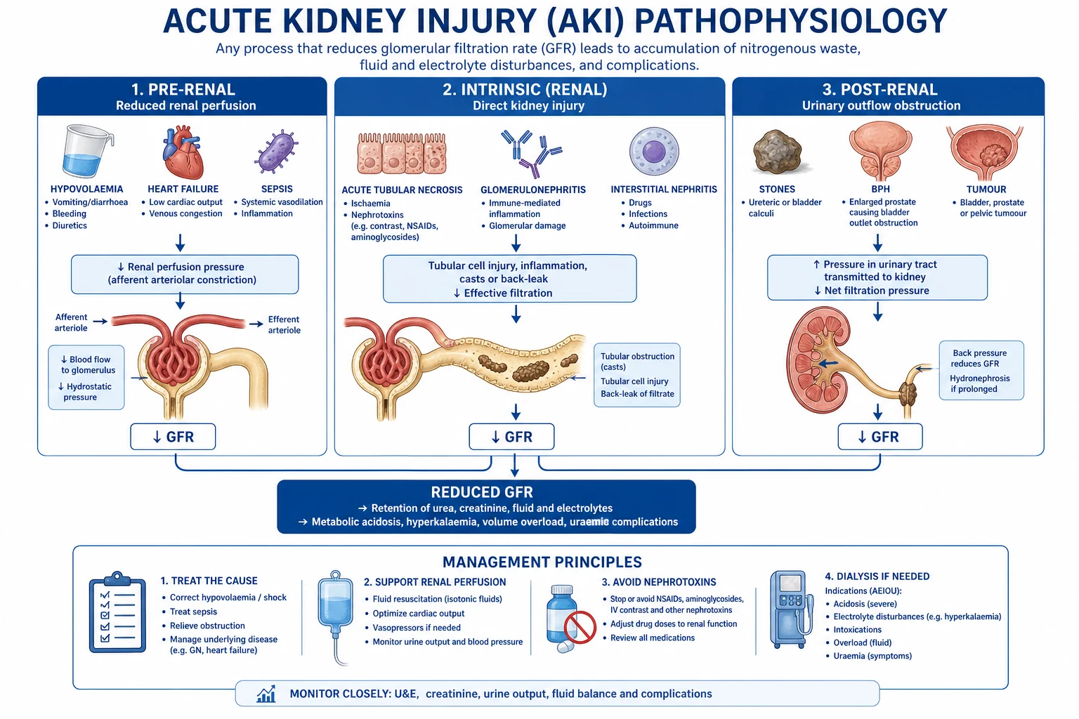
Pre-renal (commonest, ~70%)
Reversible if perfusion restored
- Reduced renal perfusion pressure: hypovolaemia (haemorrhage, dehydration, burns), low cardiac output (cardiogenic shock, heart failure), systemic vasodilation (sepsis, anaphylaxis), renal vasoconstriction (NSAIDs, hepatorenal).
- Kidney is structurally intact — restore perfusion promptly and creatinine recovers.
- KEY: afferent arteriole perfusion. NSAIDs constrict the afferent → ↓GFR. ACEi/ARB dilate the efferent → ↓GFR.
- Becomes intrinsic (ATN) if ischaemia is prolonged beyond hours — the pre-renal/ATN boundary is artificial and continuous, not binary.
Intrinsic (renal, ~25%)
Parenchymal damage
- Acute tubular necrosis (ATN): ischaemic or toxic (aminoglycosides, contrast, rhabdomyolysis-myoglobin, haemoglobinuria). The commonest intrinsic AKI in ICU.
- Acute interstitial nephritis (AIN): beta-lactams, PPIs, NSAIDs, vancomycin. Look for eosinophilia and sterile pyuria.
- Glomerular (rare in ICU): rapidly progressive GN, vasculitis — consider in pulmonary-renal syndromes.
- Vascular: malignant hypertension, cholesterol emboli, renal vein thrombosis.
- Often MULTIFACTORIAL in ICU: sepsis + nephrotoxin + hypoperfusion acting together.
Post-renal (~5%)
Obstruction — do not miss
- Bladder outlet (BPH, clot retention, neurogenic bladder), ureteric (stones, tumour, retroperitoneal fibrosis).
- ALWAYS exclude with a bladder scan / renal ultrasound — especially in anuric or elderly patients. The cheapest, fastest reversible cause.
- Easily reversible if relieved early; bilateral obstruction (or unilateral in a solitary kidney) causes AKI. Beware post-obstructive diuresis after relief.
- Residual volume measurement is part of the AKI workup in any unexplained case.
Risk factors — who to protect
Identify and protect the high-risk kidney
- Patient factors (constitutive) — Age >75 y (reduced renal reserve), pre-existing CKD (eGFR <60 — the single biggest risk factor), diabetes, heart failure, cirrhosis/ascites, cancer/haematological malignancy, chronic hypertension, peripheral vascular disease. These patients have LESS renal reserve — a small insult produces AKI.
- Acute insults (the AKI triggers) — Sepsis (commonest single cause of ICU AKI, ~50%), hypovolaemia/haemorrhage/shock, major surgery (cardiac surgery on bypass, vascular, hepatobiliary, emergency laparotomy), radiocontrast, rhabdomyolysis, massive transfusion, abdominal compartment syndrome.
- Nephrotoxin load (the PREVENTABLE burden) — NSAIDs, ACEi/ARB (especially in volume depletion/shock), aminoglycosides, amphotericin B, calcineurin inhibitors, iodinated contrast, vancomycin, tenofovir, aciclovir (crystallises in tubules), chemotherapy (cisplatin). REVIEW EVERY DRUG DAILY.
- Haemodynamic insults — Hypotension (each episode of SBP <90 doubles AKI risk), low cardiac output, venous congestion (high CVP/fluid overload impairs renal venous return → ↓GFR), abdominal compartment syndrome (pressure >20 mmHg).
- Act on risk BEFORE the insult — For elective high-risk procedures (contrast, cardiac surgery): pre-hydrate, hold nephrotoxins 24–48 h beforehand, consider remote ischaemic preconditioning and statin loading. For emergency cases: rapid resuscitation, early vasopressors, avoid nephrotoxins.
Nephrotoxins — mechanism and mitigation
NSAIDs
Afferent arteriole constriction
- Inhibit prostaglandin synthesis → loss of prostaglandin-mediated afferent arteriolar vasodilation → ↓GFR.
- Harmful in any patient with an activated RAAS/SNS (sepsis, cirrhosis, heart failure, volume depletion).
- CONTRAINDICATED in established AKI and in at-risk patients. Use paracetamol-based analgesia instead.
ACEi / ARB
Efferent arteriole dilation
- Block angiotensin II → loss of efferent arteriolar constriction → ↓intraglomerular pressure → ↓GFR.
- Beneficial long-term in CKD/HF, but cause functional AKI in volume depletion, sepsis, bilateral renal artery stenosis.
- HOLD in acute severe illness/shock; review on recovery. A mild rise (≤30%) is acceptable in stable CKD.
Aminoglycosides
Proximal tubule toxicity
- Gentamicin/tobramycin accumulate in proximal tubular cells → non-oliguric AKI after 5–7 days.
- PREVENT: once-daily extended-interval dosing (5–7 mg/kg) — higher peak, equivalent or less nephrotoxicity; monitor trough (goal <1) or AUC; limit duration ≤5–7 days; avoid concurrent loop diuretics.
- Also ototoxic. Use particular caution in CKD, older age, and existing AKI.
Iodinated contrast
Contrast-associated AKI (CA-AKI)
- Renal vasoconstriction plus direct tubular toxicity plus cast formation. Creatinine rises 24–72 h post-exposure.
- PREVENT: isotonic saline 1 mL/kg/h for 6–12 h pre and 12–24 h post (longer in CKD); minimise contrast volume; use low-/iso-osmolar agents; hold metformin and NSAIDs.
- NAC has NO benefit (PRESERVE). Statins may help. Bicarbonate is no better than saline (PRESERVE).
Vancomycin
AIN + trough-related
- Acute interstitial nephritis (dose/duration-related) — typical after ≥1 week of high troughs.
- PREVENT: AUC-guided dosing (target AUC 400–600) preferred over trough-only monitoring; avoid prolonged courses; monitor daily creatinine; stop early if possible.
- Risk is higher when combined with piperacillin-tazobactam (the VP combination) — prefer cefepime if renal risk is high.
Amphotericin B
Tubular toxicity
- Direct tubular membrane injury → distal RTA, Mg/K wasting, non-oliguric AKI.
- PREVENT: use LIPOSOMAL or lipid complex formulations (markedly less nephrotoxic); pre-hydrate; salt loading; monitor electrolytes (K, Mg).
Others
Crystalline / specific
- Aciclovir, sulphonamides, methotrexate, indinavir — crystallise in tubules. PREVENT: hydrate and dose-adjust for GFR.
- Tenofovir, cisplatin, ifosfamide — direct tubular toxicity. Monitor and dose-limit.
- Calcineurin inhibitors (ciclosporin, tacrolimus) — afferent vasoconstriction; monitor troughs.
Fluids, perfusion and venous congestion
Balanced crystalloids (Plasma-Lyte, Hartmann)
PREFERRED
- SMART trial (Semler, NEJM 2018): balanced crystalloids vs saline in critically ill adults — balanced reduced MAKE30 (Major Adverse Kidney Events: death, new RRT, or persistent renal dysfunction).
- Avoids the hyperchloraemic metabolic acidosis of saline, which itself causes renal vasoconstriction.
- First-line resuscitation fluid in ICU for patients at AKI risk.
0.9% Saline
Use with caution
- Causes hyperchloraemic acidosis → renal vasoconstriction → ↓GFR → AKI. Also promotes sodium/fluid overload.
- Acceptable for hyponatraemia, hypochloraemia, TURP syndrome, traumatic brain injury, and blood-product priming.
- SALT-ED and SMART both favour balanced crystalloids for renal outcomes.
Starches (HES)
CONTRAINDICATED
- Hydroxyethyl starch accumulates in proximal tubules → osmotic nephrosis → AKI. Also coagulopathy and pruritus.
- CONTRAINDICATED in sepsis and critical illness (CHEST, 6S, VISEP trials — increased need for RRT and increased mortality).
- Do NOT use HES for resuscitation. Crystalloids and albumin are acceptable alternatives.
Albumin 4%
Situational
- SAFE trial: albumin = crystalloid for overall mortality. No AKI benefit in general ICU.
- Reasonable in sepsis (SSC weak suggestion), severe hypoalbuminaemia with overload, and cirrhosis/spontaneous bacterial peritonitis.
- No role as routine AKI prevention.
The forgotten cause of AKI: venous congestion and fluid overload
- Renal perfusion depends on PERFUSION PRESSURE = MAP − renal venous pressure (≈CVP). A high CVP from fluid overload directly lowers the renal filtration gradient — the kidney is a pressure-driven organ wrapped in a tight capsule.
- Fluid overload → renal oedema → raised intra-abdominal/renal compartment pressure → venous congestion → ↓GFR → AKI. Cumulative positive fluid balance independently predicts AKI and mortality in multiple observational studies.
- Diagnose congestion clinically and with POCUS — raised JVP, peripheral/sacral oedema, bilateral B-lines on lung ultrasound, a plethoric IVC (>2 cm, non-collapsible), and loss of IVC variability in ventilated patients.
- Treat congestion, not just hypovolaemia — Diurese (furosemide) if overloaded; consider early RRT/CVVH if diuretic-resistant with progressive AKI. Aim for even or negative fluid balance after the resuscitation phase (CLASSIC, conservative-fluid approaches after stabilisation).
- Avoid the resuscitation–congestion–AKI spiral — Use dynamic fluid responsiveness (PLR, SVV, ΔPP) to STOP giving fluids once the patient is no longer responsive. Beyond responsiveness, escalate to vasopressors — not more fluid.
Sepsis-associated AKI (SA-AKI)
Sepsis-associated AKI — pathophysiology and prevention
- SA-AKI is NOT simply underfilling. Modern understanding: microcirculatory dysfunction (renal cortical hypoxia despite normal/global renal blood flow), mitochondrial dysfunction (cell hibernation rather than death), inflammation (PAMPs/DAMPs, cytokines, tubular activation), endothelial dysfunction, and apoptosis. The kidney is an active victim of sepsis, not just a passive bystander starved of blood.
- Diagnosis — distinguish from pre-renal — SA-AKI often has high urine sodium (FENa >2%) and muddy brown casts (ATN), but FENa is unreliable with diuretics and in early sepsis. Do NOT treat purely as pre-renal with fluid — many septic patients are NOT fluid-responsive.
- PREVENT through source control + haemodynamics, NOT more fluid — Surviving Sepsis: 30 mL/kg crystalloid is a starting suggestion (not a mandate), then reassess responsiveness. Early noradrenaline to restore MAP. Septic patients often need EARLY vasopressors to maintain renal perfusion pressure, not endless fluid.
- MAP target in sepsis — SEPSISPAM — Asfar (NEJM 2014): MAP 65 vs 80 mmHg in septic shock. No overall mortality difference. BUT in the chronic-hypertension subgroup, the higher MAP (80) reduced the need for RRT. CONCLUSION: target MAP 65; consider 75–80 in patients with chronic hypertension.
- Adjuncts that do NOT work in SA-AKI — Renal-dose dopamine (NO), fenoldopam (NO), low-dose vasopressin as renal protection (VANISH/VASST — no renal protection), furosemide for prevention (NO — may worsen). The only proven prevention is source control, antibiotics, and haemodynamic optimisation.
Biomarkers — detect AKI before creatinine
TIMP-2 × IGFBP7 (NephroCheck)
Cell cycle arrest — best validated
- Two cell-cycle arrest markers released by stressed (not yet dead) tubular cells within 1–2 h of injury. Urine test.
- Sapphire trial (Kashani, AJRCCM 2013): in critically ill patients, [TIMP-2]×[IGFBP7] >0.3 predicted AKI Stage 2–3 within 12 h with AUC 0.80 — superior to NGAL, cystatin C, KIM-1.
- Opal and Topaz validation studies confirmed: a value >2 has very high specificity (>90%) for imminent moderate–severe AKI.
- USE: risk-stratify cardiac surgery, sepsis, and contrast exposure. A high value → escalate monitoring, stop nephrotoxins, optimise perfusion.
NGAL
Neutrophil gelatinase-associated lipocalin
- Small protein produced by injured tubules and neutrophils. Rises within 2–6 h of AKI (vs 24–48 h for creatinine).
- Both plasma and urine forms. Useful in paediatric AKI, cardiac surgery, and contrast AKI.
- LIMITATIONS: elevated by sepsis (neutrophil source), systemic inflammation, chronic CKD, and malignancy — reduced specificity in mixed ICU populations.
Cystatin C
Better GFR marker, earlier rise
- Freely filtered by the glomerulus and fully reabsorbed/catabolised by the proximal tubule — NOT influenced by muscle mass, sex, or age (unlike creatinine).
- Better GFR estimate in elderly, cachexia, cirrhosis, and amputees. Rises ~24 h before creatinine in AKI.
- Increasingly used in AKI risk scores and contrast protocols; not yet universally available.
KIM-1, L-FABP, IL-18
Emerging / research
- KIM-1: proximal tubule injury marker, very specific for the kidney (vs systemic).
- L-FABP: oxidative stress marker in the proximal tubule. IL-18: inflammation marker, useful in AKI after cardiac surgery.
- Not routinely available; mostly research and multiplex panels. May feature in future composite scores.
Special populations
AKI prevention in specific high-risk contexts
- Cardiac surgery (CSA-AKI) — On-pump CPB causes haemolysis (free Hb), emboli, inflammation, and non-pulsatile flow. Prevention: minimise CPB time, avoid nephrotoxins pre-op, maintain perfusion during weaning, off-pump where feasible, remote ischaemic preconditioning (RIPC — ERICCA/RIPHeart mixed results). NephroCheck can risk-stratify post-op.
- Cirrhosis / hepatorenal physiology — HRS-AKI diagnostic pitfall: baseline creatinine underestimates renal impairment in cirrhosis (low muscle mass). Give an albumin challenge 1 g/kg (max 100 g/day) for 2 days to exclude hypovolaemia; terlipressin + albumin for confirmed HRS. AVOID NSAIDs and large-volume paracentesis without albumin cover.
- Rhabdomyolysis — Myoglobin nephrotoxicity (pigment casts). Prevention: aggressive early IV fluid expansion (goal UO 200–300 mL/h), maintain euvolaemia, alkaline diuresis (controversial), treat compartment syndrome. The CK treatment threshold is often >5000 U/L (crush); lower for smaller insults.
- Major trauma / haemorrhage — Damage-control resuscitation: brief permissive hypotension, early blood products (1:1:1 PRBC:FFP:platelets — PROPPR), minimise crystalloid, reverse coagulopathy, prevent abdominal compartment syndrome, treat rhabdomyolysis.
- Bone marrow transplant / oncology — Tumour lysis syndrome (prophylactic rasburicase/allopurinol + hydration), calcineurin-inhibitor toxicity, sepsis, chemo-nephrotoxicity (cisplatin, ifosfamide). Aggressive hydration and electrolyte monitoring.
RRT timing — when to start
AKIKI vs ELAIN vs STARRT-AKI — early vs delayed RRT
AKIKI (Gaudry, NEJM 2016, PMID 27532830): 624 patients with KDIGO Stage 2–3 AKI. Early (RRT within 6 h of Stage 2/3) vs delayed (RRT only for an urgent indication or persistent Stage 3). Result: NO difference in 60-day mortality (48% vs 50%). ~50% of the delayed group never needed RRT. CONCLUSION: no benefit of early initiation; avoid unnecessary RRT. [1]
ELAIN (Zarbock, JAMA 2016, PMID 27557141): 231 patients with KDIGO Stage 2 + a positive biomarker (NGAL >150). Early (within 8 h) vs delayed (within 12 h of Stage 3). Result: REDUCED 90-day mortality (39% vs 55%). BUT single-centre, small, biomarker-selected — not generalisable. [1]
STARRT-AKI (NEJM 2020, PMID 32937079): 3019 patients with Stage 2–3 AKI. Accelerated (early RRT) vs standard (urgent indication). Result: NO difference in 90-day mortality (43.9% vs 43.7%). MORE adverse events (hypotension, bleeding, catheter-related infection) in the accelerated group. CONCLUSION: early RRT may be HARMFUL. [1]
OVERALL CONSENSUS: Start RRT for URGENT indications — the AEIOU mnemonic: (A)cidosis pH <7.1, (E)lectrolyte K+ >6.5 refractory, (I)ngestion of a dialysable toxin (lithium, metformin, salicylate), (O)verload refractory to diuretics, (U)raemia (pericarditis, encephalopathy, bleeding). Do NOT start early for prevention.
PRESERVE — NAC and bicarbonate for contrast nephropathy
PRESERVE (Weisbord, NEJM 2018, PMID 29172640): 5177 patients at risk of CA-AKI undergoing angiography. A 2×2 factorial of IV bicarbonate vs isotonic saline AND oral N-acetylcysteine vs placebo. Primary outcome: composite of death, need for RRT, or persistent renal impairment at 90 days. Result: NO benefit of NAC over placebo AND NO benefit of bicarbonate over saline. CONCLUSION: use isotonic saline (not bicarbonate) and placebo (not NAC) — stop prescribing NAC for contrast prophylaxis. [1]
Exam implication: when asked about contrast-nephropathy prevention, the correct answer is isotonic saline hydration — NOT NAC, NOT bicarbonate. Also minimise contrast volume, use low-/iso-osmolar contrast, and hold nephrotoxins.
Additional high-yield clinical pearls
[1] [2]Additional red flags
[1] [3] [4]Exam practice — SAQs
SAQ — AKI prevention before contrast exposure in a high-risk patient (PRESERVE trial)
10 minutes · 10 marks
A 74-year-old man with type 2 diabetes, hypertension, and known CKD (baseline creatinine 160 micromol/L, eGFR 38) is admitted with an NSTEMI and is scheduled for urgent coronary angiography with likely PCI. His usual medications are ramipril 10 mg, amlodipine 10 mg, metformin 1 g twice daily, and aspirin. He weighs 82 kg. The cardiology team asks the ICU to optimise his renal risk before contrast administration.
SAQ — Renal-sparing haemodynamics in septic AKI: fluids, vasopressors, and venous congestion
10 minutes · 10 marks
A 66-year-old woman is admitted to ICU with septic shock from ascending cholangitis. She received 30 mL/kg of 0.9% saline in the ED and is now on noradrenaline 0.25 mcg/kg/min with a MAP of 64 mmHg. Her creatinine has risen from 90 to 175 micromol/L over 24 h and her urine output is 0.3 mL/kg/h. Lactate is 3.2 mmol/L. Lung ultrasound shows bilateral B-lines and her IVC is plethoric and non-collapsible. Cumulative fluid balance is +5 L. The registrar suggests giving another fluid bolus and starting a furosemide infusion to drive urine output.
References
- [1]KDIGO Acute Kidney Injury Work Group. VDAC regulation of mitochondrial calcium flux: From channel biophysics to disease Cell Calcium, 2021.PMID 33529977
- [2]Prowle JR, et al. Notum palmitoleoyl-protein carboxylesterase regulates Fas cell surface death receptor-mediated apoptosis via the Wnt signaling pathway in colon adenocarcinoma Bioengineered, 2021.PMID 34402722
- [3]Gaudry S, Hajage D, Schortgen F, et al. Closed-Loop Insulin Delivery during Pregnancy in Women with Type 1 Diabetes N Engl J Med, 2016.PMID 27532830
- [4]STARRT-AKI Investigators, Golper TA, Wald R, et al. Cardiac electrophysiological effects of ibuprofen in dog and rabbit ventricular preparations: possible implication to enhanced proarrhythmic risk Can J Physiol Pharmacol, 2021.PMID 32937079
- [5]Zarbock A, Kellum JA, Schmidt C, et al. Thyroid disorders in Brazil: time for action Sao Paulo Med J, 2016.PMID 27557141
- [6]Weisbord SD, Gallagher M, Jneid H, et al.; PRESERVE Trial Group. The art is in the delivery Aust N Z J Psychiatry, 2018.PMID 29172640
- [7]Kashani K, Al-Khafaji A, Ardiles T, et al. Economic costs of diabetes in Saudi Arabia J Family Community Med, 2013.PMID 23723724
- [8]Ronco C, Bellomo R, Kellum JA. Can sand nourishment material affect dune vegetation through nutrient addition? Sci Total Environ, 2020.PMID 32278174
- [9]Asfar P, Meziani F, Hamel JF, et al.; SEPSISPAM Investigators. Lower versus higher hemoglobin threshold for transfusion in septic shock N Engl J Med, 2014.PMID 25270275
- [10]Semler MW, Self WH, Wanderer JP, et al.; SMART Investigators and the Pragmatic Critical Care Research Group. A Phase 3 Trial of l-Glutamine in Sickle Cell Disease N Engl J Med, 2018.PMID 30021096