MedVellum
MCQsExamsAtlas
DashboardPricing
MBBS / Core medicine✳Dermatology✳ICU Fellowship (CICM)✳Anaesthesia✳Emergency Medicine✳Psychiatry Fellowship✳Paediatrics Fellowship✳Physician Medicine✳MCQs✳SAQs✳Vivas✳OSCE✳Evidence-first✳MBBS / Core medicine✳Dermatology✳ICU Fellowship (CICM)✳Anaesthesia✳Emergency Medicine✳Psychiatry Fellowship✳Paediatrics Fellowship✳Physician Medicine✳MCQs✳SAQs✳Vivas✳OSCE✳Evidence-first✳

MedVellum.

The folio

Exam-exhaustive medical education across every specialty — evidence-graded topics, engraved plates, and practice in every written and oral format. Educational content only — not medical advice.

llms.txt · psychiatry LLM catalog · sitemap

Atlas

  • Specialty atlas
  • MBBS / Core medicine
  • Dermatology
  • ICU Fellowship (CICM)
  • Anaesthesia
  • Emergency Medicine
  • Psychiatry Fellowship
  • Paediatrics Fellowship
  • Physician Medicine

Study & account

  • MCQ practice
  • Practice alias
  • Exam tools
  • Dashboard
  • Pricing
  • Sign in

© 2026 MedVellum. For education only — not a substitute for clinical judgement.

Folio edition · Set in Instrument Serif & Archivo

ICU TopicsRenal/Metabolic

ICU · Renal/Metabolic

Acute kidney injury: prevention, biomarkers, and RRT timing

Also known as AKI prevention · AKI biomarkers · NGAL · Cystatin C · RRT timing · ELAIN trial · AKIKI trial · STARRT-AKI trial · KDIGO definition · AEIOU indications · NephroCheck · TIMP-2 IGFBP7

AKI prevention and management in ICU. PREVENTION: avoid nephrotoxins (NSAIDs, aminoglycosides, contrast), maintain perfusion (adequate BP, goal-directed fluids), monitor drugs. BIOMARKERS: NGAL (neutrophil gelatinase-associated lipocalin) and cystatin C rise EARLIER than creatinine (within hours vs 24-48h) — may allow EARLIER detection and intervention. KDIGO DEFINITION: creatinine rise (≥26.5 μmol/L in 48h or ≥1.5× baseline) or urine output <0.5 mL/kg/h for ≥6h. RRT TIMING: ELAIN trial (early RRT within 8h of AKI stage 2) vs AKIKI trial (RRT only for urgent indications) vs STARRT-AKI (accelerated strategy) — current consensus favours waiting for URGENT indications (AEIOU: Acidosis, Electrolytes, Ingestion/toxin, Overload, Uraemia) rather than routine early RRT. AKIKI (2016) and STARRT-AKI (2020): no benefit of early RRT.

high13 referencesUpdated 3 July 2026
On this page & tools

Your progress

Saved locally on this device.

Target exams

CICMFFICMEDIC

Red flags

AKIKI and STARRT-AKI: EARLY RRT does NOT improve outcomes (start for URGENT indications, not routine early)Avoid nephrotoxins (NSAIDs, aminoglycosides, ACEi in shock) — most preventable AKI causeNGAL/cystatin C rise earlier than creatinine — may allow early detectionAEIOU mnemonic for urgent RRT: Acidosis, Electrolytes, Ingestion (toxin), Overload (fluid), UraemiaEven KDIGO stage 1 AKI doubles mortality — prevention is the single best strategyAKIKI 50% of 'delayed' arm never needed RRT — early initiation exposes patients unnecessarily

Your progress

Saved locally on this device.

Target exams

CICMFFICMEDIC

Red flags

AKIKI and STARRT-AKI: EARLY RRT does NOT improve outcomes (start for URGENT indications, not routine early)Avoid nephrotoxins (NSAIDs, aminoglycosides, ACEi in shock) — most preventable AKI causeNGAL/cystatin C rise earlier than creatinine — may allow early detectionAEIOU mnemonic for urgent RRT: Acidosis, Electrolytes, Ingestion (toxin), Overload (fluid), UraemiaEven KDIGO stage 1 AKI doubles mortality — prevention is the single best strategyAKIKI 50% of 'delayed' arm never needed RRT — early initiation exposes patients unnecessarily
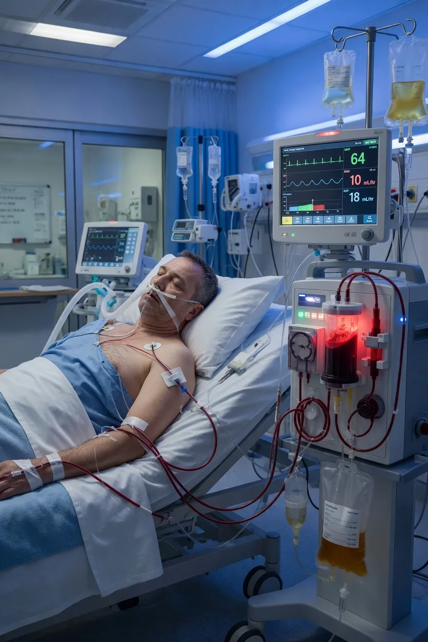
Cinematic ICU scene of a critically-ill patient on vasopressors beside a covered continuous renal replacement therapy machine, a creatinine trend on the monitor climbing, a nephrotoxin medication chart marked for review, clinical-blue lighting, medical educational, no faces, no text
FigureAKI prevention is the only intervention with proven mortality benefit — avoid nephrotoxins, maintain perfusion, use balanced crystalloids. Damage biomarkers (NGAL, TIMP-2·IGFBP7) rise hours before creatinine. RRT timing: AKIKI and STARRT-AKI showed NO benefit (and possible harm) from early RRT — start only for URGENT indications, captured by AEIOU (Acidosis, Electrolytes, Ingestion, Overload, Uraemia).

In one line

AKI prevention: avoid nephrotoxins (NSAIDs, aminoglycosides, contrast), maintain perfusion (BP, fluids), monitor drugs. Biomarkers: NGAL, cystatin C rise earlier than creatinine (hours vs 24-48h) — early detection. RRT timing: AKIKI and STARRT-AKI: NO benefit of EARLY RRT — start for URGENT indications only (AEIOU: Acidosis, Electrolytes, Ingestion/toxin, Overload, Uraemia). ELAIN suggested benefit but not confirmed. PREVENTION remains the best strategy.

[1]

KDIGO definition and staging

KDIGO (Kidney Disease: Improving Global Outcomes) defines AKI by either a functional criterion (creatinine) or a flow criterion (urine output). Either is sufficient — they do NOT have to coexist. The diagnosis is made when any ONE of the following holds: [1]

  • Creatinine rise of ≥26.5 μmol/L (≥0.3 mg/dL) within 48 h, OR
  • Creatinine rise to ≥1.5× the baseline value (known or presumed to have occurred within the prior 7 days), OR
  • Urine output <0.5 mL/kg/h for ≥6 consecutive hours. [1]

Once AKI is diagnosed it is staged 1→3 by severity. Stage 3 includes anyone who has been started on RRT, regardless of preceding creatinine or urine output.[5]

| 1 | ≥1.5–1.9× baseline OR ≥26.5 μmol/L (0.3 mg/dL) rise in 48 h | <0.5 mL/kg/h for 6–12 h | | 2 | ≥2.0–2.9× baseline | <0.5 mL/kg/h for ≥12 h | | 3 | ≥3× baseline OR rise to ≥353.6 μmol/L (4.0 mg/dL) OR initiated on RRT OR (in <18 yr) eGFR drop to <35 mL/min/1.73 m² | <0.3 mL/kg/h for ≥24 h OR anuria for ≥12 h |

KDIGO AKI staging — creatinine and urine output criteria
[{"header":"Stage"},{"header":"Serum creatinine"},{"header":"Urine output"}]
[1]

| Pre-renal (most common in ICU) | Reduced glomerular perfusion — intact tubules | Hypovolaemia, sepsis vasodilation, low CO (cardiogenic shock), ACEi/ARB efferent dilatation | FENa <1%, urine osmol >500, BUN:Cr >20, responds to fluids (if volume-responsive) | | Intrinsic (ATN dominates) | Tubular / glomerular / interstitial / vascular injury — ATN dominates in ICU (sepsis, ischaemia, nephrotoxin) | ATN (ischaemic, septic, nephrotoxic), AIN (β-lactams, PPIs, NSAIDs), AGN, vasculitis | FENa >2%, muddy-brown granular casts, fails to recover after perfusion restored | | Post-renal (rarely isolated in ICU) | Urinary tract obstruction — bilaterally or in single kidney | BPH, prostate/cloncal cancer, retroperitoneal fibrosis, blocked catheter, calculi | Hydronephrosis on POCUS/CT, anuria that resolves with catheterisation |

Pre-renal vs intrinsic vs post-renal AKI — the three mechanistic buckets
[{"header":"Type"},{"header":"Mechanism"},{"header":"Examples"},{"header":"Early clue"}]
[1]

KDIGO-aligned AKI work-up on the ICU round (ABCDE)

  1. A — Assess risk and recognise. Pull the KDIGO risk profile: sepsis, CKD (eGFR <60), diabetes, age >75, heart failure, cirrhosis, hypovolaemia, recent contrast/NSAID/aminoglycoside/vancomycin exposure. If at risk → daily creatinine + hourly urine output chart.
  2. B — Baseline. Determine the baseline creatinine (the most recent normal value in the prior 3 months). If unavailable, back-calculate from a MDRD-derived estimate assuming an eGFR of 75 mL/min/1.73 m². Without a baseline you cannot stage AKI.
  3. C — Cause. Pre-/intrinsic/post-renal screen: history (drugs, hypotension, rash, pigment), exam (volume status, bladder, prostate), urinalysis (dipstick + microscopy for casts), bedside POCUS (hydronephrosis, B-lines, IVC, bladder). Send FENa and, if available, urine microscopy.
  4. D — Drugs and diagnostics. Renally dose-adjust every renally-cleared drug; stop and review every nephrotoxin (the 'nephrotoxin time-out'). Check K⁺, Mg²⁺, phosphate, bicarbonate/venous lactate, FBC, CK (if pigmenturia), troponin, and — if diagnosis uncertain — ANA, ANCA, complements, myoglobin, free light chains.
  5. E — Escalate and exit. Decide whether RRT is needed NOW (apply AEIOU) or whether the patient can be managed conservatively (fluids if responsive, vasopressors, K⁺ binders, bicarbonate if pH 7.1–7.2 with shock). Document a recovery plan and a re-check interval.
[1]

Prevention bundle

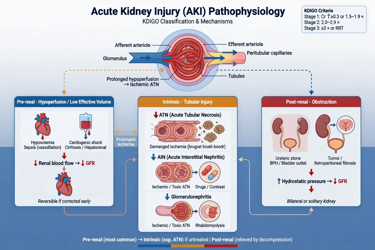
Educational diagram of AKI mechanisms: hypoperfusion, nephrotoxins, inflammation, and rising damage biomarkers before creatinine, clinical-blue educational style, no faces
FigurePathophysiology — creatinine lags; damage biomarkers and the prevention bundle target the reversible drivers of ICU-AKI.

Prevention is the only intervention with proven mortality benefit in AKI. Once AKI is established, no pharmacological agent (including dopamine, fenoldopam, nesiritide, ANP, furosemide) has been shown to alter its course. The KDIGO bundle is therefore a small set of high-yield, low-cost manoeuvres.[5][6]

| Haemodynamics | Achieve and maintain adequate MAP (≥65 mmHg, individualise in chronic HTN); use vasopressors after volume resuscitation; monitor lactate clearance | Permissive hypotension beyond the first 6 h of shock; flooding with crystalloid once patient no longer fluid-responsive | | Fluids | Use balanced crystalloids in shock; reassess responsiveness every 4–6 h; de-resuscitate once shock resolves | Sustained 0.9% saline (hyperchloraemic acidosis, AKI risk — SMART trial); hydroxyethyl starch (RENAL and CHEST trials — harm); albumin except in cirrhosis with bacterial peritonitis | | Drugs | Once-daily extended-interval aminoglycoside dosing with TDM; dose-adjust vancomycin to AUC/MIC; lipid formulations of amphotericin B | Concurrent ≥2 nephrotoxins; NSAIDs in shock/CKD/decompensated HF; ACEi/ARB initiation during shock; empiric high-dose loop diuretics to 'force urine' | | Contrast | Pre- and post-procedure isotonic saline; use lowest possible contrast volume; hold metformin/diuretics peri-procedure | Routine N-acetylcysteine (PRESERVE trial — no benefit); prophylactic RRT after contrast; intra-arterial gadolinium | | Glycaemic control | Insulin to keep glucose 6–10 mmol/L; avoid hypoglycaemia | Severe hyperglycaemia (osmotic diuresis, volume depletion) | | Source control | Drainage / debridement / removal of infected hardware within 6–12 h of septic shock recognition | Delayed source control — each hour of delay in septic shock adds ~7.6% to mortality |

KDIGO AKI prevention bundle — what to do and what NOT to do
[{"header":"Domain"},{"header":"DO"},{"header":"AVOID / MINIMISE"}]
[1]

| SMART (Semler 2018) | 15,802 | Balanced (lactated Ringer's/Plasma-Lyte) vs 0.9% saline for 30 days | Significantly less MAKE-30 (Major Adverse Kidney Events — death, new RRT, or persistent renal dysfunction): 14.3% vs 15.4% | | SPLIT (Young 2015) | 2,278 | Buffered vs saline in ICU | No difference in AKI — small, single-region, saline volume low | | SMART high-risk subgroup | 1,641 | Balanced vs saline in septic shock | MAKE-30 23.0% vs 30.0% — larger effect in sepsis | | Practical rule | — | — | Default to balanced crystalloid in shock; reserve saline for hyponatraemia, brain injury, mandatory chloride load |

ICU crystalloids and AKI — what the trials actually showed
[{"header":"Trial"},{"header":"n"},{"header":"Comparison"},{"header":"AKI signal"}]
[1]

Goal-directed vs liberal fluid therapy — the de-resuscitation phase

The kidney is a victim of both under-resuscitation (ischaemic ATN) AND of fluid accumulation (interstitial oedema raises intra-renal pressure, lowers GFR, and independently worsens survival). The CLASSIC trial (liberal vs restrictive fluids in septic shock after initial resuscitation) showed no mortality difference but the restrictive arm did no harm — supporting the 'give less after the first 4–6 h' principle. Bouchard's PICARD analysis showed patients whose cumulative fluid balance exceeded 10% of body weight had roughly double the mortality of those who stayed near net-zero. The practical arc is therefore: resuscitate aggressively to achieve responsiveness → switch to maintenance only → actively de-resuscitate with loop diuretics once shock resolves. The aim is a fluid balance that trends back toward zero within 72 h.

[1]

Biomarkers — functional vs damage markers

A central KDIGO concept is the distinction between functional markers (creatinine, cystatin C, urine output — they fall only AFTER the GFR has dropped) and damage markers (NGAL, KIM-1, IL-18, L-FABP, and the cell-cycle arrest pair TIMP-2·IGFBP7 — they rise within hours of tubular injury, BEFORE GFR falls). Damage markers therefore create a 'pre-AKI' window — patients at risk of, but not yet meeting criteria for, AKI — during which prevention is most effective.[4]

| Creatinine | Functional (GFR) | Serum | Loss of filtration (any cause) | 24–48 h after injury | | Cystatin C | Functional (GFR) | Serum | Loss of filtration — freely filtered, not muscle-mass dependent | 12–24 h (earlier than creatinine) | | Urea / BUN | Functional + volume | Serum | Filtration loss AND volume depletion (BUN:Cr >20 suggests pre-renal) | Variable | | NGAL (lipocalin-2) | Damage (tubular) | Urine or plasma | Tubular epithelial stress — ischaemia, sepsis, nephrotoxin | 2–6 h | | KIM-1 | Damage (proximal tubule) | Urine | Proximal tubule injury, especially ischaemic / toxic | 12–24 h | | IL-18 | Damage (inflammatory) | Urine | Inflammatory / ischaemic ATN (distinguishes from pre-renal) | 4–12 h | | L-FABP | Damage (oxidative) | Urine | Oxidative tubular stress | 4–12 h | | TIMP-2·IGFBP7 (NephroCheck) | Damage (cell-cycle arrest) | Urine | Cell-cycle arrest — risk of AKI stage 2–3 within 12 h | 4–12 h |

AKI biomarkers — what they tell you and when
[{"header":"Biomarker"},{"header":"Type"},{"header":"Source"},{"header":"What it detects"},{"header":"Time-to-rise"}]
[1]

NephroCheck (TIMP-2·IGFBP7) — the only FDA-cleared AKI risk biomarker

The Sapphire study (Kashani, Crit Care 2013) screened over 300 candidate biomarkers and identified the urinary product of two cell-cycle arrest proteins — tissue inhibitor of metalloproteinases-2 (TIMP-2) and insulin-like growth factor-binding protein 7 (IGFBP7) — as the most accurate predictor of moderate-to-severe AKI (KDIGO stage 2–3) within the subsequent 12 h in critically ill patients. AUC 0.80, with risk stratified by a single urinary value (cut-off 0.3 for 'at-risk'; 2.0 for 'high-risk'). It was the first AKI biomarker cleared by the FDA. Clinically it shifts the question from 'do you have AKI?' to 'are you ABOUT to have AKI?' — useful in high-risk cohorts (cardiac surgery, contrast exposure, septic shock) where a positive result justifies an aggressive prevention bundle and avoidance of nephrotoxins. It does NOT, however, change the trigger for RRT — that remains clinical.

[1]

Why creatinine is a delayed and misleading biomarker in ICU AKI

Creatinine is a lagging indicator. It is generated at a near-constant rate from muscle, freely filtered, partly secreted by the proximal tubule, and excreted in urine — so its serum level reflects the steady-state between generation and elimination. In ICU this steady-state is broken by: (1) reduced muscle mass (cachexia, burns, amputation) — underestimates AKI severity; (2) volume expansion — dilutes creatinine by up to 30% after 5 L of resuscitation; (3) blocked tubular secretion by trimethoprim, cimetidine, cobicistat — mimics AKI without injury; (4) hyperbilirubinaemia interferes with the Jaffé assay. The result is that a 'normal' creatinine in a resuscitated, oedematous, paralysed ICU patient can hide a GFR of <30 mL/min. This is exactly why KDIGO emphasises urine output trends and why damage biomarkers add value.

[1]

AEIOU — urgent indications for RRT

The mnemonic AEIOU captures the only reasons to start RRT on a patient who does not have a relative indication already met. They are absolute, not negotiable — each represents a situation where conservative therapy has failed or cannot work.[6]

| A | (refractory) Acidosis — metabolic | pH <7.1–7.15 despite buffer and source control (some use pH <7.2 with haemodynamic instability) | Bicarbonate generation by the kidney has failed and buffer alone worsens sodium/CO₂ load | | E | Electrolytes — typically hyperkalaemia | K⁺ >6.5 mmol/L with ECG changes, or >7.0 mmol/L regardless; also tumour lysis (K⁺, phosphate, Ca²⁺×P >80) and severe hypernatraemia refractory to free water | Medical therapy (calcium, insulin/dextrose, salbutamol, resonium) only shifts K⁺ intracellularly — it will rebound | | I | Ingestion / Intoxication — dialyzable toxin | Lithium (>4 mmol/L or symptomatic), metformin-associated lactic acidosis (pH <7.25), salicylate (>6.5 mmol/L or neurotoxicity), methanol/ethylene glycol (anion-gap acidosis + osmolar gap), valproate, carbamazepine | Removes the toxin directly; haemodialysis preferred for small water-soluble molecules; haemoperfusion for theophylline | | O | Overload — fluid, refractory to diuretics | Pulmonary oedema unresponsive to high-dose loop diuretic ± thiazide; cumulative fluid balance >10% body weight with rising oxygen requirement | Slow continuous ultrafiltration (SCUF) or CVVH removes 100–200 mL/h while maintaining haemodynamics | | U | Uraemia — complications of | Uraemic pericarditis (rub + effusion), uraemic encephalopathy (asterixis, seizures, coma), uraemic bleeding (platelet dysfunction), intractable uraemic pruritus or nausea | Removes middle molecules and reduces toxin load that is causing the end-organ complication |

AEIOU — the urgent (absolute) indications for RRT
[{"header":"Letter"},{"header":"Indication"},{"header":"Typical threshold"},{"header":"Why RRT"}]
[1]

Two extra 'I' triggers worth knowing — Inflammation and Intoxications caveat

Some ICU texts extend AEIOU to AEIOU + I₂: refractory Inflammation (e.g. thermoregulatory instability in malignant hyperthermia / severe heat stroke where RRT is used as a cooling adjunct) and Intoxications (the dialyzable-drug list). The dialyzability of an intoxicant depends on four properties — low molecular weight, low volume of distribution (ideally <1 L/kg), low protein binding, and water solubility. Lithium, metformin, salicylate, methanol, ethylene glycol, valproate and carbamazepine fit these criteria. Critically, the answer to 'should this be dialysed?' for an intoxication comes from the EXTRIP workgroup consensus statements, not from the drug level alone — clinical severity (coma, acidosis, end-organ failure) drives the decision.

[1]

RRT timing — the four trials

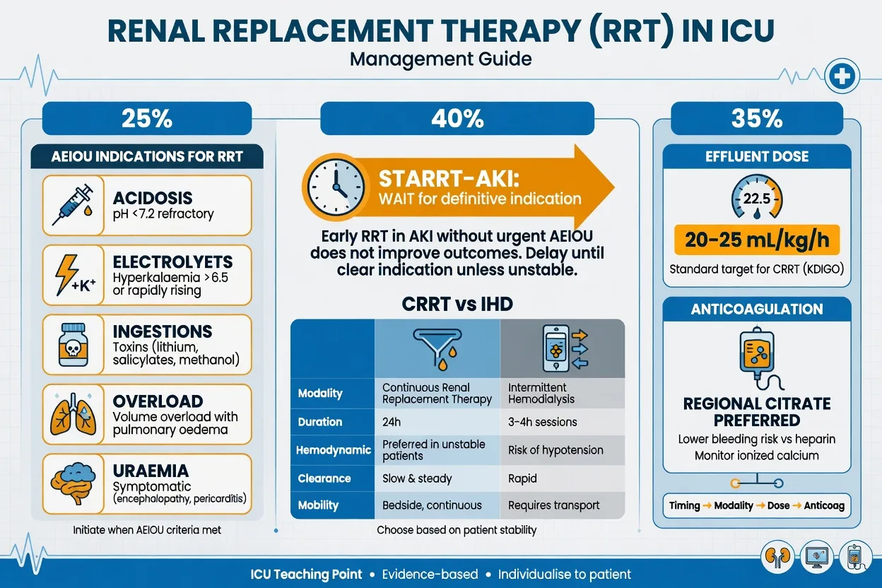
Decision pathway for RRT timing: AEIOU absolute indications, delayed strategy from AKIKI and STARRT-AKI, avoid early RRT without urgent indication
FigureRRT timing — start for AEIOU urgencies; early RRT without absolute indication did not improve survival in large RCTs.

Four multicentre randomised trials now define the evidence base for RRT initiation timing in ICU-AKI. They are unified by one conclusion: routine, accelerated initiation does not improve survival, and in STARRT-AKI may harm.[1][2][3][7]

| AKIKI (Gaudry 2016) | 620 (KDIGO stage 2) | RRT within 6 h of stage 2 (mostly within 18 h) | RRT only on urgent indication or anuria for >72 h; 49% never started RRT | 60-day mortality 48% vs 50% — no difference | | ELAIN (Zarbock 2016) | 231 (KDIGO stage 2 + NGAL >150 or stage 3) | RRT within 8 h of stage 2 | RRT within 12 h of stage 3; 91% started RRT | 90-day mortality 39% vs 55% — early better (absolute risk reduction 16%) | | IDEAL-ICU (Barbar 2018) | 488 (septic shock, RIFLE 'Failure') | RRT within 12 h of stage 3 (Failure) | RRT after 48 h delay if no recovery | 90-day mortality 58% vs 54% — no difference; stopped early for futility | | STARRT-AKI (Bagshaw 2020) | 3,019 (KDIGO stage 2/3 + shock or organ failure) | Accelerated RRT within 12 h | Standard strategy — wait for urgent indication or AKI persists >72 h; 62% started RRT | 90-day mortality 43.9% vs 43.7% — no difference; MORE adverse events (RRT dependence 10.0% vs 6.0%) |

The four RRT-timing RCTs — head-to-head
[{"header":"Trial (year)"},{"header":"n"},{"header":"Early arm"},{"header":"Delayed/Standard arm"},{"header":"Primary outcome"}]
[1]

AKIKI — Gaudry 2016 (NEJM) — early vs delayed RRT

Population: 620 French ICU patients with KDIGO stage 2 AKI (≥2× baseline creatinine OR <0.5 mL/kg/h for ≥12 h) and at least one organ failure. Design: Multicentre, open-label, parallel-group RCT. Intervention: Early RRT started immediately (median 2 h after randomisation, mostly within 6 h) vs delayed strategy in which RRT was started ONLY if one of the urgent indications developed or if anuria persisted >72 h. Result: 60-day mortality 48% (early) vs 50% (delayed) — no difference (P=0.79). Critically, 49% of the delayed group NEVER needed RRT — meaning that routine early initiation exposes half of patients to an invasive therapy they did not need. Catheter-related infections and bleeding were more common in the early arm. Take-home: In severe ICU-AKI without an urgent indication, waiting is safe. The delayed strategy is the default.

[1]

STARRT-AKI — Bagshaw 2020 (NEJM) — accelerated vs standard RRT

Population: 3,019 critically ill patients across 168 ICUs in 15 countries, with KDIGO stage 2/3 AKI AND at least one non-renal organ failure or shock. Design: International, multicentre, parallel-group RCT. Intervention: Accelerated strategy — RRT within 12 h of stage 2/3 diagnosis. Standard strategy — wait for urgent indication OR AKI persisting beyond 72 h. Result: 90-day mortality 43.9% (accelerated) vs 43.7% (standard) — no difference. Adverse events (a composite of RRT dependence at 90 days, major bleeding, catheter-related infection) were significantly MORE common with accelerated RRT — 34.6% vs 30.5%. RRT dependence at 90 days occurred in 10.0% of the accelerated group vs 6.0% of the standard group. Take-home: The largest, most generalisable RRT-timing trial to date shows that accelerating initiation confers no benefit and exposes patients to harm. The standard (delayed) strategy should be the default in the absence of an urgent indication.

[1]

ELAIN — Zarbock 2016 (JAMA) — early vs delayed RRT in KDIGO stage 2

Population: 231 German ICU patients with KDIGO stage 2 AKI plus a damage biomarker (urinary NGAL >150 ng/mL) or progression to KDIGO stage 3. Design: Single-centre, randomised, open-label. Intervention: Early RRT within 8 h of stage 2 vs delayed RRT within 12 h of progression to stage 3. Result: 90-day mortality 39% (early) vs 55% (delayed) — absolute risk reduction 16%, number-needed-to-treat 6.2. Reduced duration of RRT and shorter hospital length of stay in the early group. Caution: This is the lone positive trial. It was small, single-centre, used NGAL selection (enriching for patients with active tubular injury), and over 90% of the delayed arm eventually received RRT. AKIKI and STARRT-AKI — larger and multicentre — failed to reproduce the signal. ELAIN remains an interesting outlier but has not changed international consensus. Take-home: ELAIN is the trial that makes the 'early RRT in stage 2' question live. For exam purposes, know it AND know that AKIKI/STARRT-AKI superseded it.

[1]

IDEAL-ICU — Barbar 2018 (NEJM) — early vs delayed RRT in septic shock AKI

Population: 488 French ICU patients with septic shock and RIFLE 'Failure'-class AKI (urine output <0.3 mL/kg/h for ≥6 h OR creatinine ≥3× baseline). Design: Multicentre, randomised, open-label; stopped early for futility after second interim analysis. Intervention: Early RRT within 12 h of 'Failure' class vs delayed RRT after a 48-h waiting period (if no renal recovery). Result: 90-day mortality 58% (early) vs 54% (delayed) — no difference. The trial was underpowered due to early stopping; 38% of the delayed group never received RRT. Take-home: Even in the high-risk subgroup of septic shock with severe AKI, an early strategy does not improve survival. Strengthens the AKIKI/STARRT-AKI consensus.

[1]

Applying RRT timing at the bedside — a decision algorithm

  1. Confirm the patient meets KDIGO stage 2 or 3 criteria. Staging requires a baseline creatinine — find one (or back-calculate). Document stage on the chart.
  2. Run through AEIOU. Acidosis (pH <7.1–7.15)? Electrolytes (K⁺ >6.5 with ECG change)? Ingestion (lithium, metformin, salicylate, methanol, ethylene glycol)? Overload (pulmonary oedema unresponsive to diuretics)? Uraemia (pericarditis, encephalopathy, bleeding)? If ANY is present → start RRT now.
  3. If no urgent indication, reassess every 12–24 h. Re-check creatinine, urine output, K⁺, bicarbonate, fluid balance, and overall trajectory. The question is not 'does the patient have AKI?' but 'is the patient improving, static, or worsening?'.
  4. Declare anuria plateau. If a patient has had anuria for >72 h despite reversible causes being addressed, RRT should be initiated (the AKIKI delayed criterion) — the kidney has shown it will not recover on its own in the immediate term.
  5. Pick the modality to match the patient. Continuous (CVVHDF) for the haemodynamically unstable, fluid-overloaded, or brain-injured. Intermittent HD for stable, mobile patients needing solute clearance. Sustained low-efficiency dialysis (SLED) as a hybrid. Peritoneal for the paediatric or vascular-access-poor adult.
  6. Set the dose and reassess. KDIGO dose target: 20–25 mL/kg/h of effluent for CRRT (delivered is usually 80% of prescribed due to interruptions). Monitor daily — effluent dose, filter life, anticoagulation (citrate preferred unless citrate toxicity/accumulation in severe liver failure).
  7. Plan the wean. Watch for spontaneous rise in urine output (especially during CRRT 'stop-test' — pause for 4–6 h to assess native urine output). Stop when creatinine stable and urine output adequate without RRT for 24–48 h.
[1]

Clinical pearls

High-yield AKI prevention/RRT timing points for CICM/FFICM exam

  1. AKIKI and STARRT-AKI: EARLY RRT does NOT improve outcomes. AKIKI (Gaudry, NEJM 2016): early RRT (within 6h of KDIGO stage 2/3) vs delayed (only for urgent indications). No difference in mortality (48% vs 50%). STARRT-AKI (NEJM 2020): accelerated vs standard RRT — no benefit, possible HARM (more adverse events, more RRT dependence at 90 days). CONCLUSION: don't start RRT 'early' (preventatively) — wait for URGENT indication.[1][2]
  2. ELAIN trial suggested benefit of early RRT — but not confirmed. ELAIN (Zarbock, JAMA 2016): early RRT (within 8h of KDIGO stage 2) vs delayed (within 12h of stage 3). Early: REDUCED 90-day mortality (39% vs 55%). BUT: small (231 patients), single-centre, NGAL-selected. Not confirmed by AKIKI/STARRT-AKI (larger, multicentre). Current consensus: DON'T do routine early RRT.[3]
  3. Urgent RRT indications — AEIOU. (1) Acidosis (refractory metabolic, pH <7.1–7.15). (2) Electrolytes (K⁺ >6.5 refractory, tumour lysis). (3) Ingestion/Intoxication (lithium, metformin, salicylate, methanol, ethylene glycol). (4) Overload (pulmonary oedema unresponsive to diuretics). (5) Uraemia (pericarditis, encephalopathy, bleeding). Add anuria >72 h as a relative indication.[6]
  4. Most preventable AKI cause: NEPHROTOXINS. Top culprits: NSAIDs (constrict afferent arteriole), ACEi/ARB (dilate efferent → drop GFR — especially in volume depletion, shock), aminoglycosides (proximal tubule toxicity), contrast (contrast-induced nephropathy), vancomycin (interstitial nephritis), amphotericin B (tubular toxicity). REVIEW ALL MEDICATIONS in AKI — stop or reduce nephrotoxins (the 'nephrotoxin time-out').[5]
  5. NGAL (neutrophil gelatinase-associated lipocalin) — early AKI biomarker. Produced by kidney tubules in response to injury. Rises within 2-6h of AKI (vs 24-48h for creatinine). Urine or serum. Promising for: early detection, risk stratification. NOT routinely available in all ICUs (cost, availability). Currently RESEARCH — may become standard.[4]
  6. Cystatin C — better GFR estimate than creatinine (especially in ICU). Freely filtered by glomerulus (like creatinine) but NOT affected by muscle mass, age, sex (unlike creatinine). Better estimate of GFR in: elderly, cachexia, cirrhosis, amputees. Rises earlier than creatinine in AKI (within hours). May become standard for GFR estimation in ICU.[4]
  7. AKI increases MORTALITY — even mild AKI. Stage 1 AKI: mortality 2-3x higher than no AKI. Stage 3: 6-10x higher. Even transient, mild AKI is a marker of severity (sicker patients). ALSO: AKI independently contributes to mortality (fluid overload, electrolyte, drug toxicity). PREVENTION matters.[13]
  8. AKI → CKD progression. Survivors of severe AKI: 20-40% develop CKD within 5 years (even if creatinine normalised initially). Risk factors: severity of AKI, recurrent AKI episodes, older age, diabetes, pre-existing CKD. Long-term follow-up needed (monitor renal function annually).[5]
  9. Contrast-induced nephropathy (CIN) — preventable. After contrast (CT, angiography): creatinine rises >44 μmol/L or 25% within 48-72h. Prevention: (1) Hydrate (IV normal saline 1 mL/kg/h for 6-12h before and after). (2) Minimise contrast volume. (3) Use low/iso-osmolar contrast. (4) Hold nephrotoxins (NSAIDs, metformin) peri-procedure. (5) N-acetylcysteine: NO benefit (PRESERVE trial). (6) Statins: may reduce CIN (some evidence).[8]
  10. Volume status assessment before RRT. Before starting RRT (especially for fluid overload): assess: (1) Is patient truly OVERLOADED? (JVP, oedema, IVC, chest X-ray). (2) Try DIURETICS first (if overloaded but not uraemic) — may avoid RRT. (3) Consider: fluid removal via diuretics vs RRT (if diuretic-resistant, need RRT).[12]
  11. Drug dosing in AKI — adjust for renal function. Many ICU drugs are renally cleared (beta-lactams, vancomycin, digoxin, morphine). In AKI: REDUCE dose (to avoid toxicity) or EXTEND interval. Use: Cockcroft-Gault or CKD-EPI (estimate GFR). For: critical illness (may be inaccurate — cystatin C better). MONITOR: drug levels (vancomycin, aminoglycosides, beta-lactams if available).[5]
  12. Renal recovery — monitor and wean. After RRT started: monitor for recovery (urine output increasing, creatinine falling when RRT off). Wean: reduce RRT frequency/intensity. Stop: when stable off RRT for 24-48h (adequate urine, stable creatinine). Some need prolonged RRT (days-weeks). Few never recover (permanent dialysis).[6]
  13. Multimodal AKI: sepsis + nephrotoxin + hypoperfusion. ICU AKI often MULTIFACTORIAL: sepsis (inflammatory), hypoperfusion (shock), nephrotoxin (drug), pre-existing CKD. Each contributes. Address ALL: treat sepsis, restore perfusion, stop nephrotoxin, support kidneys. Single-cause AKI is rare in ICU.[4]
  14. KDIGO guidelines — the standard AKI definition and management. KDIGO (Kidney Disease: Improving Global Outcomes): (1) DEFINITION: creatinine or urine output criteria. (2) STAGING: 1 (mild), 2 (moderate), 3 (severe — includes RRT). (3) PREVENTION: fluids, avoid nephrotoxins, monitor. (4) RRT: start for urgent indications. (5) FOLLOW-UP: monitor for CKD progression. Accepted worldwide.[5]

Practical pearls — 14 more high-yield points for the viva and the bedside

  1. Pre-renal AKI and ATN are a continuum, not two diseases. Untreated pre-renal AKI progresses to ischaemic ATN within hours. The earlier you restore perfusion, the more likely you reverse the AKI before tubular necrosis sets in. FENa and BUN:Cr help, but they are unreliable once diuretics are given (falsely elevate FENa) or in sepsis/cirrhosis (falsely low FENa despite ATN). Treat the patient, not the FENa.
  2. Sepsis-associated AKI has a unique haemodynamic signature. Unlike classical ischaemic ATN, septic AKI often occurs with HIGH renal blood flow and only modestly reduced GFR — the 'acute kidney success' paradox of increased cortical shunting and mitochondrial dysfunction. This is why aggressive fluid resuscitation in established septic AKI does not restore urine output and worsens fluid balance.
  3. The 'two-hit' kidney in ICU. A patient with mild AKI (stage 1) who is exposed to contrast on day 3 is at 5–10× the risk of progressing to stage 3 AKI compared with a patient without prior injury. This is why KDIGO emphasises secondary prevention — once AKI is present, the kidney is exquisitely sensitive to a second insult.
  4. Urinary sodium and FENa mislead in four common ICU scenarios. (1) Pre-existing CKD (FENa high even in pre-renal). (2) Diuretics within 12 h (FENa high — use FELiA instead, fractional excretion of lithium or urea). (3) Sepsis (FENa low despite ATN due to tubular preservation of some transport). (4) Pigment nephropathy (rhabdo / haemolysis — urine dipstick is positive for 'blood' but no RBCs on microscopy).
  5. Muddy-brown granular casts = ATN. A urinary sediment showing pigmented granular casts and renal tubular epithelial cells is essentially pathognomonic of established ATN and indicates that fluid resuscitation alone will not reverse the injury. In contrast, hyaline casts and a bland sediment point toward a pre-renal picture that should respond to volume.
  6. AKI biomarkers do not change the RRT trigger. The Sapphire study and the subsequent PrevAKI and Sapphire-NGAL interventional trials showed that NephroCheck-guided care bundles reduce the incidence of moderate-to-severe AKI in cardiac surgery — but they have NOT been shown to alter the threshold for starting RRT. Biomarkers inform prevention, not initiation.
  7. Permissive hypercapnia in lung-protective ventilation causes a respiratory acidosis that masks the metabolic component of AKI. Always read the bicarbonate (and base excess / strong ion difference) in the context of the PaCO₂ — a 'normal' pH with a PaCO₂ of 60 and bicarbonate of 18 hides a significant metabolic acidosis that may be an early AKI sign.
  8. Avoid hyperchloraemia — it causes renal vasoconstriction. Large-volume 0.9% saline (chloride 154 mmol/L) induces hyperchloraemic metabolic acidosis, which causes afferent arteriolar vasoconstriction and reduces renal blood flow. SMART (Semler, NEJM 2018) confirmed that balanced crystalloids reduce MAKE-30 events compared with saline. The practical rule: in shock, default to a balanced crystalloid; reserve saline for hyponatraemia, traumatic brain injury, or a mandatory chloride load.
  9. Loop diuretics do not prevent or treat AKI. They convert oliguric AKI into non-oliguric AKI — useful for fluid management but with no effect on renal recovery, mortality, or RRT-free days. KDIGO explicitly recommends AGAINST using diuretics to prevent or treat AKI in the absence of fluid overload.
  10. The 'renal-dose dopamine' myth is dead. Low-dose dopamine (1–3 μg/kg/min) increases renal blood flow and urine output in healthy volunteers but has NO effect on renal recovery, RRT requirement, or mortality in critically ill patients with AKI. It also causes tachyarrhythmias, gut ischaemia, and pituitary suppression. Do not use it.
  11. Citrate anticoagulation for CRRT is preferred over heparin — with one caveat. Regional citrate anticoagulation reduces bleeding and prolongs filter life compared with unfractionated heparin. The caveat: citrate is metabolised by the liver, and in severe liver failure or profound shock it accumulates, causing total calcium to rise while ionised calcium falls (citrate toxicity). Monitor ionised calcium and total calcium every 6 h — a ratio of total-to-ionised calcium >2.5 indicates citrate accumulation.
  12. The 90-day mortality endpoint in AKI trials matters. AKIKI used 60-day mortality; ELAIN used 90-day; STARRT-AKI used 90-day. 90-day is now the standard because AKI-related deaths cluster in the late (post-RRT) phase, and a shorter endpoint under-captures the difference. Always read the primary endpoint before comparing trials.
  13. AKI survivors are long-term renal patients. Even after 'recovery', AKI survivors have a 10-fold increased risk of progressing to CKD and a 2–3-fold increased risk of cardiovascular events. The KDIGO follow-up recommendation is: serum creatinine and blood pressure at 3 months, then annually. If proteinuria is present at 3 months, refer to nephrology.
  14. 'Dialysis disequilibrium syndrome' is rare but real. Rapid solute shifts during the first HD session in a profoundly uraemic patient (urea >50 mmol/L) can cause cerebral oedema, seizures and coma. Prevent it by using a low-efficiency first session (BFR 150 mL/min, dialysate flow 300 mL/min, <2 h duration), or use CRRT. The risk is essentially absent with CRRT.
[1]

Pharmacology pearls — the drugs you must know cold for AKI

  1. ACEi / ARB and AKI. These drugs dilate the efferent arteriole, lowering intraglomerular pressure. In a volume-replete patient this is fine (and protective long-term in CKD). In a patient with reduced effective circulating volume (shock, sepsis, dehydration, diuretics, heart failure exacerbation) the efferent dilatation causes a precipitous GFR drop. Hold ACEi/ARB during any acute illness with haemodynamic compromise — the 'sick day rule'. Restart on recovery.
  2. NSAIDs and AKI. Three mechanisms: (1) afferent arteriolar vasoconstriction (blockade of vasodilator prostaglandins — especially dangerous in volume depletion, where the kidney relies on prostaglandins to maintain GFR); (2) acute interstitial nephritis (an idiosyncratic allergic reaction, often with eosinophiluria and a delayed onset of 1–2 weeks); (3) minimal-change nephrotic syndrome. Avoid NSAIDs in any patient with AKI, CKD, hypovolaemia, or who is on ACEi/ARB ('triple whammy' = NSAID + ACEi + diuretic → catastrophic AKI).
  3. Aminoglycoside dosing. Once-daily extended-interval dosing (5–7 mg/kg gentamicin/tobramycin) is MORE effective and LESS nephrotoxic than divided dosing, because (1) it achieves higher peak/MIC ratios, and (2) aminoglycoside uptake into the proximal tubule is saturable — a single high dose saturates uptake, leaving less drug available for tubular accumulation. Avoid aminoglycosides in pre-existing CKD, pregnancy, and avoid >5 days of therapy.
  4. Vancomycin — two toxicities. (1) AUC/MIC-driven nephrotoxicity (target AUC 400–600 mg·h/L — trough-only monitoring is obsolete; risk rises with concomitant piperacillin-tazobactam). (2) Vancomycin infusion reaction (histamine-mediated, rate-related, not a true allergy — slow the infusion). Therapeutic drug monitoring is mandatory.
  5. Amphotericin B. The deoxycholate formulation is severely nephrotoxic (proximal tubule injury, type 1 RTA with hypokalaemia and hypomagnesaemia). Liposomal amphotericin B and the echinocandins are far less nephrotoxic — reserve deoxycholate for cost-constrained settings or localised bladder instillation.
  6. Loop diuretics and ototoxicity. Furosemide, bumetanide and ethacrynic acid are ototoxic when given rapidly IV (especially at high doses, in renal failure, or with concurrent aminoglycosides). The safe rule: furosemide <4 mg/min IV (or 80 mg over 20 min). Ethacrynic acid is reserved for sulpha-allergic patients.
  7. Contrast-induced nephropathy — what actually works. Isotonic saline (1 mL/kg/h for 6–12 h pre- and post-contrast) is the only intervention with consistent evidence. The PRESERVE trial (Weisbord, NEJM 2018) definitively showed that neither intravenous bicarbonate nor N-acetylcysteine is superior to saline placebo — abandon both. Hold metformin peri-procedure (lactic acidosis risk, not CIN). Use low/iso-osmolar contrast at the lowest possible volume.
[1]

Examiner's pet points — what gets asked in CICM/FFICM vivas

  1. 'Define AKI' — be able to recite KDIGO verbatim: rise in SCr by ≥26.5 μmol/L within 48 h, OR to ≥1.5× baseline within 7 days, OR UO <0.5 mL/kg/h for ≥6 h. Either criterion suffices; staging follows.
  2. 'When do you start RRT?' — recite AEIOU and the AKIKI/STARRT-AKI conclusion that routine early RRT does not improve outcomes. The examiner wants the AEIOU list and the trial names.
  3. 'Name three risk factors' — sepsis, CKD, diabetes, age >75, heart failure, cirrhosis, nephrotoxin exposure, IV contrast. Pick three and explain why each contributes.
  4. 'What is FENa and when does it mislead?' — fractional excretion of sodium; <1% suggests pre-renal, >2% suggests ATN. Misleads with diuretics, CKD, sepsis, and pigment nephropathy.
  5. 'Compare AKIKI and STARRT-AKI' — both negative, but STARRT-AKI is the bigger and more generalisable trial (3,019 vs 620) AND it shows harm (adverse events, RRT dependence). Know the numbers: AKIKI 48% vs 50%; STARRT-AKI 43.9% vs 43.7%.
  6. 'Why was ELAIN positive when others were not?' — NGAL selection (enriched for true tubular injury), single-centre expertise, small sample, timing of the early arm (within 8 h of stage 2, before most trials would randomise). Always present ELAIN as a hypothesis-generating outlier.
  7. 'How do you dose CRRT?' — effluent dose 20–25 mL/kg/h (delivered ~80% of prescribed due to downtime). Switch to citrate anticoagulation by default; use heparin only if citrate contraindicated (severe liver failure, citrate accumulation).
  8. 'What is the follow-up for an AKI survivor?' — SCr and BP at 3 months, then annually; refer to nephrology if proteinuria or eGFR <60. Twenty to forty per cent develop CKD within 5 years.
[1]

Red flags

Critical AKI prevention/RRT timing red flags

  • EARLY RRT does NOT improve outcomes (AKIKI, STARRT-AKI) — start for URGENT indications.[1][2]
  • STARRT-AKI showed HARM — accelerated RRT caused more adverse events and more RRT dependence at 90 days. The standard (delayed) strategy is the default.[2]
  • Nephrotoxins (NSAIDs, ACEi in shock, aminoglycosides, contrast, vancomycin, amphotericin B) — most preventable AKI cause.[5]
  • 'Triple whammy' — diuretic + ACEi/ARB + NSAID — catastrophic AKI, especially in elderly/CKD.
  • AKI even stage 1 doubles mortality — prevention matters.[13]
  • Contrast-induced nephropathy — hydrate before/after; PRESERVE trial killed N-acetylcysteine and bicarbonate.[8]
  • AKI → CKD progression (20-40% after severe AKI) — long-term follow-up.[5]
  • Hyperkalaemia with ECG changes is a 'dialyse now' indication — calcium first (membrane stabilisation), insulin/dextrose and salbutamol next (intracellular shift), then RRT for definitive removal.
  • Anuria >72 h after reversible causes addressed — initiate RRT (the AKIKI delayed-strategy criterion).
  • Citrate accumulation in CRRT — total Ca rises while ionised Ca falls; ratio >2.5 is the trigger to reduce citrate or switch anticoagulation.
  • Pigment nephropathy (rhabdomyolysis) — urine dipstick positive for 'blood' but no RBCs on microscopy; treat with aggressive IV fluids targeting UO 200–300 mL/h and bicarbonate only if systemic acidosis.
  • Crystal-induced AKI — aciclovir, indinavir, methotrexate, sulphonamides, ethylene glycol → oxalate; volume-expand and alkalinise urine where appropriate (urate, methotrexate).

Prognosis

RRT timing trials — AKIKI vs ELAIN vs STARRT-AKI — synthesis

AKIKI (Gaudry 2016, NEJM): 620 patients with KDIGO stage 2/3 AKI. Early RRT (within 6h) vs delayed (urgent indication only). Result: NO mortality difference (48% vs 50%). More RRT use in early group; 49% of delayed arm never received RRT. Conclusion: Don't start early RRT routinely. ELAIN (Zarbock 2016, JAMA): 231 patients with KDIGO stage 2 + NGAL >150 or stage 3. Early RRT (within 8h) vs delayed (within 12h of stage 3). Result: REDUCED 90-day mortality (39% vs 55%). BUT: small, single-centre, NGAL-selected, >90% of delayed arm received RRT. Not confirmed by larger trials. IDEAL-ICU (Barbar 2018, NEJM): 488 patients with septic shock and RIFLE-Failure-class AKI. Early RRT within 12h vs delayed (after 48h if no recovery). Result: NO mortality difference (58% vs 54%); 38% of delayed arm never received RRT. STARRT-AKI (Bagshaw 2020, NEJM): 3,019 patients with KDIGO stage 2/3 + organ failure. Accelerated RRT (within 12h) vs standard (urgent indication or AKI >72h). Result: NO benefit (43.9% vs 43.7%). MORE adverse events with accelerated (RRT dependence 10.0% vs 6.0%). Conclusion: EARLY RRT HARMFUL (don't start routinely). [1]

OVERALL CONSENSUS: Start RRT for URGENT indications (AEIOU). Do NOT start 'early' preventatively. The single exception that some units still consider is the high-risk patient with progressive KDIGO stage 2 plus a positive damage biomarker and rising lactate — but this remains a judgement call, not standard of care.

[1]

| 1 (mild) | 10–15% (2–3× baseline) | 1–2% | 5–10% | | 2 (moderate) | 20–25% | 5–10% | 10–20% | | 3 (severe) | 40–50% (6–10× baseline) | 25–50% | 20–40% | | 3 + RRT | 50–60% | 100% (acute) | 30–50% (long-term dialysis risk in survivors) |

KDIGO stage vs prognosis — what to tell the family
[{"header":"KDIGO stage"},{"header":"In-hospital mortality"},{"header":"RRT requirement"},{"header":"CKD risk at 5 yr"}]
[1]

Discharge planning for the AKI survivor — the post-AKI pathway

  1. Document the peak stage. Record the worst KDIGO stage reached and the duration of AKI. This stratifies long-term risk.
  2. Confirm recovery. Ensure SCr has plateaued (two consecutive stable values ≥48 h apart) before discharge. If SCr is still rising, do not discharge.
  3. Drug reconciliation. Re-dose every drug to the recovery (not the AKI) GFR. Restart ACEi/ARB only if SCr is within 25% of baseline. Hold NSAIDs indefinitely if possible.
  4. Arrange follow-up. SCr + BP at 3 months. If eGFR <60 or new proteinuria, refer to nephrology. Re-counsel on the 'sick day rule' — hold ACEi/ARB, NSAIDs, diuretics (SADMANS drugs) during any acute illness.
  5. Address the underlying cause. Sepsis source, obstructive uropathy, cardiac decompensation, nephrotoxin exposure — all need a management plan to prevent recurrence. Recurrent AKI dramatically accelerates the trajectory to CKD.
  6. Lifestyle. Smoking cessation, BP <130/80, glucose control, weight reduction. AKI is now recognised as a cardiovascular risk equivalent.
[1]

SADMANS — the drugs to hold on a 'sick day' (acute illness with dehydration / hypoperfusion)

Patients with CKD are routinely prescribed drugs that become dangerous during any acute intercurrent illness (gastroenteritis, sepsis, heat stress, surgery). The SADMANS mnemonic captures the medications that should be TEMPORARILY withheld for 24–48 h until the patient is haemodynamically stable and euvoalaemic:

  • S — Sulfonylureas (hypoglycaemia in reduced oral intake)
  • A — ACE inhibitors / ARBs (efferent dilatation → GFR drop in volume depletion)
  • D — Diuretics (worsen volume depletion)
  • M — Metformin (lactic acidosis in tissue hypoperfusion)
  • A — Aldosterone antagonists (hyperkalaemia in AKI)
  • N — NSAIDs (afferent vasoconstriction → AKI)
  • S — SGLT2 inhibitors (euglycaemic ketoacidosis and volume depletion in acute illness) [1]

This is a critical safety net for any AKI-prevention conversation — patients who know to hold these drugs during illness avoid countless ICU admissions for iatrogenic AKI.

[1]

AKI in the cardiac surgery patient — a high-yield model for prevention

Cardiac surgery-associated AKI (CSA-AKI) occurs in up to 30% of patients and is the most-studied model of preventable ICU AKI because the timing of the insult (cardiopulmonary bypass) is known in advance, allowing biomarker-guided prevention. The risk factors are the 'double hit' of pre-operative vulnerability (CKD, diabetes, age, NYHA IV, anaemia) and intra-operative insults (long bypass time, low MAP, haemolysis, embolism, inflammation). The validated prevention bundle is: (1) avoid hyperchloraemic fluids (use Plasma-Lyte), (2) target MAP 70–80 mmHg on bypass, (3) minimise blood transfusion (each unit raises AKI risk), (4) off-pump surgery where feasible in high-risk patients, (5) NephroCheck-guided care bundle in the first 24 h (the PrevAKI trial showed this reduced moderate-severe AKI). What does NOT work: dopamine, fenoldopam, N-acetylcysteine, bicarbonate, remote ischaemic preconditioning (the ERRIPCA trial was negative). This is a useful template because the principles generalise — predict, protect, monitor, and do not use 'renal-protective' drugs that have failed in trials.

[1]

RRT modality choice in ICU-AKI — continuous vs intermittent vs hybrid

No RCT has shown a survival advantage of continuous (CRRT) over intermittent (IHD) for ICU-AKI (the RENAL, ATN and Hannover trials were all negative for mortality). Modality is therefore chosen on physiological grounds: (1) CRRT (CVVHDF) preferred when the patient is haemodynamically unstable (vasopressors, ECMO, IABP), has severe fluid overload or traumatic brain injury (continuous solute removal avoids fluid/ICP shifts), or needs gentle toxin removal over days; (2) IHD preferred when the patient is stable, mobile, and needs rapid solute removal (severe hyperkalaemia, intoxication) or is transitioning toward outpatient HD; (3) SLED / SLED-f (sustained low-efficiency dialysis, 6–12 h sessions) as a hybrid — gentler than IHD, more efficient than CRRT, suitable for the haemodynamically marginal patient. KDIGO recommends CRRT as the default for the haemodynamically unstable ICU patient and allows any modality for the stable patient. Dose: 20–25 mL/kg/h effluent for CRRT; single-pool Kt/V 1.2–1.4 per session for IHD.

[1]

The 7+1 questions for any ICU patient with rising creatinine

Run through these at every round on every AKI patient — they frame the management:

  1. Is this AKI or CKD? (acute change vs chronic small kidneys on imaging, anaemia, hyperphosphataemia, PTH).
  2. What stage? (KDIGO — drives prognosis and monitoring intensity).
  3. Pre-, intrinsic, or post-? (POCUS for hydronephrosis + IVC, drug history, urinalysis).
  4. Is the patient volume-responsive? (passive leg raise, IVC, fluid challenge). If yes, give 250–500 mL bolus balanced crystalloid.
  5. What nephrotoxins can I stop? (the nephrotoxin time-out — every drug, every day).
  6. Is there an urgent indication for RRT? (AEIOU — every shift).
  7. Are drug doses adjusted? (renally cleared — beta-lactams, vancomycin, morphine, digoxin).
  8. What is the trajectory? (improving, static, worsening — drives the timing decision more than any single value).
[1]

Why 'renal-dose dopamine' and high-dose furosemide persist despite the evidence

Two of the most stubborn myths in ICU AKI management:

  • Renal-dose dopamine (1–3 μg/kg/min): increases renal blood flow and urine output in healthy subjects via D1 receptor-mediated afferent vasodilatation, but multiple RCTs and a meta-analysis (Friedrich, 2005) showed no effect on mortality, RRT requirement, or renal recovery in critically ill patients. It also causes tachyarrhythmias, gut ischaemia (NE release), and suppression of TSH/prolactin. There is NO role for low-dose dopamine in AKI.
  • High-dose furosemide to 'convert' oliguric to non-oliguric AKI: improves urine output but does NOT improve renal recovery, mortality, or RRT-free days. In fluid-overloaded AKI, diuretics are appropriate; in non-overloaded oliguric AKI, they worsen volume depletion and intrarenal perfusion. KDIGO explicitly recommends against diuretics to prevent or treat AKI. [1]

Both persist because clinicians confuse urine output with kidney function — but a kidney that is forced to make urine is not a kidney that is recovering. Always ask 'why am I giving this?' and 'what outcome am I trying to change?'

[1]

Exam practice — SAQs

SAQ — Timing of RRT in severe AKI without an urgent indication (AKIKI vs STARRT-AKI)

10 minutes · 10 marks

A 68-year-old man is admitted to ICU with septic shock from a perforated viscus. He is on noradrenaline 0.35 mcg/kg/min (MAP 66), lactate 3.8 mmol/L, and has had surgical source control. Day 3: creatinine has risen from 95 to 340 micromol/L and urine output is 120 mL over the last 12 hours (KDIGO stage 3). K+ 5.6 mmol/L (no ECG change), pH 7.28, bicarbonate 17, cumulative fluid balance +6.5 L. He does NOT have an urgent indication for RRT. The registrar asks whether to start RRT now to stay ahead of the kidney.

[1]

SAQ — CRRT versus intermittent haemodialysis: modality choice and dosing

10 minutes · 10 marks

A 72-year-old woman (weight 70 kg) is in ICU on day 4 of severe septic shock from pyelonephritis. She requires noradrenaline 0.4 mcg/kg/min (MAP 64) and has oligo-anuric KDIGO stage 3 AKI: creatinine 410 micromol/L, K+ 6.9 mmol/L with peaked T-waves on ECG, pH 7.12, bicarbonate 14, and a cumulative fluid balance of +8 L with pulmonary oedema on chest X-ray. RRT is to be commenced. The team debates continuous (CVVHDF) versus intermittent haemodialysis, and the prescribed dose.

[1]

References

  1. [1]Gaudry S, Hajage D, Schortgen F, et al. Initiation Strategies for Renal-Replacement Therapy in the Intensive Care Unit N Engl J Med, 2016.PMID 27181456
  2. [2]STARRT-AKI Investigators; Bagshaw SM, Wald R, Adhikari NKJ, et al. Timing of Initiation of Renal-Replacement Therapy in Acute Kidney Injury N Engl J Med, 2020.PMID 32668114
  3. [3]Zarbock A, Kellum JA, Schmidt C, et al. Effect of Early vs Delayed Initiation of Renal Replacement Therapy on Mortality in Critically Ill Patients With Acute Kidney Injury: The ELAIN Randomized Clinical Trial JAMA, 2016.PMID 27209269
  4. [4]Ronco C, Bellomo R, Kellum JA. Acute kidney injury Lancet, 2019.PMID 31777389
  5. [5]Kellum JA, Lameire N; KDIGO AKI Guideline Work Group. Diagnosis, evaluation, and management of acute kidney injury: a KDIGO summary (Part 1) Crit Care, 2013.PMID 23394211
  6. [6]Lameire N, Kellum JA; KDIGO AKI Guideline Work Group. Contrast-induced acute kidney injury and renal support for acute kidney injury: a KDIGO summary (Part 2) Crit Care, 2013.PMID 23394215
  7. [7]Barbar SD, Clere-Jehl R, Bourredjem A, et al. Timing of Renal-Replacement Therapy in Patients with Acute Kidney Injury and Sepsis N Engl J Med, 2018.PMID 30304656
  8. [8]Weisbord SD, Gallagher M, Jneid H, et al. Outcomes after Angiography with Sodium Bicarbonate and Acetylcysteine N Engl J Med, 2018.PMID 29130810
  9. [9]Semler MW, Self WH, Wanderer JP, et al.; SMART Investigators and the Pragmatic Critical Care Research Group. Balanced Crystalloids versus Saline in Critically Ill Adults N Engl J Med, 2018.PMID 29768150
  10. [10]Kashani K, Al-Khafaji A, Ardiles T, et al. Discovery and validation of cell cycle arrest biomarkers in human acute kidney injury Crit Care, 2013.PMID 23388612
  11. [11]Susantitaphong P, Cruz DN, Cerda J, et al.; Acute Kidney Injury Advisory Group of the American Society of Nephrology. World incidence of AKI: a meta-analysis Clin J Am Soc Nephrol, 2013.PMID 23744003
  12. [12]Bouchard J, Soroko SB, Chertow GM, et al.; Program to Improve Care in Acute Renal Disease (PICARD) Study Group. Fluid accumulation, survival and recovery of kidney function in critically ill patients with acute kidney injury Kidney Int, 2009.PMID 19436332
  13. [13]Hoste EA, Bagshaw SM, Bellomo R, et al. Epidemiology of acute kidney injury in critically ill patients: the multinational AKI-EPI study Intensive Care Med, 2015.PMID 26162677