ICU · Renal/Metabolic
Renal replacement therapy in the ICU: CRRT and IHD
Also known as Continuous renal replacement therapy (CRRT) · Intermittent haemodialysis (IHD) · Sustained low-efficiency dialysis (SLED) · CVVH, CVVHD, CVVHDF, SCUF · Regional citrate anticoagulation · Peritoneal dialysis
Renal replacement therapy (RRT) in the ICU includes CRRT (continuous, gentler, preferred in haemodynamically unstable patients), IHD (intermittent, faster, preferred in stable patients), and SLED (hybrid, 6-12h). CRRT modalities: SCUF (fluid removal only), CVVH (haemofiltration — convection), CVVHD (haemodialysis — diffusion), CVVHDF (both). Timing: AKIKI and STARRT trials showed NO benefit of early RRT (KDIGO stage 2) vs delayed (KDIGO stage 3 or indication-based). Start RRT when: refractory hyperkalaemia, acidosis, fluid overload, uraemia, or specific toxin. Anticoagulation: regional citrate (preferred — no systemic anticoagulation) or heparin. Dose: effluent rate 20-25 mL/kg/h (higher doses do NOT improve outcomes — RENAL and ATN trials).
On this page & tools
Your progress
Saved locally on this device.
Target exams
Red flags
Solute-transport physics — the three mechanisms
All RRT moves solute and water across a semipermeable membrane. There are exactly three transport mechanisms, and every modality is a combination of them. Understanding these is the key to the whole topic — exam questions hinge on them. [1]
[1]The sieving coefficient (SC) determines how much of a given solute passes convectively. SC = 1 (e.g. urea, sodium, potassium) means the solute passes freely; SC < 1 (e.g. albumin, protein-bound drugs) means it is retained. Middle molecules (e.g. vitamin B12, MW ~1355 Da) have an SC near 1 on modern high cut-off filters but clear poorly by diffusion — which is why CVVH (convection) clears middle molecules better than CVVHD (diffusion). [1]
Why this matters clinically:
- Removing a small molecule (urea, K⁺, lithium, salicylate) → diffusion-based modalities (CVVHD, IHD) are efficient.
- Removing inflammatory mediators / middle molecules (sepsis, rhabdomyolysis myoglobin) → convective modalities (CVVH) are theoretically superior (no proven outcome benefit).
- Removing fluid only → SCUF or the UF component of any modality. [1]
CRRT modalities
CVVH (haemofiltration)
Convection
- Solutes removed by convection (bulk flow of water + dissolved solutes through membrane)
- Replacement fluid added pre- or post-dilution
- Good clearance of middle molecules (inflammatory mediators)
- Most common CRRT modality in many ICUs
CVVHD (haemodialysis)
Diffusion
- Solutes removed by diffusion (concentration gradient across membrane)
- Dialysate runs counter-current to blood
- No replacement fluid needed
- Good for small solutes (urea, creatinine, potassium)
CVVHDF (haemodiafiltration)
Both
- Combines convection (CVVH) + diffusion (CVVHD)
- Both replacement fluid AND dialysate used
- Maximum solute clearance
- Increasingly used as standard CRRT modality
SCUF — slow continuous ultrafiltration (fluid removal only)
SCUF (slow continuous ultrafiltration) is the simplest CRRT variant: blood is pumped through a haemofilter and only fluid (water + dissolved small solutes) is removed by ultrafiltration — there is no replacement fluid and no dialysate, so there is no meaningful solute clearance. The sole purpose is controlled volume removal in diuretic-resistant fluid overload. [1]
- Use: pure fluid overload — e.g. decompensated heart failure (cardiorenal syndrome), pulmonary oedema with oliguria, where the kidney is making some solute but cannot excrete water.
- Typical UF rate: 100-300 mL/h, titrated to haemodynamic tolerance and target fluid balance.
- NOT suitable for uraemia, hyperkalaemia, acidosis, or toxin removal — solute clearance is negligible.
- Advantage: minimal circuit complexity, very low anticoagulation requirement, gentle haemodynamics.
- Modern place: largely superseded by CVVH/CVVHDF run with high UF (you get fluid removal and clearance); SCUF is now rarely used as a standalone modality but remains a valid exam answer for "fluid overload only." [1]
CVVH — continuous veno-venous haemofiltration (convection)
CVVH removes solute purely by convection: a high ultrafiltration rate generates bulk plasma water flow across the membrane, dragging dissolved solute with it (solvent drag). The lost volume is replaced by replacement fluid, given either pre-dilution (before the filter — dilutes blood, reduces clearance by ~10-15% but prolongs filter life and reduces hemoconcentration) or post-dilution (after the filter — maximises clearance efficiency but increases filter clotting). [1]
- Clearance: middle molecules superior to CVVHD (theoretical sepsis/ mediator-removal benefit, not outcome-proven).
- Effluent = ultrafiltrate, all of which is "convectively generated," so dose = effluent flow rate.
- Best for: haemodynamically unstable, catabolic, raised ICP (slow, continuous — avoids dialysis disequilibrium), and when middle-molecule clearance is desired. [1]
CVVHD — continuous veno-venous haemodialysis (diffusion)
CVVHD removes solute purely by diffusion: dialysate runs counter-current to blood, and solute moves down its concentration gradient. No replacement fluid is used; fluid removal is controlled by adjusting the dialysate/effluent balance. [1]
- Clearance: excellent for small solutes (urea, creatinine, K⁺); less middle-molecule clearance than CVVH.
- Effluent = spent dialysate, so dose = dialysate flow rate.
- Advantage: simplest CRRT circuit (no replacement fluid), least albumin/amino-acid loss, and the easiest to dose-calculate. [1]
CVVHDF — continuous veno-venous haemodiafiltration (both)
CVVHDF combines convection (replacement fluid) + diffusion (dialysate) and gives the highest total solute clearance for a given effluent rate. It is the commonest CRRT prescription in many ICUs (used in both the RENAL and ATN trials) because it delivers reliable clearance of both small and middle molecules. [1]
- Effluent = ultrafiltrate (convective) + spent dialysate (diffusive), so dose = total effluent rate (the sum).
- Best for: the standard "do everything" CRRT modality in catabolic, unstable AKI. [1]
IHD — intermittent haemodialysis (diffusion, fast)
IHD is the standard chronic-dialysis technique adapted for AKI: high blood flow (250-400 mL/min) and high dialysate flow (500-800 mL/min) for 3-4 hours, 3-6×/week. Solute removal is purely diffusive and rapid. [1]
- Advantage: high efficiency (rapid small-solute clearance), patient mobility between sessions, no continuous anticoagulation, lower cost, standard machine.
- Disadvantage: rapid solute and fluid shifts → intradialytic hypotension (dangerous in the unstable/shocked patient) and dialysis disequilibrium (rapid osmolar shifts → cerebral oedema, risk in uraemia, raised ICP, severe acidosis).
- Best for: the haemodynamically stable ICU patient, chronic dialysis patients with AKI, and rapid toxin clearance (salicylate, lithium, metformin) where fast removal is desired. [1]
SLED — sustained low-efficiency dialysis (hybrid)
SLED is the hybrid modality: a standard IHD machine run slowly — low blood flow (150-200 mL/min) and low dialysate flow (100-300 mL/min) for an extended 6-12 hours, daily. Clearance is primarily diffusive, but the slow extended delivery gives better haemodynamic tolerance than IHD (gentler fluid/solute shifts) while using a standard, cheaper machine rather than a dedicated CRRT pump.[8]
- Best for: the haemodynamically marginal patient (too unstable for IHD, not needing full CRRT), as a bridge from CRRT to IHD, and in resource-limited settings where CRRT machines are scarce.[8]
- Advantage over CRRT: less anticoagulation, allows downtime (procedures, mobilisation), less staff-intensive, less hypothermia and nutrient loss.
- Disadvantage: intermittent (solute/fluid accumulate between sessions), less middle-molecule clearance than CVVH.
Peritoneal dialysis (PD) in AKI
Peritoneal dialysis uses the peritoneum as the semi-permeable membrane: dialysate is instilled into the peritoneal cavity, solute moves by diffusion and fluid by osmotic gradient (glucose in the dialysate). It is rarely used for AKI in the developed world but remains important in resource-limited settings, paediatrics, and where vascular access / anticoagulation is contraindicated (e.g. severe bleeding diathesis). [1]
- Advantage: no vascular access, no anticoagulation, no dedicated machine, haemodynamically gentle, cheap.
- Disadvantage: lower clearance than HD/CRRT, risk of peritonitis, protein loss, unreliable in hypercatabolic AKI, contraindicated after recent abdominal surgery.
- Modern ICU role: limited — reserved for selected cases where haemodialysis/CRRT is unavailable or contraindicated. [1]
Comprehensive comparison — all modalities
SCUF
Ultrafiltration only
- Clearance mechanism: ultrafiltration (fluid removal only)
- Solute clearance: negligible — fluid removal only
- Efficiency: low
- Haemodynamic stability: excellent (slow UF)
- Anticoagulation need: low
- Drug dosing: minimal impact (no clearance)
- Use: diuretic-resistant fluid overload / cardiorenal
CVVH
Convection
- Clearance mechanism: convection (solvent drag)
- Solute clearance: middle molecules best
- Efficiency: moderate
- Haemodynamic stability: excellent (slow, continuous)
- Anticoagulation need: moderate (citrate or heparin)
- Drug dosing: significant adjustment needed (middle molecules)
- Use: unstable, catabolic, raised ICP, sepsis
CVVHD
Diffusion
- Clearance mechanism: diffusion (concentration gradient)
- Solute clearance: small solutes best
- Efficiency: moderate
- Haemodynamic stability: excellent (slow, continuous)
- Anticoagulation need: moderate (citrate or heparin)
- Drug dosing: significant adjustment needed (small solutes)
- Use: unstable AKI, simplest CRRT circuit
CVVHDF
Convection + diffusion
- Clearance mechanism: convection + diffusion combined
- Solute clearance: highest (small + middle molecules)
- Efficiency: high
- Haemodynamic stability: excellent (slow, continuous)
- Anticoagulation need: moderate (citrate or heparin)
- Drug dosing: significant adjustment needed
- Use: the standard CRRT prescription (RENAL, ATN trials)
IHD
Diffusion (fast)
- Clearance mechanism: diffusion (rapid, high flow)
- Solute clearance: very high small solute (per session)
- Efficiency: high but rapid shifts
- Haemodynamic stability: poor (rapid shifts, hypotension, disequilibrium)
- Anticoagulation need: low-moderate (heparin or none)
- Drug dosing: high clearance during session, none between — levels fluctuate
- Use: stable patient, chronic dialysis, rapid toxin removal
SLED
Hybrid (slow diffusion)
- Clearance mechanism: diffusion (slow, extended)
- Solute clearance: moderate (small solutes)
- Efficiency: moderate, good per-session
- Haemodynamic stability: good (better than IHD)
- Anticoagulation need: low (shorter session than CRRT)
- Drug dosing: moderate adjustment (session-dependent)
- Use: marginal haemodynamics, CRRT-to-IHD bridge, resource-limited
Peritoneal
Diffusion + osmosis
- Clearance mechanism: diffusion + osmotic UF across peritoneum
- Solute clearance: low-moderate
- Efficiency: low (inadequate for catabolic AKI)
- Haemodynamic stability: excellent
- Anticoagulation need: none
- Drug dosing: variable, often less removal than HD
- Use: resource-limited, paediatrics, no vascular access / anticoagulation
Timing of RRT
When to start RRT in AKI
Absolute indications (start immediately) — AEIOU
(A) Acidosis refractory to bicarbonate (pH <7.1). (E) Electrolytes — refractory hyperkalaemia (K >6.5 despite medical therapy, or with ECG changes). (I) Intoxication — dialysable toxins (lithium, metformin, salicylate, methanol, ethylene glycol). (O) Overload — fluid overload unresponsive to diuretics (pulmonary oedema). (U) Uraemia — symptomatic (uraemic pericarditis, encephalopathy, bleeding).
AKIKI and STARRT: NO benefit of early start
AKIKI (NEJM 2016) and STARRT-AKI (NEJM 2020) trials: early/accelerated RRT (KDIGO stage 2) was NOT superior to delayed RRT (KDIGO stage 3 or indication-based) for mortality or renal recovery. STARRT-AKI showed early RRT had MORE adverse events (hypotension, bleeding, catheter infection, residual dialysis dependence). Conclusion: do NOT start RRT just because AKI exists — wait for an indication.<Cite id="1" /><Cite id="2" />
Relative indications (consider individually)
Persistent oliguria despite volume optimisation, rising creatinine, progressive acidosis, fluid balance positive >10% body weight, needing massive transfusion, severe tumour lysis syndrome. Decision is individualised based on clinical context, trajectory, and available resources.
The landmark timing trials
AKIKI
NEJM 2016
620 pts with KDIGO stage 3 AKI — early RRT (immediately) vs delayed (only if urgent indication)
Key finding
No difference in 60-day mortality (48.5% early vs 49.7% delayed). In the delayed group, 51% NEVER needed RRT.
Practice change
Waiting for an indication is acceptable — do NOT start RRT solely for AKI stage 3
STARRT-AKI
NEJM 2020
3019 pts with AKI KDIGO stage 2/3 in 168 ICUs — accelerated RRT (within 12h) vs standard (only if urgent indication)
Key finding
No difference in 90-day mortality (43.9% vs 43.7%). Accelerated group had MORE adverse events (hypotension, bleeding, catheter infection) and more residual dialysis at 90 days.
Practice change
Definitive evidence: do NOT start accelerated RRT — wait for an indication (AEIOU)
ELAIN
JAMA 2016
231 pts with KDIGO stage 2 AKI + plasma NGAL elevation — early vs delayed RRT
Key finding
Trend toward lower 90-day mortality (39.3% early vs 54.7% delayed, p=0.11) and fewer major adverse events. Small, single-centre, underpowered.
Practice change
Suggested early RRT for stage 2 + biomarker elevation — but controversial, not the standard
AKIKI-2
JAMA 2022
258 pts still on RRT at day 3-4 — "late" (immediate) vs "delayed" (until indication) strategies after initial KDIGO-3 AKI
Key finding
No difference in 60-day mortality or RRT-free days; more fluid overload in the delayed arm. Refines the AKIKI "wait" strategy.
Practice change
Confirms a watch-and-wait approach is reasonable once AKI is resolving
Bottom line: Do NOT initiate RRT solely for AKI without a clinical indication. Wait for one of the AEIOU criteria. Early RRT increases complications (catheter infection, bleeding, hypotension) without survival benefit — and in AKIKI, half of delayed patients never needed RRT at all.[1][2]
Dose (effluent rate)
[1]The dose-finding trials
RENAL
NEJM 2009
1508 critically ill adults with AKI needing CRRT (ANZ) — post-dilution CVVHDF 25 vs 40 mL/kg/h
Key finding
No difference in 90-day mortality (44.7% in both; OR 1.00, 95% CI 0.81-1.23). More hypophosphataemia with higher dose.
Practice change
Established 20-25 mL/kg/h as the standard CRRT dose
ATN (VA/NIH)
NEJM 2008
1124 critically ill adults with AKI + non-renal organ failure — intensive (CVVHDF 35 or IHD 6x/week) vs less-intensive (CVVHDF 20 or IHD 3x/week)
Key finding
No difference in 60-day mortality (53.6% vs 51.5%, p=0.47), renal recovery, or non-renal organ failure.
Practice change
Together with RENAL, set the 20-25 mL/kg/h KDIGO dose standard
Anticoagulation
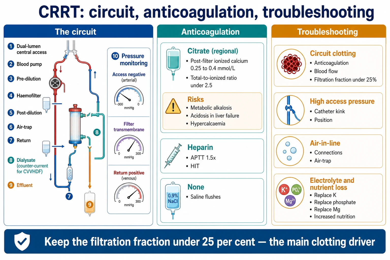
The extracorporeal circuit activates clotting. Some anticoagulation is needed to maintain filter life — but the ideal agent avoids systemic bleeding. [1]
Regional citrate (preferred)
No systemic anticoagulation
- Calcium chelated in circuit (pre-dilution) → anticoagulated circuit only
- Calcium replaced systemically (post-filter) → systemic Ca normal
- No systemic anticoagulation → lower bleeding risk
- Longer filter life vs heparin (best evidence)
- Monitor: circuit (post-filter) ionised Ca LOW (~0.25-0.35 mmol/L = anticoagulated), systemic ionised Ca NORMAL
- CAUTION: citrate toxicity — metabolic acidosis, total Ca/ionised Ca ratio >2.5
- Avoid in severe hepatic failure / severe lactic acidosis (cannot metabolise citrate)
- Metabolic alkalosis can occur (citrate → bicarbonate on metabolism)
Heparin (UFH or LMWH)
Systemic anticoagulation
- Unfractionated heparin infused into circuit (pre-filter)
- Simple, widely available, cheap, familiar
- Risk of systemic anticoagulation (bleeding)
- Risk of HIT (heparin-induced thrombocytopenia) — check platelets
- Shorter filter life than citrate
- Alternative: low molecular weight heparin (LMWH) — less monitoring but partial reversal
- Use when citrate contraindicated (severe hepatic failure, citrate accumulation)
No anticoagulation
For bleeding patients
- Used when bleeding risk is very high (recent surgery, active haemorrhage, coagulopathy)
- Very short filter life (clotting within hours)
- Pre-dilution replacement fluid helps prolong filter life (dilutes blood before filter)
- Higher blood flow rate may help reduce stasis/clotting
- Saline flushes (200 mL q30 min) can help
- Accept the trade-off: shorter circuit life vs bleeding risk
Citrate toxicity — recognition and management
Recognise
Citrate accumulates when the liver cannot metabolise it (severe hepatic failure, severe lactic acidosis/shock, post-cardiac arrest). Signs: progressive metabolic acidosis (citrate is an unmeasured anion), low systemic ionised calcium despite calcium infusion, and a rising total-to-ionised calcium ratio >2.5 (total Ca stays normal/ high because calcium is bound to citrate but ionised Ca falls).
Confirm
Calculate the ratio: total calcium / ionised calcium. Normal <2.1. Accumulation >2.5 (some units use >2.1 with rising trend + worsening acidosis). The acidosis is high-anion-gap with a rising anion gap. Systemic ionised Ca falls despite increased calcium replacement.
Manage
Reduce or stop citrate infusion. Increase systemic calcium infusion to maintain ionised Ca in the normal range. Switch anticoagulation to systemic heparin or no anticoagulation. Treat the underlying cause (improve perfusion/lactate, support liver). If severe, consider bolus calcium and bicarbonate for the acidosis.
Prevent
Avoid citrate in severe hepatic failure (Child-Pugh C) and profound shock/lactic acidosis (lactate >4-5 mmol/L relative). Monitor ionised calcium (systemic) and the total/ionised ratio at least 6-hourly. Use reduced-dose citrate protocols in moderate hepatic dysfunction. When in doubt, use heparin.
Drug dosing on CRRT
CRRT removes many drugs — especially water-soluble, low protein-binding, small-to-middle molecular weight drugs (most antibiotics, some anti-epileptics). Under-dosing is common (the DALI study found up to ~75% of ICU beta-lactam levels were sub-therapeutic) and drives treatment failure and antimicrobial resistance.[7] Clearance depends on the modality (effluent flow), filter type, residual renal function, and whether the drug is removed by convection (CVVH) or diffusion (CVVHD).
[1]Vancomycin
Glycopeptide
- Clearance: significant on CRRT (mainly convection)
- Load: 25-30 mg/kg (real body weight)
- Maintenance: 15-25 mg/kg q12-24h, OR continuous infusion 30-60 mg/kg/day
- Monitor: trough 15-20 mg/L, or AUC 400-600 mg·h/L
- TDM essential — clearance varies widely with effluent rate and filter
Beta-lactams
Time-dependent
- Clearance: high — piperacillin-tazobactam, meropenem, cefepime all significantly cleared
- Strategy: increase dose, increase frequency, OR extended/continuous infusion
- Continuous infusion (e.g. meropenem 3g/day over 24h) maximises time above MIC
- Monitor levels where available (beta-lactam TDM) — DALI: many are sub-therapeutic
- Sepsis: augmentd dosing first 24-48h, then adjust
Aminoglycosides
Concentration-dependent
- Clearance: gentamicin, tobramycin, amikacin cleared by CRRT
- Dosing: daily (once-daily) 5-7 mg/kg gentamicin, then redose when trough <1 mg/L
- Monitoring: trough levels (pre-next-dose); toxicity (renal, ototoxic) still relevant
- Avoid prolonged courses — additive nephrotoxicity
- Extended-interval dosing harder than in normal renal function — monitor closely
The CRRT circuit
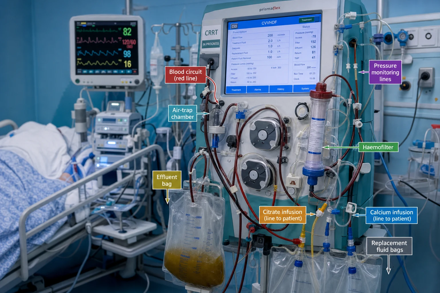
Blood leaves the patient via a dual-lumen central venous catheter (right internal jugular or femoral preferred; subclavian avoided due to stenosis risk) → blood pump → pre-dilution replacement fluid (if used) → citrate/heparin infusion (pre-filter) → haemofilter (hollow-fibre membrane) → post-filter sampling port (for circuit ionised Ca) → calcium infusion (if citrate) → air-trap/return → patient. The effluent (ultrafiltrate + spent dialysate) drains to a collection bag; its flow rate = the prescribed dose. [1]
- Access: right internal jugular (shortest, straightest) or femoral; avoid subclavian in AKI (stenosis jeopardises future AV fistula).
- Filter membrane: synthetic high-flux (polysulfone, polyethersulfone, AN69) — high sieving for middle molecules.
- Down-time: prescribed dose ≠ delivered dose — every interruption (filter change, procedure, transport, clotting) reduces delivered dose by ~20%; prescribe high to compensate. [1]
Exam practice
SAQ — Choosing the RRT modality in the unstable septic patient
10 minutes · 10 marks
A 58-year-old man is admitted to ICU with fulminant pseudomonal pneumonia and septic shock. He requires noradrenaline 0.4 mcg/kg/min and vasopressin 0.03 U/min for a MAP of 65. He has developed KDIGO stage 3 AKI (creatinine 380 umol/L, K+ 6.9 mmol/L with widened QRS, pH 7.18, HCO3 12) and is 7 L in positive fluid balance. The registrar suggests intermittent haemodialysis 'to clear the potassium quickly'.
SAQ — Citrate anticoagulation in cirrhosis
10 minutes · 10 marks
A 64-year-old woman with alcohol-related cirrhosis (Child-Pugh B, baseline INR 1.8, platelets 75) is on CVVHDF with regional citrate anticoagulation for AKI secondary to spontaneous bacterial peritonitis. Six hours after starting CRRT: post-filter ionised calcium 0.32 mmol/L, systemic ionised calcium 0.95 mmol/L, total calcium 2.55 mmol/L, pH 7.22, HCO3 14, lactate 5.2 mmol/L, anion gap 22.
Clinical pearls
Red flags
[1] [1]Key takeaways
References
- [1]Gaudry S, Hajage D, Schortgen F, et al. Initiation Strategies for Renal-Replacement Therapy in the Intensive Care Unit N Engl J Med, 2016.PMID 27181456
- [2]STARRT-AKI Investigators; Canadian Critical Care Trials Group; ANZICS CTG, et al. Timing of Initiation of Renal-Replacement Therapy in Acute Kidney Injury N Engl J Med, 2020.PMID 32668114
- [3]Tsujimoto H, Tsujimoto Y, Nakata Y, et al. Pharmacological interventions for preventing clotting of extracorporeal circuits during continuous renal replacement therapy Cochrane Database Syst Rev, 2020.PMID 33314078
- [4]Zarbock A, Kellum JA, Schmidt C, et al. Effect of Early vs Delayed Initiation of Renal Replacement Therapy on Mortality in Critically Ill Patients With Acute Kidney Injury: The ELAIN Randomized Clinical Trial JAMA, 2016.PMID 27209269
- [5]RENAL Replacement Therapy Study Investigators; Bellomo R, Cass A, et al. Intensity of continuous renal-replacement therapy in critically ill patients N Engl J Med, 2009.PMID 19846848
- [6]VA/NIH Acute Renal Failure Trial Network; Palevsky PM, Zhang JH, et al. Intensity of renal support in critically ill patients with acute kidney injury N Engl J Med, 2008.PMID 18492867
- [7]Roberts JA, Paul SK, Akova M, et al. DALI: defining antibiotic levels in intensive care unit patients: are current β-lactam antibiotic doses sufficient for critically ill patients? Clin Infect Dis, 2014.PMID 24429437
- [8]Schwenger V, Weigand MA, Hoffmann O, et al. Sustained low efficiency dialysis using a single-pass batch system in acute kidney injury - a randomized interventional trial: the REnal Replacement Therapy Study in Intensive Care Unit PatiEnts Crit Care, 2012.PMID 22839577