ICU · Renal and metabolic
Drug-induced nephrotoxicity: mechanism, offending agents, and prevention in ICU
Also known as Drug-induced nephrotoxicity · Nephrotoxic AKI · Acute tubular necrosis drugs · Acute interstitial nephritis · Contrast nephropathy · Vancomycin nephrotoxicity
Drug-induced nephrotoxicity accounts for ~20% of hospital-acquired AKI and up to 35% of ICU-acquired AKI. Five dominant mechanisms: (1) HAEMODYNAMIC (NSAIDs constrict afferent arteriole via prostaglandin inhibition; ACEi/ARB dilate efferent arteriole by blocking angiotensin II; calcineurin inhibitors constrict afferent arteriole via endothelin — the 'triple whammy' abolishes all three autoregulatory limbs). (2) ACUTE TUBULAR NECROSIS (ATN) — direct tubular toxicity: aminoglycosides accumulate in proximal tubular cells via the megalin/cubilin receptor causing lysosomal phospholipidosis; amphotericin B forms pores in the cholesterol-rich distal tubular cell membrane; tenofovir causes proximal tubular mitochondrial toxicity (Fanconi syndrome); iodinated contrast causes medullary vasoconstriction/hypoxia plus direct cytotoxicity; cisplatin enters via OCT2. (3) ACUTE INTERSTITIAL NEPHRITIS (AIN) — Type IV hypersensitivity: beta-lactams, PPIs (now 1), NSAIDs, rifampicin, sulfonamides, allopurinol. (4) CRYSTAL NEPHROPATHY — aciclovir, sulphonamides, methotrexate precipitate in distal tubules. (5) OSMOTIC NEPHROSIS — IV immunoglobulin (sucrose stabiliser), hydroxyethyl starch, mannitol cause tubular cell swelling. Prevention: identify high-risk patients (elderly, CKD, diabetes, sepsis, hypovolaemia), avoid/limit nephrotoxins, therapeutic drug monitoring (vancomycin AUC 400-600, aminoglycoside trough <1), isotonic volume expansion for contrast (PRESERVE: saline = bicarbonate, NAC no benefit), recognise and STOP offending drug early.
On this page & tools
Your progress
Saved locally on this device.
Target exams
Red flags


Glomerular autoregulation — the foundation for understanding nephrotoxicity
Every haemodynamic drug-induced AKI makes sense once the two-gate model of the glomerulus is clear. GFR is governed by the net filtration pressure across the glomerular capillary wall, which depends on the balance between afferent (inflow) and efferent (outflow) arteriolar tone: [1]
- Afferent arteriole (the IN-gate) — regulated mainly by prostaglandins (PGE2, PGI2), which keep it dilated. Block prostaglandin synthesis (NSAIDs inhibit COX-1/COX-2) and the afferent arteriole constricts → less blood reaches the glomerulus → GFR falls. The calcineurin inhibitors (cyclosporin, tacrolimus) also constrict the afferent arteriole (endothelin-mediated, reduced NO).
- Efferent arteriole (the OUT-gate) — regulated mainly by angiotensin II, which keeps it constricted. This constriction maintains glomerular capillary pressure and therefore GFR. Block angiotensin II (ACE inhibitors, or block its receptor with ARBs) and the efferent arteriole dilates → intraglomerular pressure drops → GFR falls.
- Tubuloglomerular feedback (TGF) — the macula densa senses distal NaCl delivery and adjusts afferent tone via adenosine. Tubular toxins that increase distal delivery (or contrast, which increases proximal pressure) feed back through adenosine to constrict the afferent arteriole, compounding the GFR fall. [1]
Clinical corollary — the "triple whammy": when a patient is simultaneously on an NSAID (constricts afferent) + ACEi/ARB (dilates efferent) + diuretic (depletes circulating volume), all three compensatory mechanisms that defend GFR are abolished at once → an abrupt, often severe, fall in GFR. This is the single highest-yield drug-AKI mechanism in the exam.[2]
Mechanisms of drug-induced nephrotoxicity
| Mechanism | Classic drugs | Pathophysiology | Urine findings | Onset |
|---|---|---|---|---|
| Haemodynamic (pre-renal) | NSAIDs, ACEi/ARB, calcineurin inhibitors | Afferent arteriole constriction (NSAIDs, CNI) or efferent dilation (ACEi/ARB) → reduced GFR | BUN:Cr >20, Na <20, FENa <1% | Hours-days |
| Acute tubular necrosis (ATN) | Aminoglycosides, vancomycin, contrast, amphotericin B, tenofovir, cisplatin | Direct tubular epithelial toxicity → necrosis | Muddy brown casts, FENa >2% | 5-10 days (aminoglycosides) |
| Acute interstitial nephritis (AIN) | Beta-lactams (methicillin, piperacillin), PPIs, NSAIDs, rifampicin, sulfonamides, allopurinol | Type IV hypersensitivity → interstitial inflammation | WBC casts, eosinophiluria, sterile pyuria | Days-weeks |
| Crystal nephropathy | Aciclovir, sulphonamides, methotrexate, indinavir | Drug crystallises in tubules → obstruction | Crystals on microscopy | Hours-days |
| Osmotic nephrosis | IV immunoglobulin (sucrose), mannitol, hydroxyethyl starch | Hyperosmolar agents cause tubular cell swelling (vacuolisation) | Bland urine | Variable |
| Rhabdomyolysis (indirect) | Statins (esp. with azoles/macrolides), colchicine | Muscle breakdown → myoglobin nephrotoxicity | Blood +ve on dipstick, few RBC, raised CK | Days |
Mechanisms in depth
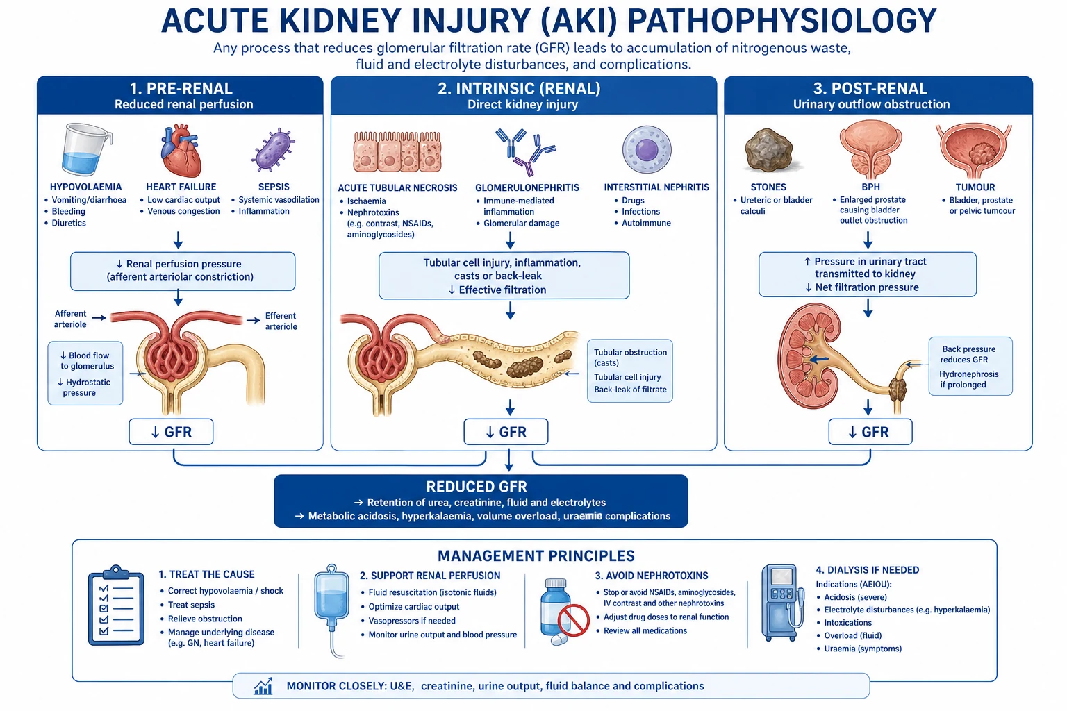
1. Haemodynamic (pre-renal) — drug-induced loss of GFR
The kidney defends GFR across a wide range of perfusion pressures through autoregulation, which depends on three endogenous systems acting on the two arterioles. Drugs that interfere with any one cause a functional (reversible) fall in GFR; drugs that interfere with several at once cause severe AKI: [1]
- NSAIDs (afferent limb) — COX inhibition depletes vasodilatory prostaglandins (PGE2, PGI2). In a well-perfused kidney this is harmless, but in any state of high vasoconstrictor tone (volume depletion, sepsis, cirrhosis, heart failure, "effective circulating volume" depletion) the prostaglandins are the only thing keeping the afferent arteriole open. Block them and GFR collapses. NSAIDs also cause AIN (a separate intrinsic mechanism — weeks after exposure).
- ACEi / ARB (efferent limb) — angiotensin II normally constricts the efferent arteriole, holding up glomerular capillary pressure. Blocking it lowers GFR. This is physiological and benign in most patients — a small creatinine rise on starting an ACEi is expected and acceptable (up to 30% baseline creatinine). It becomes pathological when: (a) the patient is volume-depleted; (b) there is bilateral renal artery stenosis (the stenotic kidney is exquisitely dependent on efferent constriction to filter); (c) combined with NSAIDs/diuretics (triple whammy); or (d) in cardiorenal or hepatorenal states.
- Calcineurin inhibitors (cyclosporin, tacrolimus) — cause afferent arteriolar vasoconstriction (endothelin release, reduced nitric oxide, increased sympathetic tone). Chronic use causes structural arteriolar hyalinosis and striped interstitial fibrosis — irreversible. CNI also cause a distal tubular toxicity (type IV RTA, hyperkalaemia, magnesium wasting) distinct from the haemodynamic effect.[2]
2. Acute tubular necrosis (ATN) — direct tubular toxicity
ATN is the common endpoint of direct tubular cell injury. The subsegment and mechanism matter because they explain the timing, urine findings and prevention: [1]
- Aminoglycosides (gentamicin, tobramycin, amikacin) — filtered freely, then re-absorbed into proximal tubular cells via the megalin/cubilin endocytic receptor on the apical membrane. Inside the cell they accumulate in lysosomes, disrupt phospholipid metabolism (inhibit phospholipase A1/A2 → phospholipidosis with characteristic myeloid bodies), and ultimately trigger apoptosis/necrosis via lysosomal rupture, mitochondrial injury and oxidative stress. Nephrotoxicity is cumulative and dose-dependent, typically appearing after 5–7 days; non-oliguric initially. The ototoxicity runs in parallel and is also concentration-dependent — irreversible (unlike the renal injury).[3][4]
- Amphotericin B — binds to ergosterol-like sterols in the cholesterol-rich distal tubular cell membrane, forming pores that allow uncontrolled electrolyte flux → potassium and magnesium wasting + type I distal RTA before frank ATN develops. The deoxycholate formulation is the most nephrotoxic; lipid formulations (liposomal amphotericin, ABLC) target the fungal cell preferentially and markedly reduce nephrotoxicity. Sodium loading (pre-hydration with saline) reduces amphotericin nephrotoxicity.[2]
- Iodinated contrast — a dual insult: (1) an initial transient vasodilation followed by sustained renal vasoconstriction causing outer medullary hypoxia (the thick ascending limb has high O2 demand and a tenuous supply); (2) direct cytotoxicity to tubular cells from the hyperosmolar iodine load, plus cast formation. Risk is highest with high-osmolar contrast, large volumes, dehydration, CKD, diabetes and concurrent nephrotoxins.[10]
- Cisplatin — enters the proximal tubule cell via the OCT2 transporter; causes mitochondrial injury, oxidative stress and DNA cross-linking → ATN with prominent magnesium and potassium wasting. Carboplatin is less nephrotoxic; amifostine and aggressive hydration mitigate.
- Tenofovir (and adefovir) — proximal tubular mitochondrial toxicity → Fanconi syndrome (glycosuria with normal serum glucose, phosphaturia, aminoaciduria, uricosuria) and occasionally full ATN. The proximal tubule expresses high levels of the transporters (OAT1) that take tenofovir up. Tenofovir alafenamide (TAF) produces lower plasma tenofovir exposure than tenofovir disoproxil fumarate (TDF) and is less nephrotoxic.
- Rhabdomyolysis (indirect nephrotoxicity) — myoglobin is filtered, taken up by proximal tubule cells and is directly cytotoxic (ferrous iron, free radicals); it also forms casts that obstruct the tubule when urine is acidic and concentrated. The drug link is anything that destroys muscle: statins (especially with CYP3A inhibitors — azoles, macrolides), colchicine, neuroleptic malignant syndrome, serotonin syndrome, ecstasy, prolonged immobilisation.
3. Acute interstitial nephritis (AIN) — immune-mediated
AIN is an immune-mediated (predominantly Type IV hypersensitivity) inflammatory infiltrate of the tubulointerstitium, not a direct toxic effect. The classic clinical tetrad — fever, rash, eosinophilia, eosinophiluria — is insensitive and is absent in the majority; AKI with sterile pyuria and WBC casts after a new drug is enough to suspect it. NSAID-AIN is distinctive: it can present with nephrotic-range proteinuria (minimal-change-like podocyte injury) and may occur only after months of exposure. Methicillin was the original culprit (classic exam answer); today PPIs and NSAIDs are the most common. Diagnosis is confirmed by kidney biopsy (interstitial oedema with eosinophils and lymphocytes, tubulitis).[5][6]
4. Crystal (intratubular) nephropathy — post-renal
Drug crystals precipitate when three conditions align: high drug concentration (high dose/rapid infusion), low urine flow (dehydration), and unfavourable urine pH. The result is intratubular obstruction plus an inflammatory foreign-body reaction: [1]
- Aciclovir — solubility is limited; high-dose IV boluses in dehydrated patients precipitate as birefringent needle crystals in the distal tubule. Prevention: slow the infusion (≥1 h), hydrate, and dose-adjust for renal function.
- Sulphonamides (sulfadiazine in cerebral toxoplasmosis, co-trimoxazole) — acetylated metabolites precipitate in acidic urine → crystals, haematuria, AKI. Prevention: alkalinise urine (sodium bicarbonate) + high fluid intake.
- Methotrexate (high-dose, >1 g/m²) — 7-OH-methotrexate precipitates in acidic urine. Prevention: alkalinisation to keep urine pH >7.0, high fluid intake, leucovorin rescue.
- Indinavir / atazanavir (HIV protease inhibitors), triamterene (diuretic), large vitamin C doses (oxalate). [1]
5. Osmotic nephrosis — tubular cell swelling
Highly osmolar or large-molecule agents are endocytosed by proximal tubular cells, where they accumulate in lysosomes and cause osmotic swelling and vacuolisation that progresses to ATN: [1]
- IV immunoglobulin (IVIG) — the sucrose stabiliser used in some formulations is the culprit; sucrose-FREE IVIG products carry much lower risk.[12]
- Hydroxyethyl starch (HES) — high-molecular-weight colloid resuscitation fluid; ICU trials showed increased RRT need vs crystalloid.
- Mannitol — usually reversible on cessation; chronic high-dose use → vacuolar nephrosis.
- Dextran and IV radiocontrast also contribute to osmotic nephrosis.
Common ICU nephrotoxins: mechanism, site and mitigation
| Drug / class | Primary site | Mechanism of injury | Pattern | Risk factors / timing | Mitigation |
|---|---|---|---|---|---|
| NSAIDs (ibuprofen, ketorolac, diclofenac) | Afferent arteriole | COX inhibition → ↓prostaglandins → afferent vasoconstriction (also AIN) | Pre-renal ± AIN | Volume depletion, CKD, cirrhosis, CHF | Avoid; if essential, limit to <5 days, avoid "triple whammy" |
| ACEi / ARB (ramipril, enalapril, valsartan) | Efferent arteriole | ↓Angiotensin II → efferent dilation → ↓GFR | Pre-renal | Bilateral RAS, volume depletion, sepsis | Hold in AKI/hypovolaemia; expect ≤30% Cr rise acceptable |
| Calcineurin inhibitors (cyclosporin, tacrolimus) | Afferent arteriole | Endothelin-mediated afferent vasoconstriction; chronic arteriolar hyalinosis | Pre-renal + distal tubular (type IV RTA, Mg wasting) | High trough levels, chronic use | Monitor trough; use lowest effective dose; CCB co-therapy |
| Aminoglycosides (gentamicin, tobramycin, amikacin) | Proximal tubule | Megalin/cubilin receptor-mediated uptake → lysosomal phospholipidosis → ATN | ATN | Course >5–7 days, high trough, elderly, CKD, sepsis | Once-daily extended-interval dosing; monitor trough (<1 mg/L); limit duration |
| Amphotericin B (deoxycholate) | Distal tubule | Membrane pore formation → electrolyte wasting → ATN | ATN (± distal RTA) | Cumulative dose >2–3 g, dehydration | Lipid formulation preferred; pre-hydration with saline |
| Iodinated contrast | Medulla + tubules | Vasoconstriction (medullary hypoxia) + direct cytotoxicity | ATN | CKD, diabetes, dehydration, high volume, multiple doses | Isotonic saline pre-hydration; low/iso-osmolar contrast; minimise volume; hold nephrotoxins |
| Vancomycin (± piperacillin-tazobactam) | Proximal tubule | Concentration-dependent oxidative/apoptotic injury (± AIN) | ATN (± AIN) | Trough >15–20, AUC high, course >7 days, concomitant pip-tazo | AUC-guided TDM (400–600 mg·h/L); prefer cefepime over pip-tazo |
| Cisplatin / carboplatin | Proximal tubule (S3) | OCT2-mediated uptake → mitochondrial/DNA injury | ATN (Mg, K wasting) | High cumulative dose | Aggressive saline hydration; amifostine; Mg/K replacement |
| Methotrexate (high dose) | Tubules | Crystal (7-OH-MTX) + direct toxicity | ATN ± post-renal crystal | Dose >1 g/m², acidic urine | High urine flow + alkalinisation (urine pH >7) + leucovorin |
| Tenofovir / adefovir | Proximal tubule | OAT1 uptake → mitochondrial toxicity → Fanconi syndrome | ATN + Fanconi | Long-term use, low body weight, CKD | Monitor phosphate/glucose; tenofovir alafenamide (TAF) less toxic |
| Aciclovir | Distal tubule | Crystal precipitation (low solubility) | Post-renal crystal | High IV dose, dehydration, rapid infusion | Slow infusion ≥1 h, hydrate, dose-adjust for eGFR |
| Sulphonamides (sulfadiazine, co-trimoxazole) | Distal tubule | Acetylated metabolite crystals in acidic urine (also AIN) | Post-renal crystal ± AIN | High dose, acidic urine, dehydration | High fluid intake; alkalinise urine |
| IVIG (sucrose formulation) | Proximal tubule | Sucrose stabiliser → osmotic nephrosis (vacuolisation) | Osmotic nephrosis | High dose, rapid infusion, CKD | Use sucrose-FREE formulation; slow infusion <4 mL/kg/hr; hydrate |
| Hydroxyethyl starch | Proximal tubule | Macromolecule pinocytosis → osmotic swelling | Osmotic nephrosis | High cumulative dose, sepsis | Avoid; prefer crystalloid |
| Statins (rhabdo-indirect) | Skeletal muscle → tubules | Myoglobin cast nephropathy | ATN | High dose, CYP3A inhibitors, elderly, hypothyroid | Check CK; avoid statin + azole/macrolide combinations |
| Chemotherapy (ifosfamide, mitomycin, gemcitabine) | Glomerulus/tubules | Endothelial injury (TMA) or direct toxicity | ATN / TMA | High cumulative dose | Hydration; dose limits; monitor |
Acute interstitial nephritis (AIN) — dedicated approach
AIN is the most reversible form of drug-induced intrinsic AKI provided the offending drug is stopped early. The examiner's high-yield facts:[5]
AIN — diagnostic and management flow
1. SUSPECT on clinical grounds
AKI + recent new drug (within days–weeks) + any of: fever, rash, eosinophilia, sterile pyuria, WBC casts, flank pain. Remember the classic tetrad is insensitive — drug history is the key. NSAID-AIN can present after MONTHS and with nephrotic-range proteinuria.
2. BUILD the differential
Distinguish from ATN (muddy brown casts, FENa >2%, no eosinophilia, no rash) and from pre-renal (BUN:Cr >20, FENa <1%, rapid recovery with volume). Urinary eosinophils (Hansel stain) historically >1% supportive but low sensitivity/specificity — not reliable alone.
3. REVIEW EVERY drug started in the last 6 months
Common culprits by frequency: PPIs, penicillins/cephalosporins, NSAIDs, sulphonamides/co-trimoxazole, rifampicin, ciprofloxacin, allopurinol, 5-ASA/mesalazine, furosemide/thiazides, omeprazole. PPI-AIN is increasingly the #1 cause.
4. BIOPSY if diagnosis uncertain or steroids contemplated
Kidney biopsy is the gold standard: interstitial oedema with eosinophil-rich inflammatory infiltrate and tubulitis. Indicated if: AKI not recovering after stopping drug, suspected concurrent glomerular disease, or steroids being considered.
5. STOP the offending drug — the single most important intervention
Withdraw the most likely culprit and any unnecessary drug. In polypharmacy, stop the most recently introduced likely offender first. Document the reaction to prevent re-exposure. Most patients begin to improve within days of withdrawal.
6. CONSIDER corticosteroids — controversial, often used
Prednisolone 1 mg/kg/day (max 60 mg) if AKI severe or biopsy-confirmed with interstitial granulomas/edema, started EARLY (within 7–14 days). Wean over 4–6 weeks if renal function recovers. Trials have NOT shown a clear mortality/renal-recovery benefit — but most nephrologists use steroids in severe/progressive AIN.
7. SUPPORTIVE care and follow-up
Manage volume status, electrolytes (hyperkalaemia is common), and dialyse if urgent indications arise (AEIOU). Monitor creatinine to recovery — incomplete recovery predicts CKD. Lifelong documentation of the causative drug as an "allergy" to prevent re-challenge.
Top causes of AIN — examiner mnemonic and frequency
| Rank | Drug class | Notes |
|---|---|---|
| 1 | PPIs (omeprazole, pantoprazole) | Increasingly the #1 cause; onset weeks–months; often subclinical until AKI noticed |
| 2 | Antibiotics — penicillins (methicillin classic), cephalosporins, rifampicin, sulphonamides, ciprofloxacin | Classic fever + rash + eosinophilia; rifampicin AIN is dose/intermittent-dose related |
| 3 | NSAIDs (including COX-2) | Distinctive: may be delayed (months) and present with nephrotic-range proteinuria (podocyte injury) |
| 4 | Diuretics — furosemide, thiazides, triamterene | Sulphonamide structural similarity; triamterene also forms crystals |
| 5 | Miscellaneous — allopurinol, 5-ASA/mesalazine, omeprazole, cimetidine, phenytoin, indinavir | Allopurinol AIN often severe with long recovery; check IgA levels / HLA-B*5801 in at-risk |
Aminoglycoside nephrotoxicity — the prototypic tubular toxin
Aminoglycosides (gentamicin, tobramycin, amikacin) remain a frequent cause of ICU ATN, and the mechanism is exam-favourite territory:[3]
- Uptake via megalin/cubilin — aminoglycosides are filtered, then bind to the megalin–cubilin complex on the apical membrane of proximal tubular cells and are endocytosed. This concentrates the drug inside the cell many-fold above plasma.
- Lysosomal accumulation — within lysosomes aminoglycosides inhibit phospholipase A1/A2 → phospholipidosis → myeloid body formation → lysosomal rupture → release of lysosomal enzymes → mitochondrial injury → apoptosis and necrosis.
- Non-oliguric onset — classically the AKI is non-oliguric for the first several days (in contrast to ischaemic ATN), appearing after 5–7 days of therapy.
- Ototoxicity runs in parallel — cochlear and vestibular hair cell damage; irreversible (unlike the renal injury). Risk relates to cumulative dose and high trough concentrations.[4]
- Prevention — once-daily extended-interval dosing — giving the entire daily dose as a single infusion produces a high peak (efficacy is concentration-dependent) and a low trough (less time for megalin-mediated uptake), so the tubular cell accumulates less drug. Meta-analyses confirm extended-interval dosing is at least as effective and less nephrotoxic than divided dosing. Limit courses to 5–7 days where possible; monitor troughs (goal <1 mg/L for gent/tobra at 18–24 h after dose).
Contrast-associated AKI (CA-AKI)
Definition (KDIGO-aligned): a rise in serum creatinine ≥0.3 mg/dL (≥26.5 µmol/L) within 48 hours of intravascular iodinated contrast administration, after excluding alternative causes. Onset typically 24–72 h, peak 3–5 days, recovery within 1–3 weeks.[10]
Pathophysiology — a dual insult: (1) initial brief vasodilation is followed by sustained renal vasoconstriction causing outer-medullary hypoxia (the S3 segment/thick ascending limb has high O2 demand and a precarious supply); (2) direct cytotoxicity of the contrast molecule to tubular cells plus osmotic diuresis-induced cast formation. High-osmolar agents are worse than low-/iso-osmolar agents. [1]
CA-AKI prevention and management
Define risk
High risk: eGFR <30, diabetes, heart failure, hypovolaemia, multiple contrast doses, high-osmolar contrast, age >75, concurrent nephrotoxins, myeloma. Low risk: eGFR >60, single dose, no comorbidities. Use the Mehran score to stratify.
Pre-hydrate
IV crystalloid before AND after contrast. PRESERVE trial (NEJM 2018): IV bicarbonate = IV saline (no difference in 90-day composite). Oral hydration alone is insufficient. Protocol: isotonic saline 1 mL/kg/hr × 6–12 h before and 6–12 h after contrast (1.5 mL/kg/hr if no CHF). Hold diuretics in high-risk patients.
Minimise contrast
Use the LOWEST possible contrast volume. Use iso-osmolar (iodixanol) or low-osmolar (iopamidol, iohexol) contrast — less toxic than high-osmolar. Consider alternative imaging (ultrasound, MRI without contrast) in high-risk patients.
Hold nephrotoxic drugs
Hold METFORMIN at the time of contrast (resume after 48 h if renal function stable — risk of lactic acidosis if AKI develops). Hold NSAIDs. ACEi/ARB: continue unless volume-depleted. Hold aminoglycosides if possible. Hold loop diuretics in high-risk patients.
Consider statins (TRACK-D)
Short-course high-dose statin (e.g. rosuvastatin) before elective contrast reduced CA-AKI in patients with CKD + diabetes (TRACK-D, JACC 2014). Benefit not confirmed in all populations — reasonable in statin-tolerant high-risk patients undergoing elective procedures.
NAC — do NOT use
N-acetylcysteine has NO benefit over placebo (PRESERVE 2018, multiple meta-analyses). Stop prescribing NAC for contrast prophylaxis — the correct answer on the exam is isotonic saline alone.
PRESERVE
NEJM 2018
5177 patients at risk of CA-AKI undergoing angiography — 2×2 factorial of IV bicarbonate vs isotonic saline AND oral NAC vs placebo
Key finding
No benefit of NAC over placebo, and no benefit of bicarbonate over saline, for the composite of death, RRT or persistent renal impairment at 90 days.
Practice change
Use isotonic saline (NOT bicarbonate) and placebo (NOT NAC) for contrast prophylaxis
TRACK-D
JACC 2014
2998 patients with diabetes + CKD undergoing coronary/contrast procedures — short-course rosuvastatin vs placebo
Key finding
Rosuvastatin reduced CA-AKI (2.4% vs 5.9%, p<0.01) with no excess hepatic/muscle adverse effects.
Practice change
Short-course statin is a reasonable adjunct in CKD + diabetes before elective contrast
Hammond 2017 (meta-analysis)
Clin Infect Dis 2017
Systematic review and meta-analysis of concomitant vancomycin + piperacillin/tazobactam vs vancomycin + cefepime
Key finding
Vancomycin + pip-tazo associated with significantly higher AKI risk than vancomycin + cefepime (OR ~2-3x).
Practice change
When MRSA + antipseudomonal cover needed, prefer cefepime over pip-tazo
Hammond 2016 (comparative)
Pharmacotherapy 2016
Comparative incidence of AKI in critically ill patients receiving vancomycin with concomitant piperacillin-tazobactam or cefepime
Key finding
Higher AKI incidence with vancomycin + pip-tazo vs vancomycin + cefepime in the critically ill.
Practice change
Use cefepime rather than pip-tazo when combining with vancomycin
Prevention strategy — a systematic approach
The cheapest, safest and most effective "treatment" for drug-induced nephrotoxicity is to not cause it in the first place. Prevention is a recurring exam theme:[2]
Preventing drug-induced nephrotoxicity — the six-step bundle
1. IDENTIFY the high-risk patient BEFORE prescribing
Risk factors: AGE >65, baseline CKD (eGFR <60), DIABETES, SEPSIS/hypovolaemia, HEART FAILURE/cirrhosis (low effective circulating volume), CONCURRENT NEPHROTOXINS (already on vancomycin — adding contrast?), HAEMODYNAMIC instability, myeloma. If high risk → AVOID nephrotoxins; if unavoidable → reduce dose, monitor, hydrate.
2. AVOID or LIMIT nephrotoxin combinations
Never combine NSAID + ACEi + diuretic (triple whammy). Avoid vancomycin + piperacillin-tazobactam (prefer cefepime — Hammond 2017 meta-analysis). Avoid aminoglycoside + loop diuretic. Avoid two nephrotoxins together unless absolutely essential — synergy of injury is real.
3. DOSE-ADJUST for renal function
Renally clear most drugs (vancomycin, aminoglycosides, gabapentin, digoxin, LMWH, many antibiotics). Use eGFR or measured CrCl to adjust; re-check renal function frequently in acute illness. A common iatrogenic cause of AKI is failing to reduce a CKD dose when AKI develops.
4. HYDRATE before contrast / crystals / amphotericin
Contrast: isotonic saline 1 mL/kg/hr × 6–12 h pre and post (PRESERVE: saline = bicarb; NAC no benefit). Aciclovir/methotrexate/sulphonamides: high urine flow + alkalinise urine (where appropriate). Amphotericin: sodium loading with saline reduces toxicity.
5. THERAPEUTIC DRUG MONITORING (TDM)
Vancomycin: AUC-guided dosing (target AUC 400–600 mg·h/L) reduces AKI vs trough-only dosing. Aminoglycosides: once-daily extended-interval dosing with trough <1 mg/L. Tacrolimus/cyclosporin: trough in target range. TDM prevents the high-exposure nephrotoxicity window.
6. MONITOR AND REVIEW DAILY
Daily creatinine + urine output while on a nephrotoxin. Stop the drug at the first sign of rising creatinine (do not "finish the course"). Renal ultrasound to exclude obstruction. If AKI persists >3–5 days after stopping → consider biopsy (AIN?). Document intolerance to prevent re-exposure.
Prevention of drug-induced nephrotoxicity (legacy bundle)
- IDENTIFY HIGH-RISK PATIENTS — before prescribing nephrotoxins, assess: (a) AGE >65. (b) CKD (baseline eGFR <60). (c) DIABETES. (d) SEPSIS/hypovolaemia (already compromised renal perfusion). (e) CONCURRENT NEPHROTOXINS (already on vancomycin + adding contrast?). (f) HAEMODYNAMIC instability (shock, low MAP). If high risk → AVOID nephrotoxins if possible; if unavoidable → reduce dose, monitor closely
- AVOID OR LIMIT NEPHROTOXINS — (a) NSAIDs: avoid in CKD, elderly, sepsis, 'triple whammy' (NSAID + ACEi + diuretic). (b) ACEi/ARB: hold in AKI, severe hypovolaemia. (c) AMINOGLYCOSIDES: use only if essential; once-daily extended-interval dosing; limit to 3-5 days. (d) VANCOMYCIN: TDM (trough 15-20 or AUC 400-600); avoid combination with piperacillin-tazobactam if possible. (e) CONTRAST: only if essential; use low-volume iso-osmolar; hold metformin
- VOLUME EXPANSION (for contrast) — PRESERVE trial (NEJM 2018): isotonic saline EQUAL to sodium bicarbonate (bicarb NOT superior). NAC: NO benefit (PRESERVE). Protocol: isotonic saline 1 mL/kg/hr for 6-12h pre and post contrast. For high-risk: hold diuretics, minimise contrast volume (<100 mL if possible)
- THERAPEUTIC DRUG MONITORING (TDM) — (a) Vancomycin: trough 15-20 mg/L (moderate-severe infection); or AUC 400-600 (newer target — Bayesian dosing). Check every 2-3 days (daily in AKI/dialysis). (b) Aminoglycosides: extended-interval dosing (5-7 mg/kg); trough <1 mg/L; limit duration. (c) Tacrolimus/ciclosporin: trough levels (C0) or AUC — nephrotoxic above target. Pharmacokinetic pharmacist input essential
- RECOGNISE AND STOP EARLY — Daily creatinine + urine output (KDIGO AKI criteria). If creatinine rises >26 μmol/L/48h or >1.5x baseline → AKI. Review ALL medications: (a) STOP nephrotoxins (NSAIDs, ACEi, aminoglycosides — switch to alternative). (b) Adjust remaining drugs for renal function. (c) If AIN suspected (eosinophiluria, rash, fever, new drug) → STOP offending drug; consider steroids if severe/prolonged
- MONITOR AND FOLLOW-UP — (a) Daily creatinine until stable. (b) Urinalysis (casts, eosinophils, crystals). (c) Renal ultrasound (exclude obstruction). (d) If AKI persists >3-5 days after stopping drug → renal biopsy (AIN? ATN? other?). (e) Drug causality assessment (Naranjo scale). (f) Document allergy/intolerance in medical record (prevent re-exposure)
SAQ — Non-oliguric AKI after prolonged gentamicin for enterococcal endocarditis
10 minutes · 10 marks
A 58-year-old man with native-valve Enterococcus faecalis endocarditis is on day 9 of intravenous ampicillin 2 g 4-hourly plus gentamicin 1 mg/kg 8-hourly (synergy dosing). His creatinine has risen from 88 to 156 µmol/L over 72 hours but urine output is preserved at 1.2 mL/kg/hr. The gentamicin trough is 2.4 mg/L. He reports new bilateral high-pitched tinnitus. Urine microscopy shows mild tubular proteinuria and a few granular casts.
SAQ — Preventing contrast-associated AKI in an emergency CT pulmonary angiogram
10 minutes · 10 marks
A 67-year-old woman with type 2 diabetes (HbA1c 64 mmol/mol), CKD stage 3b (baseline eGFR 32 mL/min/1.73 m²) and a recent STEMI presents to the emergency department with acute dyspnoea, pleuritic chest pain and a Wells score of 6.5. CT pulmonary angiography (CTPA) is requested. Her medications are metformin 1 g BD, empagliflozin 25 mg daily, ramipril 5 mg daily, furosemide 40 mg daily and aspirin. She is normotensive and euvolaemic.
Clinical pearls
Red flags
Trial evidence
Drug-induced nephrotoxicity — landmark evidence
Epidemiology: drugs cause ~20% of hospital-acquired AKI; up to 35% in ICU. The commonest culprits are NSAIDs, ACEi, contrast, aminoglycosides and vancomycin.[1]
PRESERVE (Weisbord, NEJM 2018, PMID 29130810): 5177 patients at risk of CA-AKI undergoing angiography. 2×2 factorial of IV bicarbonate vs isotonic saline AND oral N-acetylcysteine vs placebo. Primary composite (death, RRT, persistent renal impairment at 90 days): NO benefit of NAC and NO benefit of bicarbonate. Practice-changing: stop prescribing NAC; use isotonic saline.[9]
TRACK-D (Han, JACC 2014, PMID 24076297): 2998 patients with diabetes + CKD undergoing coronary procedures. Short-course rosuvastatin reduced CA-AKI (2.4% vs 5.9%, p<0.01) with no excess hepatic/muscle toxicity. Supports short-course statins in high-risk elective procedures.[11]
Contrast-associated AKI review (Mehran, NEJM 2019, PMID 31141635): defines CA-AKI, stratifies risk (Mehran score), and codifies prevention — isotonic saline hydration, minimise contrast volume, low-/iso-osmolar agents, hold nephrotoxins. NAC not recommended.[10]
Aminoglycoside mechanism review (Lopez-Novoa, Kidney Int 2011, PMID 20861826): integrates megalin/cubilin uptake, lysosomal phospholipidosis with myeloid bodies, mitochondrial injury and oxidative stress as the multi-hit mechanism of aminoglycoside nephrotoxicity.[3]
Aminoglycoside ototoxicity (Selimoglu, Curr Pharm Des 2007, PMID 17266591): cochlear and vestibular hair cell damage is IRREVERSIBLE; risk is concentration/cumulative-dose related and parallels nephrotoxicity.[4]
Drug-induced AIN (Moledina & Perazella, CJASN 2017, PMID 28893923): classic review — PPIs now the #1 cause; classic tetrad insensitive; biopsy gold standard; steroids controversial but commonly used.[5]
PPIs and kidney disease (Moledina & Perazella, J Nephrol 2016, PMID 27072818): links PPI exposure to AIN and progression to CKD; recommends periodic reassessment of long-term PPI indication.[6]
Vancomycin + pip-tazo meta-analysis (Hammond, Clin Infect Dis 2017, PMID 27940946): significantly increased AKI risk vs vancomycin + cefepime (OR ~2-3x). Prefer cefepime when both MRSA + antipseudomonal cover needed.[7]
Vancomycin + pip-tazo comparative (Hammond, Pharmacotherapy 2016, PMID 26952639): higher AKI incidence with vancomycin + pip-tazo vs vancomycin + cefepime in the critically ill.[8]
IVIG nephrotoxicity (Angeli, Minerva Gastroenterol Dietol 2008, PMID 18614975): sucrose stabiliser is the culprit in osmotic nephrosis; sucrose-free formulations carry much lower risk.[12]
Aminoglycoside dosing: meta-analyses confirm once-daily extended-interval dosing is at least as effective as divided dosing and less nephrotoxic (lower trough → less megalin-mediated tubular uptake).[3]
Vancomycin + pip-tazo: multiple cohort/meta-analyses show ~2–3× AKI risk vs vancomycin + cefepime (synergistic tubular toxicity / vancomycin AIN). Prefer cefepime; AUC-guided vancomycin TDM reduces nephrotoxicity.[7]
Renal biomarkers (NGAL, TIMP-2•IGFBP7, [TIMP-2]•[IGFBP7]): detect tubular injury 12–24 h before creatinine rises — emerging clinical role in early drug-AKI detection.[1]
Prognosis
- Pre-renal (drug-induced haemodynamic) AKI — usually fully reversible within 24–72 h of stopping the offending drug and restoring volume. No residual injury in most cases.
- ATN — recovery typically over 1–3 weeks after the insult; incomplete recovery (persistent CKD) in ~10–20% of severe cases, especially with pre-existing CKD, diabetes, older age.
- AIN — recovery over weeks; early drug withdrawal is the single biggest determinant of full recovery. Incomplete recovery / progression to CKD in ~30–40% (higher if steroids delayed or drug continued). Biopsy with granulomas or extensive interstitial fibrosis predicts worse outcome.[5]
- Crystal nephropathy — usually fully reversible with hydration and drug cessation; irreversible tubular injury if obstruction prolonged.
- Osmotic nephrosis (IVIG/HES) — usually reversible on cessation; HES-related injury in septic patients is more likely to progress to CKD.
- Mortality — drug-induced AKI carries a mortality similar to other AKI of equivalent severity (ICU AKI Stage 3 mortality ~40–50%); the underlying illness is usually the driver.[1]
- AKI → CKD — 10–30% of severe AKI survivors progress to CKD within 1 year — nephrology follow-up and avoidance of further nephrotoxins are essential.
Prognosis — outcomes at a glance
Drug-induced nephrotoxicity — outcomes by mechanism
| Mechanism | Recovery | Residual CKD risk | Key determinant |
|---|---|---|---|
| Haemodynamic (NSAID/ACEi) | 24-72 h after drug cessation | Low (<5%) | Volume restoration + drug cessation |
| ATN (aminoglycoside) | 1-2 weeks | 10-20% | Duration of therapy, baseline CKD, ototoxicity is permanent |
| ATN (contrast) | 1-3 weeks | 5-15% | Baseline eGFR, diabetes, contrast volume |
| AIN | Weeks | 30-40% | EARLY drug cessation; steroids if severe/biopsy-proven |
| Crystal nephropathy | Days | Low if prompt | Hydration + urine pH control + drug cessation |
| Osmotic nephrosis (IVIG/HES) | Days-weeks | Moderate (HES in sepsis) | Cessation; HES-related worse in septic shock |
| CNI chronic nephrotoxicity | Often incomplete | High with chronic use | TDM, dose minimisation, alternative immunosuppression |
References
- [1]Susantitaphong P, Cruz DN, Cerda J, et al. World incidence of AKI: a meta-analysis. Clinical journal of the American Society of Nephrology, 2013.PMID 23744003
- [2]Perazella MA. Pharmacology behind common drug nephrotoxicities. Clinical journal of the American Society of Nephrology, 2018.PMID 29622670
- [3]Lopez-Novoa JM, Quiros Y, Vicente L, et al. New insights into the mechanism of aminoglycoside nephrotoxicity: an integrative point of view. Kidney international, 2011.PMID 20861826
- [4]Selimoglu E. Aminoglycoside-induced ototoxicity. Current pharmaceutical design, 2007.PMID 17266591
- [5]Moledina DG, Perazella MA. Drug-induced acute interstitial nephritis. Clinical journal of the American Society of Nephrology, 2017.PMID 28893923
- [6]Moledina DG, Perazella MA. PPIs and kidney disease: from AIN to CKD. Journal of nephrology, 2016.PMID 27072818
- [7]Hammond DA, Smith MN, Painter JT, et al. Systematic review and meta-analysis of acute kidney injury associated with concomitant vancomycin and piperacillin/tazobactam. Clinical infectious diseases, 2017.PMID 27940946
- [8]Hammond DA, Smith MN, Moss CL, et al. Comparative incidence of acute kidney injury in critically ill patients receiving vancomycin with concomitant piperacillin-tazobactam or cefepime. Pharmacotherapy, 2016.PMID 26952639
- [9]Weisbord SD, Gallagher M, Jneid H, et al. Outcomes after angiography with sodium bicarbonate and acetylcysteine (PRESERVE). New England journal of medicine, 2018.PMID 29130810
- [10]Mehran R, Dangas GD, Weisbord SD. Contrast-associated acute kidney injury. New England journal of medicine, 2019.PMID 31141635
- [11]Han Y, Zhu G, Han L, et al. Short-term rosuvastatin therapy for prevention of contrast-induced acute kidney injury in patients with diabetes and chronic kidney disease. Journal of the American College of Cardiology, 2014.PMID 24076297
- [12]Angeli P, Guarda S, Fasolato S, et al. Nephrotoxicity of intravenous immunoglobulin in the setting of liver transplantation or HBV-related cirrhosis. Minerva gastroenterologica e dietologica, 2008.PMID 18614975