ICU · Renal/Metabolic
Electrolyte disturbances in the ICU
Also known as Hyponatraemia and hypernatraemia · Hyperkalaemia and hypokalaemia · Hypocalcaemia and hypercalcaemia · Hypomagnesaemia · Hypophosphataemia and refeeding syndrome · SIADH vs cerebral salt wasting
Electrolyte disturbances are ubiquitous in ICU. Sodium: hyponatraemia (correct slowly, max 8-10 mmol/L in 24h to avoid osmotic demyelination) vs hypernatraemia (free water deficit, correct over 48-72h). Potassium: hyperkalaemia (calcium gluconate for membrane stabilisation, insulin-dextrose for shift, then removal) vs hypokalaemia (correct Mg first or K will not correct). Calcium: ionised calcium is the relevant measure (not total calcium). Magnesium: essential cofactor — hypomagnesaemia causes refractory hypokalaemia and hypocalcaemia. Phosphate: hypophosphataemia in refeeding syndrome, sepsis, respiratory failure (diaphragm weakness). SIADH vs cerebral salt wasting: both cause hyponatraemia, but CSW has hypovolaemia (high urine Na + volume depletion) and requires salt + water, while SIADH is euvolaemic and needs fluid restriction.
On this page & tools
Your progress
Saved locally on this device.
Target exams
Red flags

Sodium disorders


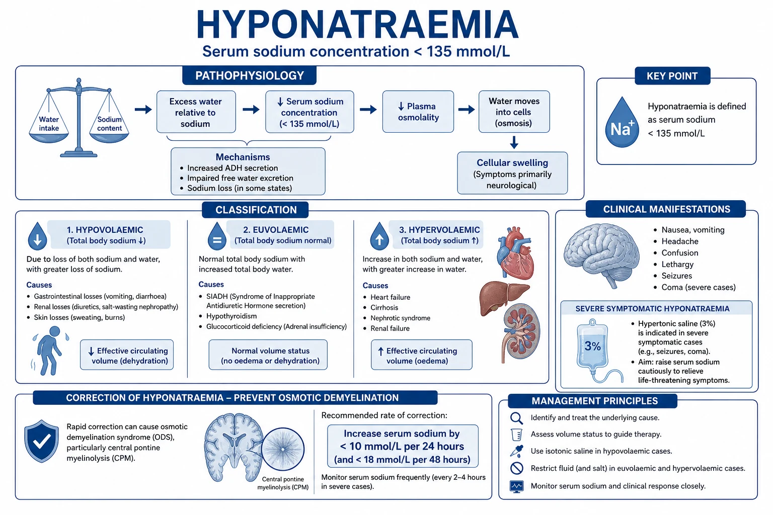
Hyponatraemia classification
The European clinical practice guideline classifies hyponatraemia by volume status and onset:[5]
Hypovolaemic hyponatraemia
Salt AND water loss
- Clinical signs of dehydration: dry mucous membranes, reduced skin turgor, tachycardia
- Causes: diuretics (thiazides), vomiting, diarrhoea, burns, pancreatitis
- Urine sodium <20 (extrarenal loss) or >20 (renal loss — diuretics)
- Treatment: normal saline (0.9% NaCl) to restore volume
Euvolaemic hyponatraemia
SIADH — water excess
- Clinically euvolaemic (no dehydration, no oedema)
- SIADH: urine osmolality >100, urine sodium >40, low uric acid
- Causes: pneumonia, brain injury, malignancy (small cell lung cancer), drugs (SSRIs, carbamazepine)
- Treatment: fluid restriction (<1 L/day), salt tablets, vasopressin antagonist (tolvaptan)
Hypervolaemic hyponatraemia
Water > sodium excess
- Clinical signs of fluid overload: oedema, raised JVP, ascites
- Causes: heart failure, cirrhosis, renal failure
- Treatment: treat underlying condition, fluid restriction, diuretics
SIADH vs cerebral salt wasting
Correction rate — osmotic demyelination
[1]Hypernatraemia
Hypernatraemia management
Assess volume status
Hypovolaemic (free water + salt loss): diarrhoea, diuretics. Give normal saline first to restore volume, then hypotonic solution. Euvolaemic (pure free water loss): DI, insensible losses. Give free water (oral, NG, or 5% dextrose IV). Hypervolaemic (sodium excess): iatrogenic NaHCO3, Conn syndrome. Remove sodium, give diuretics + 5% dextrose.
Calculate free water deficit
Water deficit = TBW x (serum Na / 140 - 1). TBW = 0.6 x body weight (men) or 0.5 x body weight (women). Example: 70kg man with Na 160: deficit = 42 x (160/140 - 1) = 42 x 0.143 = 6 L.
Correct over 48-72 hours
Maximum correction rate: 10 mmol/L/24h (chronic hypernatraemia). Rapid correction causes cerebral oedema. Give half the deficit in first 24h, then the rest over next 24-48h. Monitor Na every 4-6 hours.
Treat underlying cause
Stop diarrhoea/vomiting losses. Control fever (reduce insensible loss). Treat diabetes insipidus (desmopressin for central DI). Review medications.
Potassium disorders
Hyperkalaemia — emergency management
Hyperkalaemia severity and ECG changes (click each)
K >6.5 mmol/L
Wide QRS, sine wave, asystole. MEDICAL EMERGENCY. Calcium gluconate immediately (membrane stabilisation), then insulin-dextrose, salbutamol, and definitive removal (diuretics if urine output, RRT if anuric).
Hyperkalaemia emergency treatment algorithm
Step 1 — Calcium gluconate 10% 10 mL IV (over 2-5 min)
MEMBRANE STABILISATION. Does NOT lower potassium — stabilises cardiac membrane to prevent arrhythmias. Give FIRST if any ECG changes (peaked T waves, wide QRS, sine wave). Onset within minutes, lasts 30-60 min. Can repeat. Monitor ECG continuously.
Step 2 — Insulin-dextrose (shift K into cells)
10 units rapid-acting insulin IV + 25-50 g glucose (50-100 mL of 50% dextrose). Lowers K by 0.5-1.0 mmol/L within 15-30 min. Lasts 4-6 hours. Monitor blood glucose (risk of hypoglycaemia — check at 15, 30, 60, 120 min).
Step 3 — Salbutamol (shift K into cells)
10-20 mg nebulised salbutamol (5x the bronchodilator dose). Beta-2 agonist drives K into cells via Na-K ATPase. Lowers K by 0.5-1.0 mmol/L. Works synergistically with insulin-dextrose. Caution in cardiac disease (tachycardia).
Step 4 — Remove potassium from body
Diuretics: furosemide 40-80 mg IV (only if urine output adequate). Potassium binders: sodium zirconium cyclosilicate, patiromer, calcium resonium (slow — hours to days). RRT (haemodialysis or CRRT): definitive removal if anuric or refractory.
Step 5 — Identify and treat cause
AKI/CKD (most common), rhabdomyolysis, tumour lysis syndrome, acidosis, medications (ACEi, ARBs, K-sparing diuretics, NSAIDs, heparin), Addison disease. Stop contributing medications. Correct acidosis.
Hypokalaemia
Calcium disorders
Hypocalcaemia
Ionised Ca <1.1 mmol/L
- Use IONISED calcium, not total calcium (albumin-corrected Ca is unreliable in ICU)
- Causes: critical illness, sepsis, AKI (phosphate retention), pancreatitis, hypomagnesaemia, post-thyroid/parathyroid surgery
- Symptoms: paraesthesia, tetany, Chvostek/Trousseau signs, seizures, prolonged QT, hypotension
- Treatment: calcium gluconate 10% 10 mL IV (severe/symptomatic). Correct Mg first.
Hypercalcaemia
Ionised Ca >1.3 mmol/L
- Causes: malignancy (most common in ICU), hyperparathyroidism, immobilisation, vitamin D intoxication
- Symptoms: confusion, constipation, polyuria, dehydration, shortened QT
- Treatment: aggressive normal saline (volume expansion + calciuresis), bisphosphonates (zoledronate), calcitonin (rapid but short-lived)
Magnesium
Magnesium in critical illness
Phosphate and refeeding syndrome
Exam practice
SAQ — Electrolyte emergencies
10 minutes · 10 marks
A 65-year-old woman is admitted to ICU with severe community-acquired pneumonia. Day 2: Na 118 mmol/L (was 134 on admission), K 6.8, creatinine 220. ECG shows peaked T waves and wide QRS (140 ms). She is on amoxicillin-clavulanate and clarithromycin.
Clinical pearls
Red flags
Trial evidence
Balanced crystalloids vs saline — SMART trial (Self/Semler 2018, NEJM)
A cluster-randomised, multiple-crossover trial of 15,752 adults in 5 ICUs comparing balanced crystalloids (lactated Ringer's or Plasma-Lyte) with 0.9% saline.[16] Key finding: balanced crystalloids reduced the primary composite outcome of major adverse kidney events within 30 days (MAKE-30: 14.3% vs 15.4%, OR 0.90). Patients in the saline arm had higher mean chloride (108 vs 105 mmol/L) and more hyperchloraemia (≈14% vs 9%), and a higher rate of new RRT. Practice point: for routine ICU fluid resuscitation prefer a balanced crystalloid — the chloride load from 0.9% saline produces a non-anion-gap (hyperchloraemic) metabolic acidosis that can be misread as ongoing shock/lactic acidosis, prompting MORE saline and worsening AKI. Reserve 0.9% saline for genuine hyponatraemia, brain injury (where tonicity matters), severe hypochloraemia, and the resuscitation phase of DKA when K-containing balanced fluids are unsuitable.
Refeeding syndrome incidence — Friedli 2021 systematic review and meta-analysis (Clin Nutr)
Pooled analysis of the incidence of refeeding syndrome across 53 studies in mixed inpatient populations.[3] Key findings: refeeding hypophosphataemia occurs in up to 30–50% of high-risk patients started on standard nutrition. Mortality attributable to refeeding syndrome ranged from 0–9%, with the highest risk in the first 72 hours of feeding and in patients with the lowest BMI, longest period of starvation, and lowest pre-feed phosphate. Practice point: screen EVERY ICU patient for refeeding risk before feed initiation (NICE criteria: BMI <16, unintentional weight loss >15%, little intake >10 days, low pre-feed K/Mg/PO4). Start at 10–15 kcal/kg/day, increase over 4–7 days, give thiamine 200–300 mg before first feed, and replace phosphate/potassium/magnesium aggressively.
Hypomagnesaemia and ICU mortality — Upala 2016 meta-analysis (QJM)
Systematic review and meta-analysis of observational studies reporting serum magnesium at ICU admission.[12] Key findings: hypomagnesaemia at ICU admission was independently associated with increased all-cause mortality (pooled RR ≈ 1.4), longer ICU stay, and higher requirement for mechanical ventilation. The association held across heterogeneous populations (sepsis, trauma, cardiac, mixed ICU). Practice point: measure serum magnesium on every ICU admission and replete aggressively — Mg is a cofactor for >300 enzymatic reactions, regulates K/Ca homeostasis through Na-K ATPase and PTH release, and low Mg predicts refractory dyskalaemias, arrhythmia, and poor outcome.
Hypokalaemia — safe IV correction rates
Safe IV potassium replacement
Confirm safe peripheral concentration (max 40 mmol/L)
Peripheral IV KCl: maximum concentration 40 mmol/L (e.g. 40 mmol in 1 L of 0.9% saline or 5% dextrose). Maximum rate 10 mmol/h via peripheral line with continuous cardiac monitoring. Pain/phlebitis is common at higher concentrations — never exceed 40 mmol/L peripherally.
Central line: up to 20 mmol/h for severe symptomatic disease
Central venous KCl (max concentration 40 mmol in 100 mL = 400 mmol/L) can be infused at 10–20 mmol/h for severe symptomatic hypokalaemia (K <2.5 with ECG changes or arrhythmia). MUST have continuous ECG monitoring and central access secured. NEVER give concentrated KCl as a bolus — fatal arrhythmia.
Correct Mg first or alongside K
Replete magnesium (MgSO4 2–4 g IV over 1–2 h) BEFORE or alongside KCl — without Mg, the distal nephron wastes K through ROMK and the K will not correct. Add 1 g MgSO4 every 1–2 days until serum Mg >0.8 mmol/L.
Calculate total deficit
A serum K drop from 4.0 to 3.0 mmol/L reflects a total body deficit of 100–200 mmol; from 3.0 to 2.0 a further 200–400 mmol. Replace over 24–72 h. Oral KCl (40–80 mmol/day, slow-release) is preferred once oral route available — less phlebitis, sustained repletion, lower arrhythmia risk.
Identify and treat ongoing losses
Stop diuretics if possible. Treat diarrhoea/vomiting. Correct alkalosis (each 0.1 pH rise shifts K intracellularly by ~0.3 mmol/L). Renal K loss (urine K >20 mmol/day in hypokalaemia) suggests diuretics, hyperaldosteronism, RTA, or Mg depletion — investigate.
Potassium ECG changes — a forensic tool
Hyperkalaemia ECG progression
K >5.5 → sine wave
- Mild (5.5–6.0): tall "tented" T waves (narrow, peaked, symmetric), shortened QT
- Moderate (6.0–6.5): loss of P waves, prolonged PR interval, widening of QRS, ST depression
- Severe (6.5–7.5): sine wave (QRS merges with T wave), bradycardia, AV block
- Pre-arrest/asystole (>7.5): wide QRS progressing to ventricular fibrillation or asystole
- NOTE: the ECG is INSENSITIVE for hyperkalaemia — up to ~50% of K >6.5 mmol/L have subtle or absent changes. Treat the number, not just the tracing.
Hypokalaemia ECG progression
K <3.5 → arrhythmia
- Mild (3.0–3.5): flattening of T waves, prominent U waves (best seen in V2–V4)
- Moderate (2.5–3.0): ST depression, T–U fusion, QU prolongation, apparent long QT
- Severe (<2.5): ventricular ectopics, torsades de pointes (U wave falls on T of preceding beat), VF
- Risk amplified by concurrent hypomagnesaemia (long QT) and by digoxin (digoxin toxicity precipitated by hypokalaemia)
- AVOID long QT and check digoxin in any hypokalaemic patient with arrhythmia.
Corrected calcium — when and how
[1]Calcium disorders in depth
Severe hypocalcaemia
Ionised Ca <0.8 mmol/L
- Causes in ICU: sepsis (cytokine-mediated), massive transfusion (citrate chelation), AKI (phosphate retention), acute pancreatitis (saponification), neck surgery (post-thyroidectomy/parathyroidectomy — hungry bone), rhabdomyolysis, alkalosis (increases Ca binding to albumin)
- Clinical: circumoral paraesthesia, carpopedal spasm, Chvostek/Trousseau signs, laryngospasm, bronchospasm, seizures, hypotension, prolonged QT → torsades de pointes
- Treatment: calcium gluconate 10% 10 mL IV (each 10 mL = 2.2 mmol Ca) over 10 min, repeat as needed. Infusion: 10 ampoules in 500 mL over 6–12 h. Correct Mg FIRST or Ca will not improve. AVOID rapid IV Ca in digoxin-toxic patients (Ca²⁺ + digoxin = "stone heart").
Severe hypercalcaemia
Ionised Ca >1.4 mmol/L
- Causes: malignancy (PTHrP, bony metastases, myeloma), primary hyperparathyroidism, immobilisation (especially young paraplegic), vitamin D intoxication, granulomatous disease (sarcoid, TB), milk-alkali syndrome, thiazide diuretics
- Clinical: "stones, bones, abdominal groans, psychic moans" — confusion/coma, polyuria/polydipsia (nephrogenic DI from renal concentrating defect), constipation, nephrolithiasis, dehydration, shortened QT
- Treatment: (1) aggressive normal saline 4–6 L/day to expand volume and calciuresis; (2) furosemide 10–20 mg IV once euvolaemic (NOT before — worsens hypercalcaemia if hypovolaemic); (3) bisphosphonate zoledronate 4 mg IV (onset 24–72 h, lasts weeks); (4) calcitonin 4 IU/kg SC every 12 h (rapid onset, short-lived, tachyphylaxis); (5) glucocorticoids for vitamin D-mediated hypercalcaemia (sarcoid, lymphoma, vitamin D toxicity)
Magnesium — the forgotten cation
Hypomagnesaemia severity and management
Mg <0.3 mmol/L or symptomatic
Seizures, torsades de pointes, refractory hypokalaemia, atrial and ventricular arrhythmia, spasms. IV MgSO4 2 g bolus over 5–10 min (in arrest/torsades — 2 g IV push), then infusion 1–2 g/h. Continuous cardiac monitoring.
Magnesium pharmacology — ICU dosing
Phosphate disorders — beyond refeeding
Hypophosphataemia
PO4 <0.8 mmol/L (severe <0.3)
- Causes in ICU: refeeding syndrome (insulin-driven cellular uptake), sepsis, respiratory alkalosis, DKA recovery (insulin), CRRT/dialysis clearance, diuretics, antacids, phosphate binders, hepatic resection, severe burns
- Consequences: muscle weakness (diaphragm → respiratory failure, failure to wean from ventilator), rhabdomyolysis, impaired tissue oxygen delivery (left-shifted O2 curve via depleted 2,3-DPG), impaired neutrophil function, cardiac failure, haemolysis.
- Treatment: sodium phosphate or potassium phosphate IV (0.08–0.24 mmol/kg over 4–6 h, max 0.6 mmol/kg/24 h). Oral 20–30 mmol/day if mild. Re-check PO4, Ca, Mg, K every 6–12 h. AVOID rapid IV phosphate — fatal arrhythmia, hypocalcaemia, AKI.
Hyperphosphataemia
PO4 >1.5 mmol/L
- Causes in ICU: AKI/CKD (reduced excretion), tumour lysis syndrome, rhabdomyolysis, lactic acidosis, ketoacidosis (cellular shift), bowel preparation (sodium phosphate enemas), hypoparathyroidism, respiratory or metabolic acidosis
- Consequences: hypocalcaemia (precipitation of CaPO4), metastatic calcification (especially if Ca × PO4 >4.4 mmol²/L²), pruritus, secondary hyperparathyroidism (chronic)
- Treatment: (1) treat the cause (e.g. dialysis for AKI, hydration + rasburicase for tumour lysis); (2) phosphate binders (calcium acetate, sevelamer, lanthanum) — for chronic management; (3) CRRT or haemodialysis for severe symptomatic hyperphosphataemia; (4) restrict dietary phosphate
Chloride — the silent regulator of acid–base
Hyperchloraemia (acidosis)
Cl⁻ high, SID low
- Causes: 0.9% saline resuscitation, parenteral nutrition, uretero-enteric fistula (GI loss of HCO3⁻), acetazolamide, renal tubular acidosis type 1 (distal) and type 2 (proximal), resolution phase of diabetic ketoacidosis (Ketones cleared, Cl⁻ retained)
- Findings: metabolic acidosis (low pH, low HCO3⁻), normal anion gap, hyperchloraemia. Compensatory respiratory alkalosis (low PaCO2)
- Treatment: stop saline → switch to balanced crystalloid (Ringer's lactate, Plasma-Lyte); treat cause (RTA — bicarbonate; diarrhoea — rehydrate); CRRT/dialysis in renal failure
Hypochloraemia (alkalosis)
Cl⁻ low, SID high
- Causes: vomiting/prolonged NG suction (loss of HCl), thiazide and loop diuretics, post-hypercapnia, congenital chloride-losing diarrhoea, Bartter and Gitelman syndromes
- Findings: metabolic alkalosis (high pH, high HCO3⁻), hypochloraemia, often hypokalaemia (concurrent)
- Treatment: replace chloride (0.9% saline — Cl⁻ 154 mmol/L is ideal here; or KCl); correct K; stop diuretics; acetazolamide in volume-overloaded alkalosis
Electrolytes in common ICU scenarios
DKA / HHS
Total body deficit, normal/high serum
- Insulin deficiency → intracellular K, Mg, PO4 depletion despite normal/high serum levels
- When insulin starts: K plummets (cellular shift) — replace at 5–10 mmol/h IV once K <5.5 mmol/L
- PO4 falls during recovery (refeeding-like state) — replace if <0.5 mmol/L, especially if respiratory failure
- Mg co-depleted — replace alongside K
- Sodium: PSEUDOHYPONATRAEMIA from hyperglycaemia — correct Na for glucose (add 2.4 mmol/L per 10 mmol/L glucose >5.5)
AKI
Failure of excretion
- Hyperkalaemia (failure of renal K excretion) — calcium gluconate, insulin-dextrose, salbutamol, dialysis
- Hyperphosphataemia → hypocalcaemia (CaPO4 precipitation)
- Metabolic acidosis (high AG — sulphate, phosphate)
- Hyponatraemia (dilutional, or osmotic with hyperglycaemia)
- CRRT clears K, PO4, Mg — MONITOR and replace (Mg, PO4 especially)
CRRT
Iatrogenic clearance
- Hypophosphataemia (cleared by CRRT) — common, predicts prolonged ventilation; replace IV
- Hypomagnesaemia — replete 2–4 g MgSO4/24 h; check daily
- Hypokalaemia — adjust CRRT replacement fluid K concentration
- Hypocalcaemia — citrate regional anticoagulation chelates Ca; monitor ionised Ca q6h
- Hypernatraemia — if replacement fluid or dialysate is high in Na; check daily Na
Diuretics
Iatrogenic renal loss
- Loop (furosemide): hypokalaemia, hypocalcaemia, hypomagnesaemia, hypochloraemic alkalosis, hyperglycaemia, hyperuricaemia
- Thiazides: hyponatraemia (SIADH-like pattern), hypokalaemia, hypercalcaemia, hyperglycaemia
- Spironolactone/amiloride: HYPERkalaemia (especially with AKI or ACEi/ARB)
- Acetazolamide: hypokalaemia, hyperchloraemic metabolic acidosis (bicarbonate wasting)
Sepsis
Capillary leak + shifts
- Hypocalcaemia (ionised) common in septic shock — prognostic of mortality
- Hypomagnesaemia common — replete aggressively; predicts mortality
- Hypophosphataemia (redistribution + decreased intake) — replete; predicts prolonged ventilation
- Hypokalaemia (intracellular shift from stress catecholamines + alkalosis)
- SIADH pattern (especially Legionella, Mycoplasma pneumonia)
- Stress hyperglycaemia with osmotic diuresis → sodium derangement
BRASH syndrome — bradycardia + hyperkalaemia
[1]Additional clinical pearls — beyond the basics
Additional red flags — pitfalls and traps
References
- [1]Leonardi-Bee J, et al. Cerebral Salt Wasting Syndrome 2026.PMID 30521276
- [2]Verbalis JG, et al. Syndrome of inappropriate antidiuresis/hyponatremia in COVID-19 Pituitary, 2024.PMID 39196447
- [3]Friedli N, et al. The incidence of the refeeding syndrome. A systematic review and meta-analyses of literature Clin Nutr, 2021.PMID 34134001
- [4]Reinhart RA. Magnesium deficiency: pathophysiologic and clinical overview Am J Kidney Dis, 1994.PMID 7977315
- [5]Spasovski G, Vanholder R, Allolio B, et al. Clinical practice guideline on diagnosis and treatment of hyponatraemia Eur J Endocrinol, 2014.PMID 24569125
- [6]Sterns RH. Disorders of plasma sodium--causes, consequences, and correction N Engl J Med, 2015.PMID 25551526
- [7]Adrogue HJ, Madias NE. Hyponatremia N Engl J Med, 2000.PMID 10824078
- [8]Adrogue HJ, Madias NE. Hypernatremia N Engl J Med, 2000.PMID 10816188
- [9]Kovesdy CP. Management of hyperkalaemia in chronic kidney disease Nat Rev Nephrol, 2014.PMID 25223988
- [10]Palmer BF, Clegg DJ. Hyperkalemia across the Continuum of Kidney Function Clin J Am Soc Nephrol, 2018.PMID 29114006
- [11]Farkas JD, Long B, Koyfman A. BRASH Syndrome: Bradycardia, Renal Failure, AV Blockade, Shock, and Hyperkalemia J Emerg Med, 2020.PMID 32565167
- [12]Upala S, et al. Hypomagnesemia and mortality in patients admitted to intensive care unit: a systematic review and meta-analysis QJM, 2016.PMID 27016536
- [13]Fiaccadori E, et al. Hypophosphatemia and phosphorus depletion in respiratory and peripheral muscles of patients with respiratory failure due to COPD Chest, 1994.PMID 8181325
- [14]Geerse DA, et al. Approach to hypophosphataemia in intensive care units - a nationwide survey Neth J Med, 2012.PMID 23123542
- [15]Simon DB, et al. Bartter's syndrome, hypokalaemic alkalosis with hypercalciuria, is caused by mutations in the Na-K-2Cl cotransporter NKCC2 Nat Genet, 1996.PMID 8640224
- [16]Semler MW, Self WH, Wanderer JP, et al. Balanced Crystalloids versus Saline in Critically Ill Adults N Engl J Med, 2018.PMID 29485925
- [17]Kaplan LJ, Kellum JA. Fluids, pH, ions and electrolytes Curr Opin Crit Care, 2010.PMID 20613504
- [18]Simon DB, et al. Gitelman's variant of Bartter's syndrome, inherited hypokalaemic alkalosis, is caused by mutations in the thiazide-sensitive Na-Cl cotransporter Nat Genet, 1996.PMID 8528245