ICU · Renal/Metabolic
Acute severe hyperkalaemia: emergency management algorithm
Also known as Hyperkalaemia · High potassium · Potassium emergency · Hyperkalemia
Hyperkalaemia (K+ >5.5 mmol/L): common ICU emergency. SEVERE (K+ >6.5 or ECG changes): life-threatening arrhythmia. Causes: AKI/CKD, rhabdomyolysis, tumour lysis, drugs (ACEi/ARB, K-sparing diuretics, TMP-SMX, heparin), Addison's, acidosis, massive transfusion. ECG changes: peaked T waves → widened QRS → sine wave → asystole/VF. Management: (1) STABILISE cardiac membrane (calcium gluconate 10 mL 10% IV). (2) SHIFT K+ into cells (insulin/dextrose, salbutamol, sodium bicarbonate). (3) REMOVE K+ from body (loop diuretics, GI cation exchange — patiromer/zirconium, dialysis). ECG changes = EMERGENCY.
On this page & tools
Your progress
Saved locally on this device.
Target exams
Red flags

ECG changes — recognise the progression
The ECG is the single most important bedside test in suspected hyperkalaemia. ECG changes reflect the progressive depolarisation of cardiac myocytes as extracellular K+ rises, and they correlate roughly (not perfectly) with severity. Any ECG change attributable to hyperkalaemia mandates calcium before anything else.[11]
Hyperkalaemia severity and ECG changes
| K+ (mmol/L) | Severity | Typical ECG changes | Action |
|---|---|---|---|
| 5.5-5.9 | Mild | Usually none | Treat cause, K-restricted diet, binders. Stop K+-sparing drugs |
| 6.0-6.4 | Moderate | Peaked T waves (may appear) | Shift K+ (insulin/dextrose, salbutamol), binders, consider dialysis. Continuous cardiac monitor |
| 6.5-6.9 | Severe | Peaked T, PR prolongation, early QRS widening | CALCIUM first (if ECG changes), shift K+, REMOVE (diuretics/dialysis). Emergency |
| >7.0 | LIFE-THREATENING | Wide QRS, sine wave, VF, asystole | CALCIUM immediately + shift + urgent dialysis. Prepare for arrest |
Emergency management algorithm

Emergency management of severe hyperkalaemia (K+ >6.5 or ECG changes)
- ECG immediately — peaked T waves? Wide QRS? Sine wave? If ANY ECG changes → CALCIUM FIRST. Attach continuous cardiac monitor and establish IV access. Take a venous/arterial gas for rapid K+ (lab serum K+ may lag)
- STABILISE cardiac membrane (if ECG changes) — CALCIUM GLUCONATE 10 mL of 10% IV over 2-5 min (or calcium chloride 10 mL of 10% via central line — 3x more ionised Ca²⁺). Onset 1-3 min. Duration 30-60 min. May repeat once after 5 min if ECG changes persist. Does NOT lower K+ — only prevents arrhythmia. Exclude digoxin toxicity first (give Fab, not calcium)[3]
- SHIFT K+ into cells — INSULIN/DEXTROSE: 10 units rapid-acting insulin + 25 g (50 mL of 50%) dextrose IV over 15 min (onset 15 min, lowers K+ 0.5-1.0 mmol/L for 4-6 h). PLUS SALBUTAMOL 10-20 mg nebulised (or 500 mcg IV over 15 min) — synergistic, adds 0.5-1.0 mmol/L. ADD SODIUM BICARBONATE 50 mmol of 8.4% IV (especially if acidotic or arrest) — modest additional shift.[4]
- REMOVE K+ from body — LOOP DIURETICS (frusemide 40-80 mg IV — only if kidneys functioning and producing urine). GI BINDERS (sodium zirconium cyclosilicate 10 g, or patiromer 8.4-16.8 g — slower, hours). DIALYSIS (definitive — intermittent haemodialysis fastest; CVVHDF slower). Begin arranging dialysis EARLY if K+ >7 despite therapy, ongoing K+ release, or anuric renal failure[2]
- Identify and treat cause — AKI/CKD, rhabdomyolysis, tumour lysis, drugs (ACEi/ARB, spironolactone, amiloride, TMP-SMX, heparin, beta-blockers, calcineurin inhibitors), Addison's, acidosis (DKA, lactic), GI bleed (digested blood = K+ load), massive transfusion. REVIEW THE MEDICATION CHART and stop all K+-sparing/raising agents
- Monitor — continuous ECG, repeat K+ at 1-2 h (then 2-4 h), CHECK GLUCOSE at 0, 30, 60 min and then hourly for 4-6 h after insulin/dextrose (hypoglycaemia risk). Document calcium given. Once K+ <6.0 and ECG normal → step down to oral binders + cause treatment
Stepwise pharmacology — what each drug actually does (exam framework)
- CALCIUM GLUCONATE 10% — membrane stabiliser. Mechanism: raises the threshold potential of myocardial myocytes (restores the gap between resting and threshold potential that hyperkalaemia has narrowed). Dose: 10 mL IV over 2-5 min (2.2 mmol Ca²⁺), repeat once after 5 min if ECG changes persist. Onset 1-3 min, duration 30-60 min. No effect on serum K+. Indication: ANY ECG change attributable to hyperkalaemia. Contra-indication/caution: digoxin toxicity (give Fab instead)[3]
- INSULIN + DEXTROSE — intracellular shift (the workhorse). Mechanism: insulin directly stimulates the Na/K ATPase → drives K+ into cells. Dextrose prevents hypoglycaemia. Dose: 10 units rapid-acting insulin (e.g., Actrapid) + 25 g dextrose (50 mL of 50%) IV over 15 min. Lowers K+ 0.5-1.0 mmol/L within 15-30 min, effect lasts 4-6 h. Most reliable shifting therapy. MUST monitor glucose hourly for 4-6 h — hypoglycaemia in up to 20-30%, the leading complication[5]
- SALBUTAMOL — intracellular shift (add-on). Mechanism: beta-2 agonist → cAMP → stimulates Na/K ATPase → K+ into cells. Dose: 10-20 mg nebulised (this is 5-10x the asthma dose) OR 250-500 mcg IV over 15 min. Onset 15-30 min, lowers K+ 0.5-1.0 mmol/L, lasts 2-4 h. Synergistic with insulin/dextrose (give both). Cautions: tachycardia, tremor, ischaemia (beta-1 effect), avoid in tachyarrhythmia/active ischaemia. Less effective in beta-blocked patients[8]
- SODIUM BICARBONATE — shift (mainly if acidotic). Mechanism: corrects acidosis → H+ stops exiting cells → K+ no longer needs to exit to maintain electroneutrality → K+ shifts in. Dose: 50 mmol of 8.4% IV over 5-15 min (or 500 mL of 1.26%). Minimal effect in non-acidotic patients — do not rely on it alone. Useful in DKA/lactic acidosis and in cardiac arrest (see below). Cautions: hypernatraemia, volume overload, alkalosis, hypocalcaemia (lowers ionised Ca²⁺)[8]
- SODIUM ZIRCONIUM CYCLOSILICATE / PATIROMER — gut removal. SZC: 10 g orally/NG, exchanges Na+/H+ for K+ in the gut, lowers K+ ~0.5-0.7 mmol/L over 2-4 h. Patiromer: 8.4-16.8 g, exchanges Ca²+ for K+, slower. Both are for subacute/chronic control — too slow for an emergency but useful as bridge to dialysis or to prevent rebound. Avoid SPS (sodium polystyrene sulfonate) in the acute setting — slow and associated with bowel necrosis[9]
- HAEMODIALYSIS / CVVHDF — definitive removal. Intermittent haemodialysis: 1-1.5 mmol/L drop in 1-2 h (FASTEST). CVVHDF: slower, gentler, lower rebound risk. Indications: K+ >7 despite medical therapy, ongoing endogenous K+ release (rhabdo, TLS), end-stage renal failure, severe acidosis, refractory arrest. REBOUND is common — K+ diffuses out of cells and from dialysate gradients; recheck within 1-2 h of finishing[2]
Calcium gluconate vs calcium chloride
| CALCIUM GLUCONATE 10% | CALCIUM CHLORIDE 10% | |
|---|---|---|
| Ionised Ca²⁺ per 10 mL | 2.2 mmol | 6.8 mmol (≈3x more) |
| Route | Peripheral or central | CENTRAL line only |
| Extravasation risk | Low (less tissue necrosis) | High — severe necrosis if extravasated |
| Hepatic metabolism | Requires hepatic conversion to release Ca²⁺ (slower in shock/hepatic failure) | Immediately bioavailable |
| Preferred use | First-line in awake/non-arrest patients, peripheral access | Cardiac ARREST (central line) — more Ca²⁺ delivered fast |
| Dose | 10 mL IV over 2-5 min, repeat once after 5 min | 10 mL IV via central line (rapid), repeat once |
| Onset/duration | 1-3 min / 30-60 min | 1-3 min / 30-60 min |
K+-shifting therapies — insulin/dextrose vs salbutamol vs bicarbonate
| Therapy | Dose | K+ fall (mmol/L) | Onset | Duration | Key cautions |
|---|---|---|---|---|---|
| Insulin + dextrose | 10 U insulin + 25 g dextrose IV | 0.5-1.0 | 15 min | 4-6 h | Hypoglycaemia (20-30%) — monitor glucose hourly ×6 h |
| Salbutamol (neb) | 10-20 mg nebulised | 0.5-1.0 | 15-30 min | 2-4 h | Tachycardia, tremor, ischaemia; less effective if beta-blocked |
| Salbutamol (IV) | 250-500 mcg over 15 min | 0.6-1.0 | 15 min | 2-4 h | As above; avoid in active ischaemia/VT |
| Sodium bicarbonate | 50 mmol 8.4% IV (or 500 mL 1.26%) | 0.2-0.5 (acidotic) / <0.2 (non-acidotic) | 30-60 min | 2-4 h | Only reliable if ACIDOTIC; hypernatraemia, alkalosis, hypocalcaemia |
| Insulin + salbutamol | Both together | Additive (~1.0-1.5) | 15 min | 4-6 h | COMBINE for moderate-severe hyperkalaemia |
K+-removal therapies — diuretics vs binders vs dialysis
| Therapy | Speed | Use in oliguria/anuria | Setting | Notes |
|---|---|---|---|---|
| Loop diuretic (frusemide) | Hours | Ineffective if anuric | Functioning kidneys | Dose 40-80 mg IV; add thiazide for sequential blockade. Also treats volume overload |
| Sodium zirconium cyclosilicate | 2-4 h | Works (gut, not kidney) | Subacute, bridge, chronic | 10 g oral/NG; rapid onset among binders; may cause oedema (Na+ load) |
| Patiromer | 4-7 h | Works | Chronic, heart-failure patients on RAASi | 8.4-16.8 g; exchanges Ca²+ (mild hypomagnesaemia/hypocalcaemia) |
| SPS (Kayexalate/resonium) | 12-24+ h | Works | Largely AVOIDED | Slow; bowel necrosis/fibrosis reported — not for acute use |
| Haemodialysis (intermittent) | 1-2 h (FASTEST) | Indicated if anuric | Definitive | 1-1.5 mmol/L drop; beware rebound — recheck within 1-2 h |
| CVVHDF/SLED | Hours (gentler) | Indicated if haemodynamically unstable | ICU, unstable | Slower, lower rebound, continuous removal |
GI potassium binders — patiromer vs sodium zirconium cyclosilicate vs SPS
| PATIROMER | SODIUM ZIRCONIUM CYCLOSILICATE | SPS (RESONIUM/KAYEXALATE) | |
|---|---|---|---|
| Exchange ion | Calcium | Sodium / hydrogen | Sodium (or calcium prep) |
| Onset | 4-7 h | 2-4 h | 12-24+ h |
| K+ reduction | ~0.2-0.4 mmol/L per dose | ~0.5-0.7 mmol/L per dose | Variable, modest |
| Approved for | Chronic hyperkalaemia (CKD, on RAASi) | Acute and chronic hyperkalaemia | Older; now largely avoided |
| Safety concerns | Hypomagnesaemia, hypocalcaemia, GI upset; binds some drugs (separate dosing) | Oedema/heart failure exacerbation (Na+ load); avoid if Na+ restricted | Bowel necrosis, perforation — FDA warning; not for acute use |
| Take-home | Good for chronic CKD/HF patients needing RAASi | Best binder for subacute ICU control | AVOID in the acute setting |
Causes — organised by mechanism
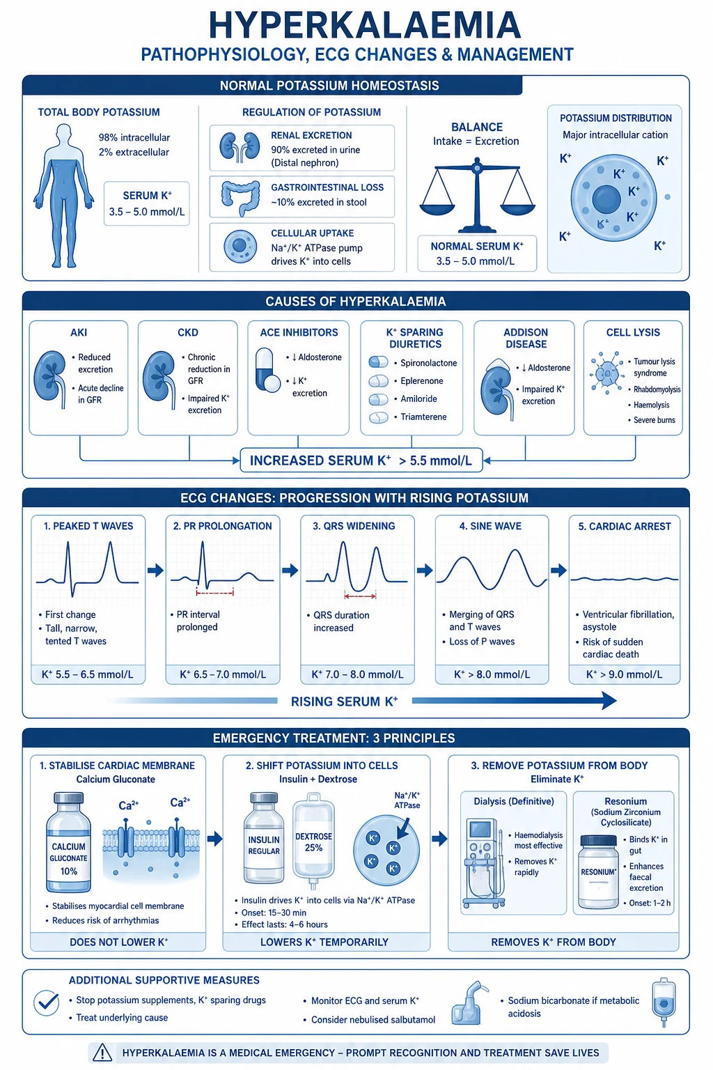
Hyperkalaemia always reflects an imbalance between K+ intake, distribution (cellular shift), and excretion. In the ICU, impaired renal excretion + transcellular shift + drug effect usually combine. Structure your cause hunt around three buckets: impaired excretion, transcellular shift, increased intake/release.[2]
[1]Drug causes of hyperkalaemia — mechanism at a glance
| Drug | Mechanism | Typical context |
|---|---|---|
| ACEi / ARB | ↓ aldosterone (block RAAS) | CKD, HF — 'RAASi hyperkalaemia' |
| Spironolactone / eplerenone / finerenone | Aldosterone receptor antagonism | HF, cirrhosis, resistant HTN |
| Amiloride / triamterene | Block ENaC in collecting duct → ↓ K+ secretion | K-sparing diuretics; kombiglyze |
| Trimethoprim / pentamidine | Block ENaC (like amiloride) | UTI/pneumocystis prophylaxis in renal failure |
| Heparin (any route) | Inhibits aldosterone synthesis | Surprisingly common in ICU patients on prophylaxis |
| NSAIDs | ↓ renin → ↓ aldosterone | AKI + hyperkalaemia in elderly |
| Calcineurin inhibitors (cyclosporin, tacrolimus) | Type 4 RTA-like effect | Transplant patients |
| Beta-blockers | Block beta-2 mediated K+ uptake into cells; also suppress renin | Any patient; BRASH syndrome with AV node blockers |
| Digoxin | Inhibits Na/K ATPase (acute toxicity → hyperkalaemia) | Acute overdose; check level |
| Succinylcholine | Depolarising agent → K+ efflux from muscle | Burns >24 h, crush, denervation, immobilisation |
| Mannitol / hypertonic saline | Osmotic shift / acid-base effect | Ongoing infusion |
| K+-containing IV fluids / Penicillin G | Direct K+ load | Large-volume KCl-containing fluids |
Pseudohyperkalaemia — don't treat a false alarm
Pseudohyperkalaemia vs true hyperkalaemia
| Feature | PSEUDOHYPERKALAEMIA | TRUE HYPERKALAEMIA |
|---|---|---|
| Mechanism | K+ released from cells DURING/after venepuncture | Genuinely high plasma/serum K+ in vivo |
| Causes | Haemolysis, fist clenching, prolonged tourniquet (>1 min), small-gauge needle, delayed transport, severe thrombocytosis (>1000) or leucocytosis (>100) leaking K+ ex vivo | (see cause list above) |
| ECG | Normal (no peaked T, no wide QRS) | Often abnormal (but may be normal — see pitfall) |
| Clinical picture | Asymptomatic, no reason to be hyperkalaemic | Fits the clinical context (AKI, drugs, acidosis, rhabdo) |
| Confirmation | Repeat sample: ARTERIAL or free-flowing venous (no tourniquet), rapid draw, on ice, analyse promptly. Consider plasma K+ (heparin tube) vs serum (clots → more release) | Repeat K+ confirms persistent elevation |
| Action | Recheck before treating — avoid iatrogenic hypokalaemia | Treat per algorithm |
Insulin/dextrose — getting it right and avoiding hypoglycaemia
Insulin/dextrose is the most effective and most reliable K+-shifting therapy — but hypoglycaemia after a 10-unit + 25 g regimen occurs in up to 20-30% of patients, and is the single most important iatrogenic complication of hyperkalaemia treatment. The risk is highest 1-3 h after administration, in patients with renal failure (reduced insulin clearance), hepatic failure, low body weight, fasting, or a baseline glucose <7 mmol/L.[5]
Safe insulin/dextrose administration (anti-hypoglycaemia bundle)
- Check a baseline glucose before giving insulin/dextrose. If glucose <7 mmol/L, raise it first (give additional dextrose or run the dextrose before the insulin)
- Dose to the patient: 10 units rapid-acting insulin (Actrapid/Humulin R) + 25 g dextrose IV (50 mL of 50%) over 15 min. Consider 5 units + 50 g dextrose (or 25 g then 25 g over 1-2 h) in high-risk patients (low weight, fasting, dialysis, baseline glucose <7)
- Monitor glucose at 0, 30, 60 min, then hourly for 6 h. Most hypoglycaemia occurs at 1-3 h — do NOT stop checking at 1 h
- Keep 50% dextrose at the bedside and treat any glucose <4 mmol/L (or symptomatic) with 25-50 g IV dextrose, then a dextrose infusion
- Warn the ward team — patients are often transferred to a ward where the insulin/dextrose effect persists but glucose checks stop. Hand over the monitoring plan explicitly
- Document insulin dose, dextrose dose, baseline glucose, and the monitoring schedule in the chart
Hyperkalaemic cardiac arrest
Hyperkalaemia is one of the reversible causes of cardiac arrest (the H's) and should be sought in EVERY arrest — especially PEA with bradycardia, asystole, or VF/pVT that is refractory to standard ALS. A venous or arterial gas at the point of arrest gives a K+ within minutes.[7]
Hyperkalaemic cardiac arrest bundle (in addition to standard ALS)
- Suspect hyperkalaemia in every arrest — send a VBG/ABG immediately (K+, Na+, glucose, lactate, pH). Especially suspect with: PEA/bradycardia, wide-complex arrest, known renal failure, dialysis patient who missed a session, diabetic in DKA, crush injury, drug overdose (digoxin, beta-blocker)
- CALCIUM CHLORIDE 10 mL of 10% via CENTRAL line (preferred in arrest — 6.8 mmol Ca²⁺, immediately bioavailable). If no central line: calcium gluconate 10 mL of 10% via large peripheral line. Repeat every 5-10 min (up to 3-4 doses). Calcium stabilises the membrane and may restore a perfusing rhythm if hyperkalaemia is the cause[7]
- INSULIN/DEXTROSE — 10 units insulin + 25 g dextrose IV bolus (push the dextrose 50%, then insulin, in arrest). Even in arrest, this shifts K+ and is given alongside ongoing CPR
- SODIUM BICARBONATE 50 mmol of 8.4% IV bolus — given during CPR in suspected hyperkalaemic arrest. Recent randomised data (Eggertsen 2024) suggest a survival benefit from combined calcium chloride + bicarbonate in hyperkalaemic arrest. May repeat
- SALBUTAMOL — limited role in arrest (IV beta-agonism competes with ongoing CPR); consider if ROSC achieved or as adjunct
- EMERGENCY HAEMODIALYSIS if refractory — arrange while CPR continues if a viable cause and reversible context (e.g., missed dialysis). ECPR/extracorporeal support may buy time
- Post-ROSC: recheck K+, treat cause (restart dialysis, stop offending drugs, treat rhabdo/TLS), continuous monitoring, plan definitive removal (HD). Beware rebound
Sodium bicarbonate + calcium chloride in hyperkalaemic cardiac arrest — Eggertsen 2024 (RCT)
Design: randomised, blinded, placebo-controlled, prehospital + in-hospital cardiac arrest with suspected hyperkalaemia (K+ >6.0 mmol/L). Intervention: calcium chloride 5 mmol + sodium bicarbonate 50 mmol IV vs placebo, in addition to standard ALS.[7] Key finding: combined calcium + bicarbonate improved surrogate outcomes (return of spontaneous circulation, sustained ROSC) versus placebo in hyperkalaemia-attributed arrest. Practice point: this is the first RCT-grade evidence for specific metabolic therapy in hyperkalaemic arrest and supports giving both calcium AND bicarbonate during CPR for suspected hyperkalaemia — not calcium alone. Give early, in parallel with standard ALS, and send a gas to confirm. Caveats: prehospital setting; surrogate outcomes; treat the patient in front of you — if K+ confirmed >6.5 in arrest, give the full bundle (calcium chloride + insulin/dextrose + bicarbonate).
BRASH syndrome — a hyperkalaemia-specific trap
BRASH syndrome — recognise and break the loop
- Recognise the cluster: Bradycardia + Renal failure + AV blockade (medication) + Shock + Hyperkalaemia. The patient on a beta-blocker or non-DHP calcium-channel blocker (or digoxin) who develops renal impairment and hyperkalaemia slips into a self-reinforcing loop[6]
- Mechanism of the loop: AV-nodal blocker → bradycardia + hypotension → renal hypoperfusion → worsening AKI + acidosis → worsening hyperkalaemia → worsening bradycardia/AV block → more hypoperfusion. Each element feeds the next[6]
- Why standard ALS fails: atropine and transcutaneous pacing often do NOT work because the driver is hyperkalaemia and drug toxicity, not primary conduction disease. The bradycardia resolves when K+ is corrected and the AV-blocker is antagonised/cleared[6]
- Treatment bundle: (a) CALCIUM to stabilise the membrane; (b) INSULIN/DEXTROSE + SALBUTAMOL to shift K+; (c) treat the AV-blocker — beta-blocker: glucagon/high-dose insulin euglycaemia therapy; calcium-channel blocker: calcium (high dose); digoxin: Fab fragments; (d) vasopressors/inotropes for shock; (e) treat the AKI (fluids if volume-depleted, then dialysis if refractory); (f) treat acidosis[6]
- Don't be fooled by a 'reasonable' heart rate. A beta-blocked patient may not be profoundly bradycardic, but the AV-nodal blockade still drives the loop. The combination of AV-blocker + AKI + hyperkalaemia + hypoperfusion is the syndrome.[6]
High-yield clinical pearls (examiner favourites)
Exam practice — SAQs
SAQ — Severe hyperkalaemia with ECG changes in a CKD patient on RAAS inhibitors
10 minutes · 10 marks
A 72-year-old man with CKD stage 4 (baseline creatinine 280 micromol/L) on perindopril, spironolactone and trimethoprim-sulfamethoxazole for a urinary tract infection presents with progressive weakness and presyncope. He is bradycardic (HR 44) with a BP of 92/58. Venous gas: K+ 7.4 mmol/L, pH 7.28, bicarbonate 16. ECG shows a wide QRS (134 ms), peaked and tented T waves in V2-V4, PR prolongation and loss of P waves.
SAQ — Hypokalaemia with Torsades de Pointes and cardiac arrest
10 minutes · 10 marks
A 58-year-old woman with a history of alcohol misuse and bulimia nervosa is admitted with three days of severe vomiting and diarrhoea. She has a nasogastric tube on free drainage and has received frusemide 80 mg IV for pulmonary oedema. Her regular medications include methadone and ondansetron. Bloods: K+ 2.1 mmol/L, Mg 0.4 mmol/L, ionised Ca 0.9 mmol/L. ECG shows QTc 560 ms with a run of polymorphic VT that twists around the baseline (Torsades de Pointes); she becomes pulseless.
Red flags
Prognosis and evidence
UKKA / KDIGO approach to hyperkalaemia (Clase 2020, Kidney Int)
KDIGO Controversies Conference conclusions on potassium homeostasis and dyskalaemia management (Clase 2020):[1]
- Mild (5.5-5.9): cause-specific treatment, dietary advice, K+ binders (patiromer/SZC for chronic). No emergency treatment
- Moderate (6.0-6.4): shift K+ (insulin/dextrose if symptomatic or ECG changes), binders, review meds. Monitor closely
- Severe (≥6.5): EMERGENCY — calcium (if ECG changes), insulin/dextrose, salbutamol, consider dialysis
- Insulin/dextrose dose: 10 units + 25 g dextrose (adult). Monitor glucose every hour for 4-6 h (hypoglycaemia risk emphasised)
- Calcium: gluconate 10 mL 10% (peripheral) or chloride 10 mL 10% (central — 3x more Ca²⁺)
- RAAS inhibitors should generally be CONTINUED in heart failure/CKD where possible, using binders to manage K+ (stopping RAASi worsens long-term outcomes) Mortality: hyperkalaemia-caused cardiac arrest mortality is high (>50% if K+ was the proximate cause). With prompt treatment: arrhythmia prevented, K+ lowered → survival depends on underlying cause.
Insulin/dextrose evidence — Harel 2016 systematic review (PLoS One)
Systematic review of optimal insulin dose/method for emergency hyperkalaemia:[5]
- Insulin/dextrose reliably lowers K+ by 0.5-1.0 mmol/L within 15-30 min — the most consistent shifting therapy
- Hypoglycaemia is common — across studies, any hypoglycaemia up to 20-30%, severe hypoglycaemia (glucose <3.0 mmol/L) 5-10%, peaking 1-3 h post-dose
- Lower insulin doses (5 units) with adequate dextrose achieve a similar K+ fall with less hypoglycaemia in some studies — but 10 units remains the standard adult dose
- Practice point: give insulin/dextrose, then commit to ≥6 h of glucose monitoring with bedside dextrose available. Identify high-risk patients (renal/hepatic failure, low body weight, fasting, baseline glucose <7) and consider reduced-dose regimens.
Bicarbonate/insulin/albuterol synergy — Allon 1996 (Am J Kidney Dis)
Classic dialysis-patient crossover study:[8]
- Insulin/dextrose lowered K+ by ~1.0 mmol/L; albuterol by ~1.0 mmol/L; combined by ~1.3-1.5 mmol/L (additive)
- Bicarbonate alone had a trivial K+-lowering effect in these (non-acidotic) dialysis patients — confirming bicarbonate is useful mainly in acidosis
- Practice point: combine insulin/dextrose + salbutamol for moderate-severe hyperkalaemia; reserve bicarbonate for the acidotic patient or the arrest bundle.
Sodium zirconium cyclosilicate and patiromer — long-term efficacy and safety
Sodium zirconium cyclosilicate (Roger 2021, NDT): long-term (up to 12 months) SZC maintained normokalaemia across mild/moderate and severe/end-stage CKD groups; efficacy was rapid (K+ fall within 2-4 h of first dose) and sustained. Oedema (Na+ load) was the main caution — relevant in heart failure.[9] Patiromer (Pitt 2018, ESC Heart Fail): in heart-failure patients with diabetic nephropathy on RAAS inhibitors, long-term patiromer enabled patients to remain on RAASi while controlling K+. Main adverse effects: mild hypomagnesaemia and hypocalcaemia.[10] Practice point: both novel binders allow continuation of guideline-directed RAASi in HF/CKD. SZC has a role in subacute ICU control (faster onset among binders); patiromer suits chronic outpatient management. Avoid SPS (sodium polystyrene sulfonate) in the acute setting due to bowel-necrosis risk.
Recent evidence and outcomes — Geldermann 2026 (Emerg Med J)
Contemporary evidence-based review of acute hyperkalaemia in emergency care:[11]
- A normal ECG does NOT exclude severe hyperkalaemia — up to ~50% of K+ >6.5 mmol/L have subtle or absent changes; treat the number, not just the tracing
- Pseudohyperkalaemia remains under-recognised — always confirm with a non-haemolysed or arterial sample before treating an unexpected value in a well patient
- Combined shifting therapy (insulin/dextrose + salbutamol) outperforms monotherapy
- Hypoglycaemia is the leading iatrogenic complication of insulin/dextrose — reinforce the 6-hour monitoring bundle
- Novel binders and dialysis timing continue to evolve — start removal planning EARLY in severe hyperkalaemia rather than waiting for refractory K+
Putting it together — exam one-liners
References
- [1]Clase CM, Carrero JJ, Ellison DH, et al. Potassium homeostasis and management of dyskalemia in kidney diseases: conclusions from a Kidney Disease: Improving Global Outcomes (KDIGO) Controversies Conference Kidney Int, 2020.PMID 31706619
- [2]Palmer BF, Carrero JJ, Clegg DJ. Clinical Management of Hyperkalemia Mayo Clin Proc, 2021.PMID 33160639
- [3]Long B, Warix JR, Koyfman A. Hyperkalemia in the Emergency Department: Yes, a Need for Further Evidence, but Do Not Discount What We Have J Emerg Med, 2019.PMID 31326003
- [4]Moussavi K, Fitter S, Gabrielson SW. Management of Hyperkalemia With Insulin and Glucose: Pearls for the Emergency Clinician J Emerg Med, 2019.PMID 31084947
- [5]Harel Z, Kamel KS. Optimal Dose and Method of Administration of Intravenous Insulin in the Management of Emergency Hyperkalemia: A Systematic Review PLoS One, 2016.PMID 27148740
- [6]Farkas JD, Long B, Koyfman A. BRASH Syndrome: Bradycardia, Renal Failure, AV Blockade, Shock, and Hyperkalemia J Emerg Med, 2020.PMID 32565167
- [7]Eggertsen MA, Munch Johannsen C, Kovacevic A, et al. Sodium Bicarbonate and Calcium Chloride for the Treatment of Hyperkalemia-Induced Cardiac Arrest: A Randomized, Blinded, Placebo-Controlled Animal Study Crit Care Med, 2024.PMID 37921685
- [8]Allon M, Shanklin N. Effect of bicarbonate administration on plasma potassium in dialysis patients: interactions with insulin and albuterol Am J Kidney Dis, 1996.PMID 8840939
- [9]Roger SD, Lavin PT, Lerma EV, et al. Long-term safety and efficacy of sodium zirconium cyclosilicate for hyperkalaemia in patients with mild/moderate versus severe/end-stage chronic kidney disease: comparative results from an open-label, Phase 3 study Nephrol Dial Transplant, 2021.PMID 32030422
- [10]Pitt B, Bakris GL, Weir MR, et al. Long-term effects of patiromer for hyperkalaemia treatment in patients with mild heart failure and diabetic nephropathy on angiotensin-converting enzymes/angiotensin receptor blockers: results from AMETHYST-DN ESC Heart Fail, 2018.PMID 29767459
- [11]Geldermann N, Dzimiera J, Fischer H, et al. Acute hyperkalaemia in emergency care: evidence-based approaches Emerg Med J, 2026.PMID 41506858
- [12]Sumida K, Biruete A, Kistler BM, et al. New Insights Into Dietary Approaches to Potassium Management in Chronic Kidney Disease J Ren Nutr, 2023.PMID 37610407