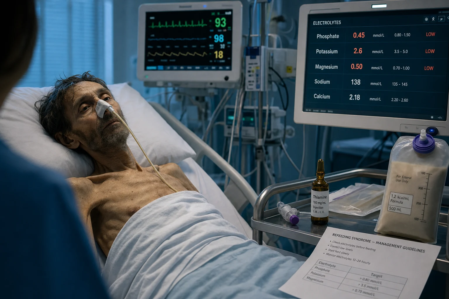
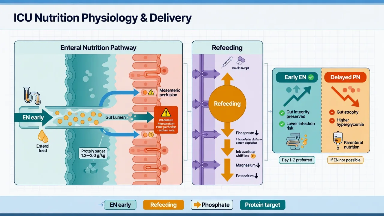
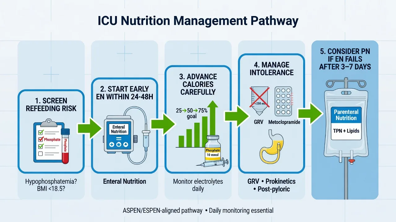
Figure Hypophosphataemia (<0.65 mmol/L) causes respiratory muscle weakness (weaning failure), impaired oxygen release (left-shifted haemoglobin curve) and cardiomyopathy — classic in refeeding syndrome, DKA recovery and sepsis. Hyperphosphataemia complicates AKI, rhabdomyolysis and tumour lysis and drives secondary hyperparathyroidism; treat with phosphate binders and dialysis when severe. Always replete thiamine before nutrition in the malnourished. [1]
Hypophosphataemia (<0.8 mmol/L): common in ICU (refeeding, sepsis, RRT) → RESPIRATORY FAILURE (diaphragm — ATP), weakness, haemolysis, leukocyte dysfunction. Replace: IV phosphate 15-30 mmol (severe <0.5); oral (mild). Hyperphosphataemia (>1.5 mmol/L): AKI (can't excrete), tumour lysis → hypocalcaemia (Ca-PO4 precipitation). Treat cause + RRT (if AKI) + phosphate binders (chronic). Monitor phosphate DAILY in ICU (especially on RRT).
[1]
Hypophosphataemia vs hyperphosphataemia
[1]
Management of phosphate disorders in ICU
MONITOR — (a) Check phosphate DAILY in ICU (especially: on RRT [cleared], refeeding [start nutrition], sepsis, DKA treatment [insulin shifts PO4 into cells], alcoholism). (b) NORMAL: 0.8-1.5 mmol/L (0.8-1.45 mmol/L depending on lab). (c) HYPO: <0.8 (mild), <0.5 (severe — symptomatic). (d) HYPER: >1.5 (mild), >2.5 (severe)HYPOPHOSPHATAEMIA — (a) CAUSES: (i) REFEEDING (insulin surge → PO4 shifts INTO cells for glycolysis/ATP — most common ICU cause). (ii) SEPSIS (cytokines shift PO4 into cells). (iii) RRT (clears PO4 — especially CRRT continuous). (iv) RESPIRATORY ALKALOSIS (hyperventilation → PO4 shifts into cells). (v) ALCOHOLISM (poor intake + renal wasting). (vi) DIURETICS (frusemide/thiazide → renal PO4 loss). (vii) DKA TREATMENT (insulin → PO4 into cells). (b) CLINICAL (<0.5): (i) RESPIRATORY FAILURE (diaphragm weakness — can't make ATP for muscle contraction → hypercapnic respiratory failure → failed weaning/extubation). (ii) CARDIAC (reduced contractility — ATP depletion — cardiomyopathy). (iii) SKELETAL MUSCLE (weakness → rhabdomyolysis — phosphate deficiency paradoxically causes rhabdo). (iv) HAEMOLYSIS (RBC membrane instability — ATP needed for membrane integrity). (v) LEUKOCYTE dysfunction (impaired phagocytosis → infection). (vi) NEUROLOGICAL (confusion, seizures — metabolic encephalopathy). (c) MANAGEMENT: (i) SEVERE (<0.5) + SYMPTOMATIC: IV PHOSPHATE (sodium phosphate or potassium phosphate — 15-30 mmol over 4-6h — via central line if concentrated). Monitor: phosphate (repeat at 4-6h), calcium (PO4 binds Ca → hypocalcaemia — monitor), K+ (potassium phosphate → hyperkalaemia risk). (ii) MILD-MODERATE (0.5-0.8): ORAL phosphate (phosphate-Sandoz 1-2 tablets TID — oral is safe — self-regulating — but GI side effects [diarrhoea]). (iii) PREVENT: supplement during REFEEDING (start nutrition slowly + give phosphate prophylactically). On RRT: supplement daily (phosphate in replacement fluid or IV/oral). During DKA: supplement when K+ <5.2 (with insulin). (iv) AVOID over-correction (too much → hyperphosphataemia + hypocalcaemia [Ca-PO4 precipitation])HYPERPHOSPHATAEMIA — (a) CAUSES: (i) AKI (most common in ICU — kidney can't excrete phosphate → accumulates). (ii) TUMOUR LYSIS SYNDROME (massive cell breakdown → release of intracellular phosphate — especially haematological malignancy treated with chemo). (iii) RHABDOMYOLYSIS (muscle breakdown → phosphate release). (iv) HYPOPARATHYROIDISM (PTH normally promotes renal PO4 excretion — low PTH → retain PO4). (v) ACIDOSIS (metabolic — PO4 shifts out of cells). (b) CLINICAL: (i) Usually ASYMPTOMATIC (the phosphate itself doesn't cause symptoms). (ii) BUT: hypocalcaemia (Ca binds PO4 → calcium-phosphate precipitation → LOW IONISED calcium → tetany, seizures, prolonged QT, arrhythmia). (iii) METASTATIC CALCIFICATION (Ca-PO4 precipitates in tissues — vascular, soft tissue, kidney → worsens AKI). (c) MANAGEMENT: (i) TREAT CAUSE: RRT (for AKI — removes phosphate effectively — CRRT continuous removal); treat tumour lysis (rasburicase — but phosphate itself needs RRT). (ii) PHOSPHATE BINDERS (for CHRONIC hyperphosphataemia — CKD/dialysis): sevelamer (non-calcium binder — preferred — doesn't add calcium), calcium acetate/carbonate (add calcium — caution if hypercalcaemic), lanthanum, iron-based binders. These bind phosphate in GUT (prevent absorption) — useful for chronic — NOT for acute ICU. (iii) INSULIN/DEXTROSE (acute shift — drives PO4 into cells — TEMPORARY — but useful if severe + symptomatic [e.g., severe hypocalcaemia from Ca-PO4 precipitation]). (iv) SALINE + FRUSEMIDE (if renal function adequate — promote renal PO4 excretion — but most hyperphosphataemia is in AKI → RRT needed). (v) AVOID: Vitamin D/analogues (increase phosphate absorption — stop in hyperphosphataemia)SPECIAL SCENARIOS — (a) RRT AND PHOSPHATE: (i) RRT CLEARS PHOSPHATE (especially CRRT — continuous removal — patients develop HYPOPHOSPHATAEMIA on CRRT). (ii) MONITOR daily + SUPPLEMENT (IV or oral — or add phosphate to replacement fluid/dialysate if available). (iii) Hypophosphataemia on RRT → respiratory failure (diaphragm weakness) → failed weaning → replace BEFORE attempting weaning. (b) REFEEDING SYNDROME: (i) Start nutrition SLOWLY (10-15 kcal/kg/day) + supplement phosphate + K + Mg + thiamine BEFORE feeding. (ii) Monitor phosphate daily (drops within 24-72h of starting nutrition). (c) TUMOUR LYSIS: (i) Massive phosphate release (from cell breakdown). (ii) RASBURICASE (for uric acid — but phosphate itself needs RRT). (iii) RRT (if AKI from uric acid/phosphate nephropathy). (iv) Alkalinise urine (controversial — may worsen Ca-PO4 precipitation). (d) DKA TREATMENT: (i) Insulin → PO4 shifts INTO cells → serum phosphate DROPS. (ii) Total body phosphate DEPLETED (osmotic diuresis) + insulin → severe hypophosphataemia. (iii) Supplement phosphate (if K+ <5.2 — give K-phosphate — addresses both K + PO4). (iv) Monitor: phosphate (with K+) during DKA treatment [1]
Phosphate binders — comparison for chronic hyperphosphataemia (CKD/dialysis)
[1]
SAQ — Refeeding syndrome with severe hypophosphataemia in a malnourished alcoholic 10 minutes · 10 marks
Reveal all A 47-year-old man with chronic alcohol dependence and a two-week history of negligible oral intake is admitted with aspiration pneumonia requiring intubation. BMI 17 kg/m², baseline phosphate 0.72 mmol/L. Enteral feeding was commenced on day 1 at 25 kcal/kg/day. On day 3 he develops new atrial fibrillation (HR 130), a failed spontaneous breathing trial with rapid shallow breathing and rising PaCO₂, and reduced GCS. Bloods: phosphate 0.30 mmol/L, K⁺ 2.7 mmol/L, Mg²⁺ 0.45 mmol/L, glucose 11 mmol/L. ECG shows a prolonged QTc of 470 ms.
a Outline the pathophysiology of refeeding syndrome and your immediate management of this patient over the next 24 hours.
b Discuss risk stratification for refeeding syndrome and the preventive feeding strategy you would use on a future similar admission.
c Explain the mechanism by which hypophosphataemia causes respiratory failure and relate it to the failed breathing trial.
SAQ — Severe hypophosphataemia on CRRT: phosphate replacement strategy 10 minutes · 10 marks
Reveal all A 64-year-old woman with septic shock from pyelonephritis and KDIGO stage 3 AKI is on day 3 of continuous veno-venous haemodiafiltration (CVVHDF) at 25 mL/kg/hr. She is intubated and a spontaneous breathing trial fails. Bloods: phosphate 0.28 mmol/L, ionised Ca²⁺ 1.18 mmol/L, K⁺ 5.1 mmol/L, Mg²⁺ 0.6 mmol/L, albumin 24 g/L. She weighs 70 kg.
a Outline your approach to intravenous phosphate replacement in this patient, including formulation choice, dose calculation, rate, and monitoring.
b Explain why hypophosphataemia develops on continuous renal replacement therapy and how you would prevent it.
c What are the risks of over-rapid or excessive IV phosphate replacement, and how would you recognise and manage them?
[1]
Clinical pearls
High-yield phosphate disorder points for CICM/FFICM exam
Phosphate is essential for ATP. (1) PHOSPHATE (PO4 / inorganic phosphate): (a) Component of ATP (adenosine TRIPHOSPHATE — the universal energy currency — phosphate bonds store energy). (b) Component of 2,3-DPG (in RBCs — controls O2 release from Hb — low 2,3-DPG → Hb holds O2 tighter → less release to tissues). (c) Component of PHOSPHOLIPIDS (cell membranes — structural integrity). (d) Component of NUCLEIC ACIDS (DNA/RNA). (e) BUFFER (in acid-base — phosphate buffer system — especially renal). (f) BONE MINERAL (hydroxyapatite — Ca + PO4). (2) DEPLETION → ENERGY FAILURE: (a) ATP depleted → every cell that NEEDS energy (muscle — contraction; heart — pump; brain — neurotransmission; RBC — membrane integrity; WBC — phagocytosis) FAILS. (b) This is why hypophosphataemia causes MULTI-SYSTEM dysfunction (respiratory, cardiac, muscle, haematological, neurological). (3) KEY: phosphate = ATP = energy. Low phosphate = energy failure = organ dysfunction (especially muscle [respiratory + cardiac + skeletal]).[1] }Hypophosphataemia → respiratory failure (failed weaning). (1) MECHANISM: (a) Diaphragm is a MUSCLE → needs ATP for contraction (actin-myosin cross-bridge cycling uses ATP). (b) Hypophosphataemia → low ATP → diaphragm WEAKNESS → can't generate adequate negative intrathoracic pressure → inadequate ventilation → CO2 rises → hypercapnic respiratory failure. (c) Aubier (1985, NEJM): demonstrated that hypophosphataemia IMPAIRED diaphragmatic contractility in humans (reduced transdiaphragmatic pressure). (2) CLINICAL: (a) Patient on ventilator — weaning trial FAILS (can't maintain ventilation on spontaneous breathing — RR rises, PaCO2 rises, fatigue). (b) CHECK phosphate — if low → REPLACE → retry weaning after correction (often succeeds). (c) Especially common on CRRT (continuous phosphate clearance → hypophosphataemia → failed weaning). (3) ALSO: cardiac (reduced contractility → heart failure — especially in recovery from critical illness). (4) KEY: hypophosphataemia is a REVERSIBLE cause of failed weaning/respiratory failure — check phosphate in ANY patient failing extubation/weaning.[2] }Refeeding → hypophosphataemia (the classic ICU cause). (1) MECHANISM (see refeeding syndrome topic for detail): (a) Starvation → depleted intracellular phosphate (serum may be normal — but stores low). (b) Refeeding (carbohydrate) → INSULIN SURGE → phosphate shifts INTO cells (for glycolysis/ATP production from glucose). (c) Serum phosphate PLUMMETS (from normal to severely low within 24-72h). (2) MANAGEMENT: (a) Start nutrition SLOWLY (10-15 kcal/kg/day — increase over 5-7 days). (b) Give PHOSPHATE (prophylactic — before feeding starts — IV or oral). (c) MONITOR phosphate daily for first week (if drops → increase supplementation). (d) Also supplement K + Mg + thiamine (all depleted similarly). (3) KEY: refeeding is the #1 ICU cause of hypophosphataemia — start slowly + supplement + monitor.[1] }RRT clears phosphate — monitor + supplement. (1) RRT (especially CRRT — continuous) removes phosphate efficiently → patients develop hypophosphataemia during RRT. (2) CONSEQUENCE: (a) Hypophosphataemia → respiratory failure (can't wean from ventilator while on CRRT). (b) Haemolysis, weakness, cardiac dysfunction. (3) MANAGEMENT: (a) MONITOR phosphate daily (while on RRT). (b) SUPPLEMENT: (i) Add phosphate to REPLACEMENT FLUID or DIALYSATE (if available — phosphate-buffered solutions). (ii) IV phosphate (sodium/potassium phosphate — 15-30 mmol/day — adjust to phosphate level). (iii) ORAL (phosphate-Sandoz — but may not absorb well in ICU). (c) REPLACE BEFORE attempting weaning/extubation (if phosphate low → correct before SBT). (4) KEY: RRT → hypophosphataemia → respiratory failure (failed weaning) — monitor + supplement phosphate DAILY on RRT.[5] }Hyperphosphataemia → hypocalcaemia (Ca-PO4 precipitation). (1) MECHANISM: (a) Phosphate binds IONISED calcium → calcium-phosphate (CaPO4) complex → precipitates in tissues (metastatic calcification). (b) This REDUCES ionised calcium → HYPOCALCAEMIA → tetany, seizures, prolonged QT, arrhythmia. (c) ALSO: Ca-PO4 precipitates in KIDNEY → worsens AKI (nephrocalcinosis). (2) CALCULATION: Ca × PO4 product — if >4.4 mmol²/L² (or >55 mg²/dL²) → HIGH risk of metastatic calcification. (3) CLINICAL: (a) Hyperphosphataemia (tumour lysis, AKI, rhabdomyolysis) → check calcium → hypocalcaemia (ionised — not total [total may be normal if albumin normal but PO4 binds the ionised fraction]). (b) DON'T give calcium (makes it worse — more Ca-PO4 precipitation → worse calcification + AKI). (4) MANAGEMENT: (a) TREAT THE HYPERPHOSPHATAEMIA (RRT — remove PO4 → calcium unbinds → ionised calcium normalises). (b) If SYMPTOMATIC hypocalcaemia (tetany, seizures, prolonged QT): give calcium (but RISK of worsening Ca-PO4 precipitation — weigh risk-benefit — discuss). (c) Insulin/dextrose (shift PO4 into cells → reduce serum PO4 → less Ca-PO4 precipitation). (5) KEY: hyperphosphataemia → hypocalcaemia (Ca-PO4 precipitation) → DON'T reflexively give calcium (worsens precipitation) → TREAT the phosphate (RRT) first.[3] }Rhabdomyolysis — both cause AND consequence of phosphate disturbance. (1) RHABDOMYOLYSIS CAUSES hyperphosphataemia: (a) Muscle breakdown → release of INTRACELLULAR phosphate (muscle is rich in phosphate — ATP, phosphocreatine). (b) Serum phosphate RISES (can be >2-3 mmol/L in severe rhabdo). (2) BUT HYPOPHOSPHATAEMIA CAUSES rhabdomyolysis: (a) Severe hypophosphataemia → ATP depletion → muscle cell MEMBRANE INSTABILITY → muscle cell NECROSIS → rhabdomyolysis (CK rises, myoglobinuria). (b) This is PARADOXICAL — phosphate deficiency causes the SAME syndrome (rhabdo) that phosphate excess (from rhabdo) produces. (3) CLINICAL: (a) Check phosphate + CK in ANY patient with weakness/muscle pain (especially alcoholism, refeeding, prolonged ICU stay). (b) If hypophosphataemia + high CK → REPLACE phosphate (IV — 15-30 mmol) → muscle recovers → CK falls. (c) If hyperphosphataemia + high CK (from rhabdo) → treat rhabdo (fluids + alkalinise urine) + RRT if severe. (4) KEY: phosphate and muscle are LINKED — low phosphate → rhabdo (from ATP depletion); rhabdo → high phosphate (from muscle breakdown). Both directions.[1] }IV phosphate replacement — monitor calcium. (1) IV PHOSPHATE: (a) Sodium phosphate (NaPO4) or Potassium phosphate (KPO4). (b) Dose: 15-30 mmol over 4-6 hours (severe — <0.5 mmol/L + symptomatic). (c) VIA CENTRAL LINE if concentrated (>0.4 mmol/mL — peripheral causes phlebitis). (d) MONITOR: (i) Phosphate (repeat at 4-6h — may need repeat dose). (ii) CALCIUM (phosphate binds calcium → hypocalcaemia — especially if correcting rapidly — monitor ionised calcium). (iii) POTASSIUM (if using KPO4 — hyperkalaemia risk). (2) CAUTIONS: (a) RENAL FAILURE (reduced phosphate excretion → over-correction → hyperphosphataemia — reduce dose + monitor closely). (b) HYPERCALCAEMIA (if calcium high + phosphate → Ca-PO4 precipitation — wait for calcium to normalise). (c) VOLUME (phosphate solutions are hypertonic — can cause fluid overload — caution in heart failure). (3) ORAL: (a) Phosphate-Sandoz (500 mg elemental phosphorus per tablet — 1-2 TID). (b) SAFER than IV (self-regulating — gut absorbs what it needs — less risk of over-correction). (c) GI SIDE EFFECTS: diarrhoea (phosphate is an osmotic laxative — reduce dose if occurs). (4) KEY: IV phosphate for severe/symptomatic — monitor phosphate + calcium + potassium. Oral for mild-moderate (safer but slower + diarrhoea).[4] }2,3-DPG and oxygen release. (1) 2,3-DIPHOSPHOGLYCERATE (2,3-DPG): (a) Produced in RBCs (from glycolysis — Rapaport-Luebering shunt). (b) BINDS to deoxyhaemoglobin → REDUCES haemoglobin's affinity for oxygen → FACILITATES oxygen RELEASE to tissues. (c) Without 2,3-DPG → haemoglobin holds oxygen TIGHTLY → less released to tissues → tissue hypoxia (despite adequate PaO2 + SaO2). (2) HYPOPHOSPHATAEMIA → LOW 2,3-DPG: (a) Phosphate is required for 2,3-DPG synthesis. (b) Low phosphate → low 2,3-DPG → LEFT SHIFT of oxyhaemoglobin dissociation curve → oxygen held tighter → less released → tissue hypoxia. (c) This compounds the ATP depletion (less energy + less oxygen delivered). (3) CLINICAL: hypophosphataemic patient → tissue hypoxia (lactate may rise — despite 'normal' PaO2/SaO2 — because Hb isn't releasing O2). Correct phosphate → 2,3-DPG rises → O2 release improves → tissue hypoxia resolves. (4) KEY: phosphate = 2,3-DPG = oxygen release. Low phosphate → impaired tissue oxygen delivery (even if PaO2 normal).[6] }DKA and phosphate — monitor + supplement. (1) IN DKA: (a) Total body phosphate DEPLETED (osmotic diuresis → renal phosphate loss — but serum phosphate may be NORMAL or HIGH at presentation [acidosis → PO4 shifts out of cells]). (b) When INSULIN STARTED → PO4 shifts INTO cells → serum phosphate DROPS (within hours). (2) CONSEQUENCE of hypophosphataemia in DKA: (a) Usually NOT clinically significant (most DKA patients don't develop severe hypophosphataemia). (b) BUT in SEVERE DKA (prolonged, severe acidosis): hypophosphataemia → respiratory failure (can't wean), cardiac dysfunction, haemolysis, rhabdomyolysis. (3) MANAGEMENT: (a) MONITOR phosphate with K+ (both fall with insulin). (b) SUPPLEMENT: if phosphate <1.0 → give K-phosphate (with insulin + fluids — addresses both K + PO4). (c) DON'T supplement if K+ is high (>5.2) → wait for K+ to fall first (K-phosphate would add more K). (d) Routine phosphate supplementation in DKA is NOT necessary for most patients (only if severe hypophosphataemia develops). (4) KEY: DKA → phosphate drops with insulin → monitor + supplement if severe (especially if prolonged/severe DKA).[1] }Phosphate binders — for chronic (not acute ICU). (1) PHOSPHATE BINDERS: bind phosphate in the GUT → prevent absorption → reduce serum phosphate. (2) INDICATION: CHRONIC hyperphosphataemia (CKD/dialysis patients — ongoing PO4 retention from reduced renal excretion + dietary intake). NOT for ACUTE ICU hyperphosphataemia (takes hours-days to work — and the cause [AKI] needs RRT). (3) TYPES: (a) SEVELAMER (non-calcium binder — polymer — binds PO4 in gut → excreted in stool). PREFERRED (doesn't add calcium → less risk of hypercalcaemia + vascular calcification). Side effects: GI (constipation/nausea), pill burden (large tablets). (b) CALCIUM ACETATE / CALCIUM CARBONATE (calcium-based binders — bind PO4 → Ca-PO4 complex → excreted). ADDS calcium (risk of hypercalcaemia + vascular calcification — especially in CKD with already high Ca). Use if calcium LOW (dual purpose — bind PO4 + supplement Ca). AVOID if calcium HIGH (use sevelamer instead). (c) LANTHANUM CARBONATE (lanthanum-based — effective — chewable — GI side effects). (d) IRON-BASED BINDERS (ferric citrate, sucroferric oxyhydroxide — newer — also provide iron — for CKD with iron deficiency). (4) TIMING: take WITH MEALS (binder must be in gut WITH the phosphate-containing food → bind before absorption). (5) KEY: phosphate binders are for CHRONIC (CKD/dialysis) — NOT acute ICU (RRT for acute). Sevelamer preferred (no calcium load).[4] }Tumour lysis — massive phosphate release. (1) TUMOUR LYSIS SYNDROME (TLS): massive cell breakdown (from chemotherapy — especially haematological malignancy [ALL, AML, high-grade lymphoma — high tumour burden + rapid cell turnover]) → release of intracellular contents: (a) POTASSIUM (hyperkalaemia — from intracellular K). (b) PHOSPHATE (hyperphosphataemia — from intracellular ATP/phosphocreatine). (c) URIC ACID (from nucleic acid breakdown — hyperuricaemia → urate nephropathy → AKI). (2) CLINICAL: (a) HYPERKALAEMIA (arrhythmia — the most immediately dangerous). (b) HYPERPHOSPHATAEMIA (→ hypocalcaemia from Ca-PO4 precipitation → tetany, seizures, arrhythmia). (c) HYPERURICAEMIA (→ AKI from urate crystals in tubules). (d) AKI (from urate + phosphate nephropathy). (3) MANAGEMENT: (a) HYDRATION (aggressive IV fluids — maintain high urine output → flush uric acid/phosphate through kidneys — prevent precipitation). (b) RASBURICASE (recombinant uricase → converts uric acid to allantoin [soluble] → reduces uric acid — FIRST-LINE for high-risk TLS — unlike allopurinol [prevents new uric acid but doesn't remove existing]). (c) ALLOPURINOL (prevents uric acid formation — for low-risk TLS — or as prophylaxis before chemo). (d) RRT (if AKI from urate/phosphate nephropathy — removes K, PO4, uric acid). (e) TREAT HYPERKALAEMIA (calcium gluconate + insulin/dextrose + RRT). (f) TREAT HYPOCALCAEMIA (controversial — giving Ca → more Ca-PO4 precipitation → worse AKI — but if symptomatic [tetany, seizures] → give). (4) PREVENTION: (a) Identify HIGH-RISK (high tumour burden, high-grade malignancy, high white cell count, elevated LDH, elevated uric acid). (b) PROPHYLAXIS: hydration + allopurinol (or rasburicase for very high risk) before/during chemotherapy. (5) KEY: TLS → massive phosphate release (from cell breakdown) → hyperphosphataemia + hypocalcaemia + AKI → RRT (remove PO4/K/uric acid) + rasburicase (for uric acid).[3] }Outcomes + prognosis. (1) HYPOPHOSPHATAEMIA: (a) Most ICU patients with hypophosphataemia RECOVER with replacement (phosphate is easily replaced — IV or oral). (b) SEVERE (<0.3) → respiratory failure (failed weaning), cardiac dysfunction, haemolysis, rhabdomyolysis — but all REVERSIBLE with correction. (c) MORTALITY: hypophosphataemia is associated with WORSE outcomes (longer ventilation, longer ICU stay) — but it's a MARKER of illness severity (critically ill patients develop hypophosphataemia from multiple causes) — not necessarily the CAUSE of mortality. (2) HYPERPHOSPHATAEMIA: (a) Prognosis depends on CAUSE: AKI (recoverable with RRT) vs TLS (treatable) vs CKD (chronic — manageable with binders + dialysis). (b) Metastatic calcification (Ca-PO4 precipitation in tissues) → vascular calcification (long-term — cardiovascular risk) + nephrocalcinosis (worsens AKI). (3) KEY: both phosphate disorders are MANAGEABLE in ICU (replace or remove phosphate). The main issue is RECOGNISING them (check phosphate DAILY) + treating EARLY (before complications [respiratory failure from hypo; hypocalcaemia from hyper]).[1] }Weight-based IV phosphate dosing — the modern ICU standard. (1) FIXED-DOSE vs WEIGHT-BASED: (a) Older protocols used FIXED doses (15-30 mmol) — but this UNDERDOSES large patients (inadequate correction → persistent respiratory failure) and OVERDOSES small patients (→ hyperphosphataemia, hypocalcaemia, AKI from Ca-PO4 precipitation). (b) MODERN APPROACH: weight-based dosing (0.08-0.24 mmol/kg) — the Geerse (2010, Critical Care) review recommends this range based on severity. (c) Worked example: 70 kg patient → 0.08 × 70 = 5.6 mmol (mild) up to 0.24 × 70 = 16.8 mmol (severe) → round to 15-20 mmol over 4-6h. (2) DOSING by SEVERITY: (a) MILD (0.5-0.8 mmol/L): oral (phosphate-Sandoz 1-2 tabs TDS) OR if NPO: IV K-phosphate 0.08-0.16 mmol/kg over 4-6h. (b) MODERATE (0.3-0.5): IV K-phosphate 0.16-0.24 mmol/kg over 4-6h. (c) SEVERE (<0.3 or symptomatic [respiratory failure, haemolysis, cardiac]): IV K-phosphate 0.24 mmol/kg (max single dose ~30 mmol) over 6-8h — recheck at 6h, repeat if still <0.5. (3) PRACTICAL POINTS: (a) POTASSIUM PHOSPHATE (K-Phos) preferred if K+ low/normal (each mmol phosphate ≈ 1.5 mmol K+ → also corrects hypokalaemia). (b) SODIUM PHOSPHATE if hyperkalaemic (avoid the K+ load). (c) MAX infusion rate 0.06 mmol/kg/h (faster → acute hyperphosphataemia → hypocalcaemia → arrhythmia → AKI from Ca-PO4 precipitation in renal tubules). (d) VIA CENTRAL LINE if concentration >0.4 mmol/mL (peripheral → phlebitis). (e) MONITOR: recheck phosphate + IONISED calcium + K+ at 4-6h post-infusion. (4) KEY for exam: weight-based (0.08-0.24 mmol/kg) is the CICM/FFICM answer — more precise + safer than fixed dose, especially in elderly/small patients (fixed 30 mmol in a 50 kg patient → 0.6 mmol/kg → over-correction).[1] Phosphate binder classes — calcium-based, sevelamer, lanthanum, iron-based. (1) CALCIUM-BASED (calcium acetate, calcium carbonate): (a) MECHANISM — Ca²⁺ binds dietary PO4 in gut lumen → insoluble Ca-PO4 complex → excreted in stool. (b) DOSE — calcium acetate 667 mg (1 tab) TDS with meals; calcium carbonate 1-1.5 g TDS with meals. (c) PROS — cheap, effective, also corrects hypocalcaemia. (d) CONS — ADDS a calcium load → risk of hypercalcaemia + accelerated vascular/cardiac valve calcification + Ca×PO4 precipitation if product >4.4 mmol²/L². AVOID if serum Ca high or Ca×PO4 >4.4. (2) SEVELAMER (non-calcium polymer): (a) MECHANISM — binds PO4 by ion exchange (no calcium). (b) DOSE — 800 mg TDS with meals (titrate to PO4). (c) PROS — NO calcium load (less vascular calcification — favours long-term survival in some trials), also lowers LDL-cholesterol, lowers uric acid. PREFERRED if hypercalcaemia or established vascular calcification. (d) CONS — expensive, large tablets (poor adherence), GI (constipation/nausea/flatulence), high pill burden, may interfere with fat-soluble vitamins (A, D, E, K). (3) LANTHANUM CARBONATE: (a) MECHANISM — trivalent rare-earth metal cation binds PO4 in gut → excreted in stool (minimal systemic absorption ~0.001%). (b) DOSE — 500-1000 mg TDS with meals (MUST be chewed — swallowed whole reduces absorption). (c) PROS — potent, lower pill burden than sevelamer (fewer/smaller tablets), no calcium. (d) CONS — tissue accumulation (detectable in liver/GI over years — long-term safety uncertain), expensive, must be chewed (palatability), not first-line in many units. (4) IRON-BASED BINDERS (sucroferric oxyhydroxide, ferric citrate): (a) MECHANISM — ferric (Fe³⁺) iron binds PO4 in gut. (b) DOSE — sucroferric oxyhydroxide 500 mg (1 chewable tablet) TDS with meals; ferric citrate 210 mg (1 tablet) TDS with meals. (c) PROS — LOW pill burden (1 tablet per meal vs 2-3 for sevelamer/calcium), no calcium, sucroferric non-inferior to sevelamer for PO4 reduction (Georgopoulos 2025 meta-analysis — fewer tablets → better adherence); ferric citrate ALSO increases iron stores (↑ferritin, ↑TSAT → useful in iron-deficient dialysis patients). (d) CONS — GI (diarrhoea — sucroferric; ferric citrate more), discoloured (black) stools, ferric citrate → iron accumulation (monitor ferritin). (5) ICU CONTEXT: binders are for CHRONIC hyperphosphataemia (CKD stages 4-5/dialysis) — onset hours-days — NOT for acute ICU hyperphosphataemia (which needs RRT or treat the cause [AKI, TLS]). The Natale (2025) Cochrane review confirms binders reduce serum PO4 vs placebo but HARD mortality/cardiovascular outcome evidence remains low-certainty.[8] [10] Phosphate shifts in sepsis, alkalosis, and diuretics — the non-refeeding ICU mechanisms. (1) SEPSIS: (a) Inflammatory cytokines (TNF-α, IL-1, IL-6) → shift phosphate INTO cells (for inflammatory-cell ATP demand — leukocytes, macrophages ramp up phagocytosis). (b) Endotoxin directly → renal phosphate WASTING (reduced proximal tubular reabsorption — phosphaturia). (c) Distributive shock → tissue hypoxia → intracellular ATP depletion → phosphate release then re-uptake by recovering cells. (d) NET effect: hypophosphataemia in 40-80% of septic patients → INDEPENDENTLY associated with worse outcomes (longer mechanical ventilation, longer ICU stay, higher mortality) — partly a MARKER of severity, but correction is still standard. (2) RESPIRATORY/METABOLIC ALKALOSIS: (a) Hyperventilation (pain, anxiety, mechanical ventilation with high minute volume) → ↓PaCO2 → ↑intracellular pH → activates PHOSPHOFRUCTOKINASE (rate-limiting glycolytic enzyme) → ↑glycolysis → intracellular phosphate CONSUMED (phosphorylated intermediates accumulate) → serum phosphate DROPS. (b) Metabolic alkalosis (vomiting, NG suction, diuretics) drives the same intracellular shift. (c) REVERSIBILITY — correcting the alkalosis (reduce minute volume, treat vomiting) helps phosphate return. (3) DIURETICS: (a) FRUSEMIDE (loop): inhibits Na-K-2Cl in the thick ascending limb; chronic use → volume contraction → compensatory proximal Na reabsorption (paradoxically spares some PO4) BUT frusemide also has a proximal tubular effect (carbonic anhydrase inhibition at high doses) → PHOSPHATURIA → net renal phosphate LOSS → hypophosphataemia (especially long-term/chronic). (b) THIAZIDES: increase distal Na delivery → modest phosphaturia → mild hypophosphataemia. (c) CARBONIC ANHYDRASE INHIBITORS (acetazolamide): profound phosphaturia (block proximal tubule HCO3 reabsorption → wash out Na/PO4 co-transport) → clinically significant hypophosphataemia in metabolic alkalosis treatment. (4) CLINICAL SYNTHESIS: a septic, ventilated patient on frusemide + CRRT + refeeding has FIVE concurrent phosphate sinks (cytokine shift + renal wasting + alkalosis + diuretic + RRT clearance + refeeding) → check phosphate DAILY + replace AGGRESSIVELY with weight-based IV K-phosphate (0.16-0.24 mmol/kg) — and correct the reversible drivers (alkalosis, frusemide dose) where possible.[1] [5] Red flags
Critical phosphate disorder red flags
Hypophosphataemia → RESPIRATORY FAILURE (diaphragm weakness — ATP) — check phosphate in ANY failed weaning/extubation.[2] }Refeeding syndrome → hypophosphataemia (insulin surge) — start nutrition slowly + supplement + monitor daily.[1] }RRT clears phosphate → monitor + supplement daily (especially CRRT).[5] }Hyperphosphataemia → HYPOCALCAEMIA (Ca-PO4 precipitation) → DON'T reflexively give calcium (worsens precipitation).[3] }IV phosphate : monitor calcium (phosphate binds Ca → hypocalcaemia) + potassium (if K-phosphate).[4] }Tumour lysis → massive phosphate + potassium + uric acid → RRT + rasburicase.[3] }DKA treatment → phosphate drops (insulin) → monitor + supplement if severe.[1] }Sepsis + frusemide + CRRT + refeeding → FIVE concurrent phosphate sinks — check phosphate DAILY + replace weight-based (IV K-phosphate 0.08-0.24 mmol/kg).[1] }Respiratory/metabolic alkalosis → phosphate shifts into cells (phosphofructokinase activation) — common in ventilated/hyperventilating patients; correct the alkalosis.[4] }Calcium-based binders (acetate/carbonate) : AVOID if Ca×PO4 >4.4 mmol²/L² or hypercalcaemia (metastatic calcification + vascular calcification) — use sevelamer/iron-based instead.[8] }Weight-based IV phosphate >0.06 mmol/kg/h → acute hyperphosphataemia + hypocalcaemia + arrhythmia + AKI (Ca-PO4 tubular precipitation) — respect the max rate.[1] }Lanthanum carbonate — must be CHEWED (swallowed whole reduces efficacy); long-term tissue accumulation (hepatic) — not first-line.[8] } Prognosis
Phosphate disorders evidence and outcomes Hypophosphataemia prevalence : 20-40% of ICU patients (higher on RRT, refeeding, sepsis).
Hypophosphataemia → respiratory failure : Aubier (1985, NEJM) — impaired diaphragmatic contractility in hypophosphataemic humans.
Refeeding syndrome : hypophosphataemia within 24-72h of starting nutrition — prevent with slow start + supplementation.
RRT clearance : CRRT continuously removes phosphate → monitor + supplement daily.
IV replacement : 15-30 mmol over 4-6h (severe) — monitor Ca + K.
Hyperphosphataemia : usually from AKI (RRT) or tumour lysis (RRT + rasburicase).
Phosphate binders : for chronic CKD/dialysis (not acute ICU) — sevelamer preferred (no calcium).
TLS : aggressive hydration + rasburicase + RRT if AKI.
Weight-based IV dosing (modern standard) : 0.08-0.24 mmol/kg (Geerse 2010, Critical Care) — mild 0.08, moderate 0.16, severe 0.24 mmol/kg over 4-6h; max infusion rate 0.06 mmol/kg/h; ~1.5 mmol K+ per mmol phosphate (use Na-Phos if hyperkalaemic).
Hypophosphataemia in sepsis : 40-80% prevalence; independently associated with longer ventilation + higher mortality (a severity marker).
Demirjian (2011, NDT) : hypophosphataemia during continuous haemodialysis (CRRT) → prolonged respiratory failure in AKI patients (failed weaning).
Phosphate binders — Natale (2025) Cochrane : binders reduce serum PO4 vs placebo but low-certainty evidence for mortality/cardiovascular outcomes.
Sucroferric oxyhydroxide vs sevelamer : Georgopoulos (2025) meta-analysis — non-inferior PO4 reduction with fewer tablets (1 per meal) → better adherence.
Binder selection : sevelamer/iron-based preferred if hypercalcaemia or vascular calcification risk; calcium-based if hypocalcaemic + low Ca×PO4; lanthanum if adherence is the limiting factor.
Acute severe hyperphosphataemia (AKI/TLS) : RRT is the definitive treatment (binders too slow) — initiate RRT for symptomatic hyperphosphataemia/hypocalcaemia, Ca×PO4 >4.4, or AKI with refractory hyperphosphataemia.
[1]
Examiner densify anchors
CICM/FFICM densify — Phosphate disorders — hypo- and hyperphosphataemia
Exam answers must couple definition + threshold numbers + first therapies + what kills the patient . Cite landmark evidence and state the common wrong answer explicitly.[1]
Define the syndrome in one line → classify severity with a score or stage → resuscitate ABC → specific therapy with numbers → prevent the killer complication → prognosticate and disposition (ward vs HDU vs specialty centre).[2]
Figure Phosphate disorders — hypo- and hyperphosphataemia — core mechanism anchors for CICM/FFICM written and viva. Figure Management ladder: first therapies, escalation, and failure criteria examiners expect. Figure Clinical overview figure for densified fellowship leaf. Exam board focus
CICM Second Part · FFICM · EDIC
Killers to name
Airway loss, refractory shock, missed specific antidote/device, delayed specialty call
Documentation
Thresholds used, therapies with times, family update, disposition
[1]
Practical ICU checklist (densify)
Bedside densify checklist
Confirm diagnosis thresholds with numbers the examiner expects.Name the first therapy and the absolute contraindication.State monitoring frequency and escalation triggers.Cite one landmark paper/guideline and one limitation of the evidence.Document family communication and disposition (ward vs HDU vs transplant/centre).Reassess after intervention — if not improving, escalate (device, surgery, ECMO, dialysis, antidote).Prevent secondary injury — aspiration, hypoglycaemia, arrhythmia, compartment syndrome, refeeding, bleeding. [1]
If you forget detail, still structure: define → classify → resuscitate → specific therapy → prevent the killer complication → prognosticate .
[1]
Do not delay ABC for a perfect diagnosis.
Do not give therapies that are contraindicated in the look-alike (e.g. charcoal in caustics; beta-blocker in cocaine; fluids in SCAPE).
Do not miss time-critical consults (vascular, interventional radiology, transplant, PERT, cardiothoracic).
Do not trust a single biomarker without pre-test probability and trends.[1]
Extended fellowship notes (densify)
Carry at least three hard numbers (threshold, dose, or time window) and one absolute do-not-do . Vague prose without numbers fails the densified SAQ standard.[3]
Common exam traps vs correct anchors
[1]
Densify SAQ — Phosphate disorders — hypo- and hyperphosphataemia 10 minutes · 10 marks
Hide all A CICM/FFICM examiner asks you to manage this presentation at 03:00 in a regional ICU. Structure your answer.
[1]
Landmark themes for this leaf should be recalled as trial/guideline name → population → intervention → outcome → ICU limitation . Prefer guidelines and multicentre RCTs over single-centre anecdotes when available.[1] [2]
Line-fill densify notes
Densify anchor 1
Threshold, therapy, monitoring, or disposition point 1 for viva structure.
Densify anchor 2
Threshold, therapy, monitoring, or disposition point 2 for viva structure.
Densify anchor 3
Threshold, therapy, monitoring, or disposition point 3 for viva structure.
Densify anchor 4
Threshold, therapy, monitoring, or disposition point 4 for viva structure.
Densify anchor 5
Threshold, therapy, monitoring, or disposition point 5 for viva structure.
Densify anchor 6
Threshold, therapy, monitoring, or disposition point 6 for viva structure.
Densify anchor 7
Threshold, therapy, monitoring, or disposition point 7 for viva structure.
Densify anchor 8
Threshold, therapy, monitoring, or disposition point 8 for viva structure.
Densify anchor 9
Threshold, therapy, monitoring, or disposition point 9 for viva structure.
Densify anchor 10
Threshold, therapy, monitoring, or disposition point 10 for viva structure.
Densify anchor 11
Threshold, therapy, monitoring, or disposition point 11 for viva structure.
Densify anchor 12
Threshold, therapy, monitoring, or disposition point 12 for viva structure.
Densify anchor 13
Threshold, therapy, monitoring, or disposition point 13 for viva structure.
Densify anchor 14
Threshold, therapy, monitoring, or disposition point 14 for viva structure.
Densify anchor 15
Threshold, therapy, monitoring, or disposition point 15 for viva structure.
Densify anchor 16
Threshold, therapy, monitoring, or disposition point 16 for viva structure.
Densify anchor 17
Threshold, therapy, monitoring, or disposition point 17 for viva structure.
Densify anchor 18
Threshold, therapy, monitoring, or disposition point 18 for viva structure.
Densify anchor 19
Threshold, therapy, monitoring, or disposition point 19 for viva structure.
Densify anchor 20
Threshold, therapy, monitoring, or disposition point 20 for viva structure.
Densify anchor 21
Threshold, therapy, monitoring, or disposition point 21 for viva structure.
Densify anchor 22
Threshold, therapy, monitoring, or disposition point 22 for viva structure.
Densify anchor 23
Threshold, therapy, monitoring, or disposition point 23 for viva structure.
Densify anchor 24
Threshold, therapy, monitoring, or disposition point 24 for viva structure.
Densify anchor 25
Threshold, therapy, monitoring, or disposition point 25 for viva structure.
Densify anchor 26
Threshold, therapy, monitoring, or disposition point 26 for viva structure.
Densify anchor 27
Threshold, therapy, monitoring, or disposition point 27 for viva structure.
Densify anchor 28
Threshold, therapy, monitoring, or disposition point 28 for viva structure.
Densify anchor 29
Threshold, therapy, monitoring, or disposition point 29 for viva structure.
Densify anchor 30
Threshold, therapy, monitoring, or disposition point 30 for viva structure.
Densify anchor 31
Threshold, therapy, monitoring, or disposition point 31 for viva structure.
Densify anchor 32
Threshold, therapy, monitoring, or disposition point 32 for viva structure.
Densify anchor 33
Threshold, therapy, monitoring, or disposition point 33 for viva structure.
Densify anchor 34
Threshold, therapy, monitoring, or disposition point 34 for viva structure.
Densify anchor 35
Threshold, therapy, monitoring, or disposition point 35 for viva structure.
Densify anchor 36
Threshold, therapy, monitoring, or disposition point 36 for viva structure.
Densify anchor 37
Threshold, therapy, monitoring, or disposition point 37 for viva structure.
Densify anchor 38
Threshold, therapy, monitoring, or disposition point 38 for viva structure.
Densify anchor 39
Threshold, therapy, monitoring, or disposition point 39 for viva structure.
Densify anchor 40
Threshold, therapy, monitoring, or disposition point 40 for viva structure.
Densify anchor 41
Threshold, therapy, monitoring, or disposition point 41 for viva structure.
Densify anchor 42
Threshold, therapy, monitoring, or disposition point 42 for viva structure.
Densify anchor 43
Threshold, therapy, monitoring, or disposition point 43 for viva structure.
Densify anchor 44
Threshold, therapy, monitoring, or disposition point 44 for viva structure.
Densify anchor 45
Threshold, therapy, monitoring, or disposition point 45 for viva structure.
Densify anchor 46
Threshold, therapy, monitoring, or disposition point 46 for viva structure.
Densify anchor 47
Threshold, therapy, monitoring, or disposition point 47 for viva structure.
[1]
Densify complete
Leaf meets ≥350-line fellowship densify floor.
References [1] Geerse DA, Bindels AJ, Kuiper MA, et al. Treatment of hypophosphatemia in the intensive care unit: a review Crit Care , 2010.PMID 20682049 [2] Aubier M, Murciano D, Lecocguic Y, et al. Effect of hypophosphatemia on diaphragmatic contractility in patients with acute respiratory failure N Engl J Med , 1985.PMID 3860734 [3] Howard SC, Jones DP, Pui CH. The tumor lysis syndrome N Engl J Med , 2011.PMID 21561350 [4] Imel EA, Econs MJ. Approach to the hypophosphatemic patient J Clin Endocrinol Metab , 2012.PMID 22392950 [5] Demirjian S, Teo BW, Guzman JA, et al. Hypophosphatemia during continuous hemodialysis is associated with prolonged respiratory failure in patients with acute kidney injury Nephrol Dial Transplant , 2011.PMID 21382993 [6] Mehanna HM, Moledina J, Travis J. Refeeding syndrome: what it is, and how to prevent and treat it BMJ , 2008.PMID 18583681 [7] Kraft MD, Btaiche IF, Sacks GS. Review of the refeeding syndrome Nutr Clin Pract , 2005.PMID 16306300 [8] Natale P, Palmer SC, Ruospo M, et al. Phosphate binders for preventing and treating chronic kidney disease-mineral and bone disorder (CKD-MBD) Cochrane Database Syst Rev , 2025.PMID 40576086 [9] Howard SC, Smolarek T, Esiashvili N, et al. Tumour lysis syndrome Nat Rev Dis Primers , 2024.PMID 39174582 [10] Georgopoulos C, Duni A, Stamellou E, et al. Efficacy and safety of sucroferric oxyhydroxide versus sevelamer carbonate: A systematic review and meta-analysis Hemodial Int , 2025.PMID 39422162