ICU · renal-metabolic
Pulmonary-Renal Syndrome in the ICU
Also known as Pulmonary renal syndrome · Diffuse alveolar haemorrhage · Rapidly progressive glomerulonephritis · ANCA-associated vasculitis · Anti-GBM disease · Goodpasture syndrome · Granulomatosis with polyangiitis · Microscopic polyangiitis · Crescentic glomerulonephritis · Pauci-immune glomerulonephritis · Pulmonary vasculitis with renal involvement
Pulmonary-renal syndrome (PRS) — the combination of diffuse alveolar haemorrhage (DAH) and rapidly progressive glomerulonephritis (RPGN) — caused by small-vessel vasculitis: ANCA-associated vasculitis (granulomatosis with polyangiitis [GPA/Wegener], microscopic polyangiitis [MPA], eosinophilic granulomatosis with polyangiitis [EGPA/Churg-Strauss]), anti-glomerular basement membrane disease (Goodpasture syndrome), and immune-complex-mediated diseases (lupus nephritis, cryoglobulinaemia, IgA nephropathy). ICU presentation: respiratory failure from DAH (haemoptysis, diffuse bilateral infiltrates, hypoxaemia) + acute kidney injury from RPGN (rapidly rising creatinine, active urinary sediment, proteinuria). Diagnostic workup: ANCA (MPO-ANCA/p-ANCA, PR3-ANCA/c-ANCA), anti-GBM antibodies, ANA/dsDNA/complement, cryoglobulins, renal biopsy (crescentic GN), bronchoscopy (progressively bloody lavage). ICU management: plasma exchange (for anti-GBM and severe ANCA), high-dose corticosteroids (methylprednisolone pulses 500-1000 mg/day x 3), cyclophosphamide or rituximab, renal replacement therapy, lung-protective ventilation for DAH. Mortality 10-30% (higher with DAH requiring ventilation).
On this page & tools
Your progress
Saved locally on this device.
Target exams
Red flags
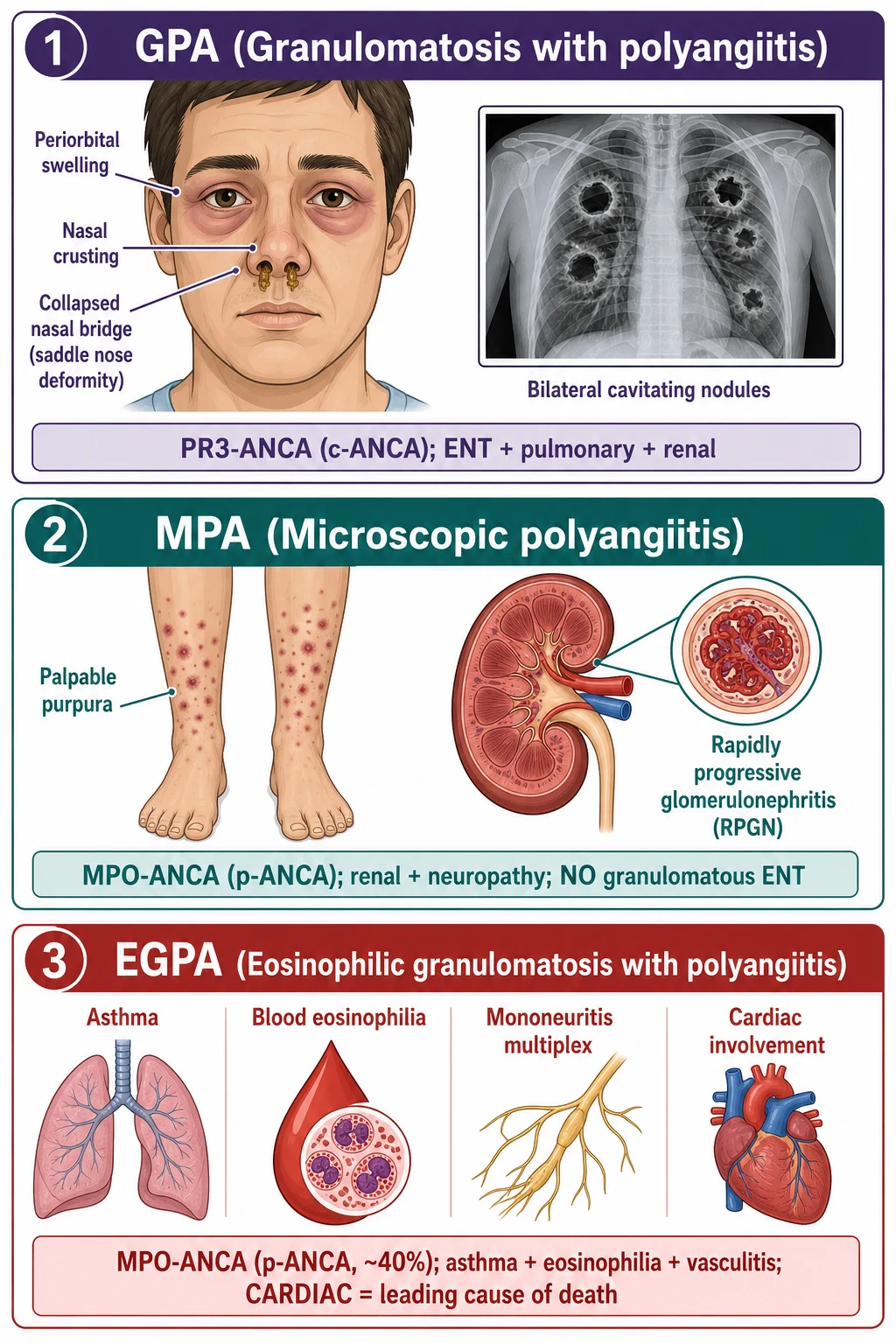
Overview
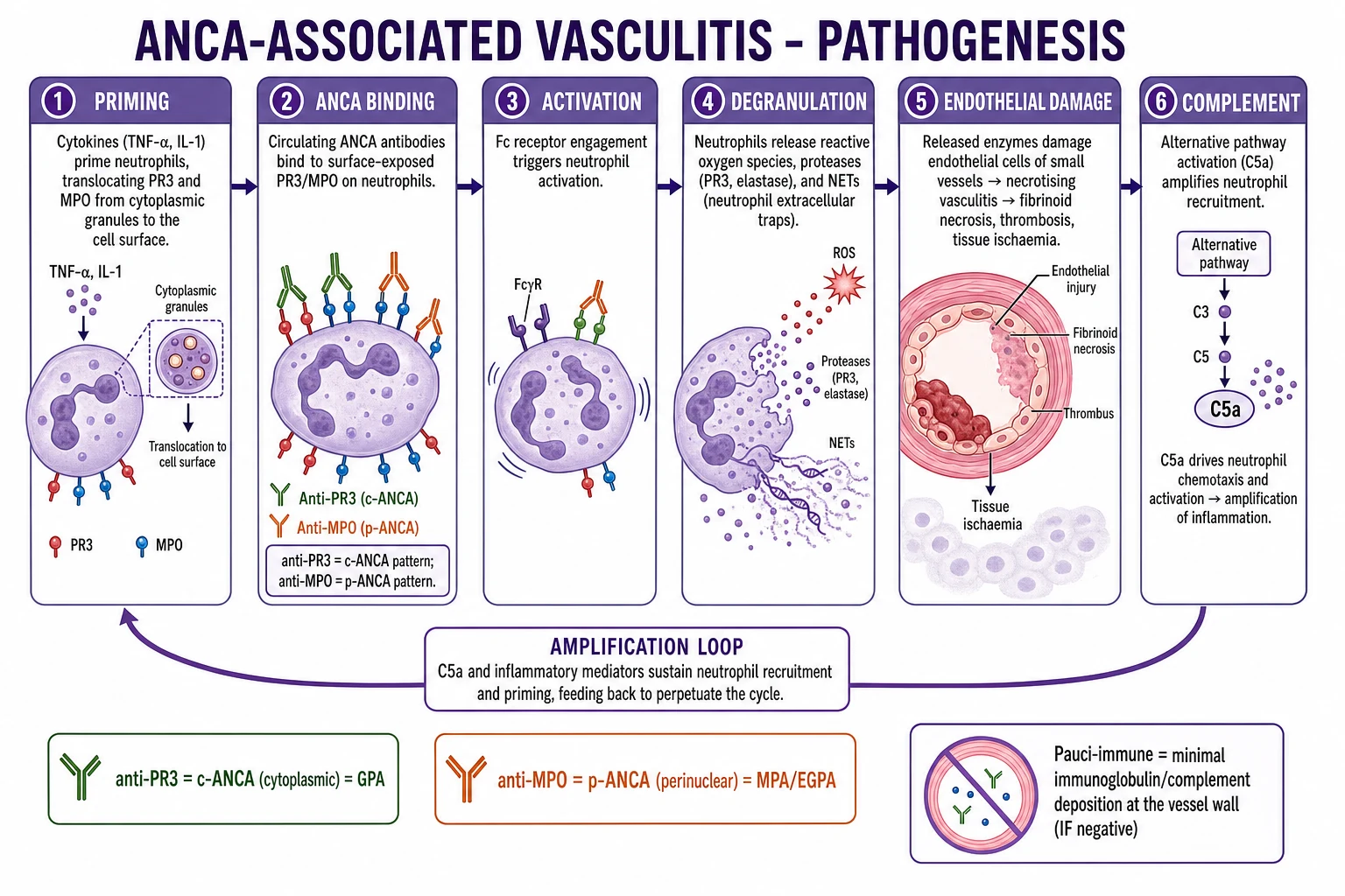
The pulmonary-renal syndrome is one of the most time-critical diagnoses in ICU medicine. The window for preserving kidney function is measured in days — delay in diagnosis and treatment can mean the difference between a patient who recovers renal function and one who is dialysis-dependent for life. The intensivist must recognise the pattern (lung bleeding + kidney failure), order the serology urgently (ANCA, anti-GBM), and initiate treatment empirically while awaiting biopsy confirmation. [1]
The mechanism is small-vessel vasculitis: immune-mediated inflammation of capillaries in the pulmonary alveolar septa and the renal glomerular tuft. In ANCA vasculitis and anti-GBM disease, the injury is pauci-immune (minimal immune complex deposition — the damage is from neutrophil-mediated capillaritis). In lupus and cryoglobulinaemia, the injury is immune-complex-mediated (granular deposition of immune complexes activates complement). Both pathways lead to capillary wall necrosis → alveolar bleeding (DAH) + glomerular necrosis (crescentic GN).[4][6]
Causes and classification
Classification of pulmonary-renal syndrome by immunopathology
| Category | Immune pattern | Specific diseases | Serology | Biopsy finding |
|---|---|---|---|---|
| Anti-GBM disease | Linear IgG on GBM | Goodpasture syndrome | Anti-GBM antibodies (positive) | Linear IgG on IF; crescentic GN |
| ANCA-associated vasculitis (AAV) | Pauci-immune (minimal deposition) | GPA (Wegener), MPA, EGPA (Churg-Strauss) | PR3-ANCA (c-ANCA) or MPO-ANCA (p-ANCA) | Pauci-immune crescentic GN; necrotising granulomas (GPA) |
| Immune-complex-mediated | Granular immune complex deposition | SLE/lupus nephritis, cryoglobulinaemia, IgA nephropathy/HSP, post-streptococcal GN | ANA+, dsDNA+, low C3/C4 (lupus); cryoglobulins+ (cryo); high IgA (IgA nephropathy) | Granular deposits on IF; "full house" (lupus); crescentic GN |
| Dual-positive | Anti-GBM + ANCA both positive | "Double-positive" disease | Anti-GBM+ AND ANCA+ | Features of both — treat as anti-GBM (plasma exchange) + ANCA (immunosuppression) |
ANCA-associated vasculitis — distinguishing features
| Feature | GPA (Wegener) | MPA | EGPA (Churg-Strauss) |
|---|---|---|---|
| ANCA type | PR3-ANCA (c-ANCA) — 75-90% positive | MPO-ANCA (p-ANCA) — 50-70% positive | MPO-ANCA — 40-50% positive |
| Upper respiratory | STRONGLY involved (sinusitis, nasal crusting, saddle nose, subglottic stenosis, otitis media) | SPARINGLY involved (less ENT) | Nasal polyps, allergic rhinitis |
| Lungs | Nodules, cavitation, DAH | DAH (common), interstitial fibrosis | Asthma (nearly universal), eosinophilic pneumonia, DAH (rare) |
| Kidneys | RPGN (pauci-immune) | RPGN (most common cause of renal-limited AAV) | RPGN (less severe than GPA/MPA) |
| Nervous system | Mononeuritis multiplex (10-20%) | Mononeuritis multiplex (30-50%) | Mononeuritis multiplex (COMMON — 60-70%) |
| Bloods | PR3+, normal eosinophils | MPO+, normal eosinophils | MPO+, eosinophilia >1.5 × 10^9/L (mandatory criterion) |
| Histology | Necrotising granulomatous inflammation | Necrotising vasculitis (NO granulomas) | Eosinophilic infiltration + granulomas + vasculitis |
The clinical presentation — recognising PRS in the ICU
The PRS presentation has three components — the intensivist must identify all three: [1]
1. Diffuse alveolar haemorrhage (DAH) — the pulmonary component:
- Haemoptysis (present in 70% — but ABSENT in 30% — do NOT exclude DAH because there is no haemoptysis)
- Dyspnoea, hypoxaemia, cough (can progress to respiratory failure rapidly)
- Bilateral diffuse infiltrates on CXR/HRCT (ground-glass or consolidation — alveolar filling pattern)
- Falling haemoglobin (unexplained drop — 1-3 g/dL over 24-48 hours — blood is pooling in alveoli)
- Bronchoscopy: progressively bloodier lavage on sequential aliquots (the hallmark — not just blood from airway trauma, but blood welling up from distal alveoli)
- HRCT: bilateral ground-glass opacities ± crazy-paving pattern [1]
2. Rapidly progressive glomerulonephritis (RPGN) — the renal component:
- Rapidly rising creatinine (doubling within days to weeks)
- Active urinary sediment: dysmorphic RBCs, RBC casts (pathognomonic for glomerular bleeding), proteinuria (1-3 g/day — nephritic range, not nephrotic)
- Oliguria/anuria (as disease progresses)
- Renal biopsy: crescentic GN (cellular crescents in >50% of glomeruli = severe RPGN) [1]
3. Systemic features of vasculitis:
- Constitutional: fever, malaise, weight loss, arthralgia
- Skin: palpable purpura (lower limbs), livedo reticularis, ulcers
- Neurological: mononeuritis multiplex (wrist drop, foot drop — classic vasculitic sign)
- Ocular: scleritis, episcleritis, orbital pseudotumour (GPA)
- ENT: sinusitis, epistaxis, oral ulcers, saddle nose deformity (GPA) [1]
Diagnostic workup — first 24 hours
- BLOODS — (a) FBC (anaemia from DAH + RPGN, eosinophilia in EGPA, thrombocytopenia in lupus). (b) U&E (rising creatinine), LFTs (hepatitis B/C co-infection in cryoglobulinaemia), CRP (elevated in active vasculitis). (c) ANCA (PR3-ANCA = c-ANCA = GPA; MPO-ANCA = p-ANCA = MPA/EGPA). (d) Anti-GBM antibodies (Goodpasture). (e) ANA + anti-dsDNA + complement C3/C4 (lupus — C3/C4 LOW in active lupus/cryoglobulinaemia, NORMAL in ANCA/anti-GBM). (f) Cryoglobulins (must be transported WARM — clump if cold). (g) Hepatitis B surface antigen, hepatitis C antibody (associated with cryoglobulinaemia and polyarteritis nodosa). (h) HIV. (i) Urine MC&S + ACR/PCR
- IMAGING — (a) CXR: bilateral diffuse alveolar infiltrates (DAH pattern). (b) HRCT chest: bilateral ground-glass opacities ± crazy-paving ± nodules (GPA). (c) AVOID IV CONTRAST — the patient already has AKI — contrast nephropathy can precipitate irreversible renal failure. Use ultrasound or non-contrast MRI. (d) Sinus CT if GPA suspected (sinusitis, bony destruction)
- BRONCHOSCOPY — sequential BAL aliquots (3-5 aliquots of 20 mL saline): progressively bloodier lavage = DAH (normal = clear throughout). Also: exclude infection (bacterial, fungal, viral PCRs — immunocompromised patient). Differential: infection, pulmonary oedema, coagulopathy
- RENAL BIOPSY — the gold standard (within 24-48 hours if feasible). Light microscopy: crescentic GN (cellular crescents in Bowman's space — indicates severe glomerular injury). Immunofluorescence: (a) LINEAR IgG along GBM = anti-GBM disease. (b) PAUCI-IMMUNE (minimal deposition) = ANCA vasculitis. (c) GRANULAR deposits = immune-complex disease (lupus = "full house", IgA nephropathy = mesangial IgA)
- EMPIRIC TREATMENT — DO NOT WAIT for biopsy or serology results in a critically ill patient with suspected PRS. Start methylprednisolone pulses immediately. Add plasma exchange if anti-GBM suspected (or if ANCA with severe AKI/DAH). Add cyclophosphamide or rituximab once diagnosis confirmed
ICU management — the three-pillar approach
Pulmonary-renal syndrome treatment protocol
-
PLASMA EXCHANGE (PLEX) — the most time-critical intervention for anti-GBM disease:
- Anti-GBM disease: MANDATORY — removes circulating anti-GBM antibodies. Daily or alternate-day exchanges (1-1.5 plasma volumes) for 14 days or until anti-GBM antibody undetectable. This is the single most important intervention for preserving kidney function in Goodpasture syndrome.[5]
- Severe ANCA vasculitis (creatinine >5.7 mg/dL/500 umol/L, or DAH requiring ventilation): BENEFICIAL — MEPEX trial showed plasma exchange improved renal recovery at 3 months vs methylprednisolone pulses (76% vs 54% dialysis-free).[3]
- Lupus nephritis / cryoglobulinaemia: consider PLEX for severe DAH or rapidly progressive renal failure
- Technique: 1-1.5 plasma volume exchange with 5% albumin ± fresh frozen plasma (FFP if bleeding risk). Central venous access. Anticoagulation: citrate (regional) or heparin. Monitor: fibrinogen, calcium (citrate chelates), platelets
- Risks: bleeding (coagulopathy from plasma removal + immunosuppression + thrombocytopenia), infection (central line), hypocalcaemia (citrate), hypotension (volume shifts)
-
HIGH-DOSE CORTICOSTEROIDS — universal first-line for ALL causes of PRS:
- Methylprednisolone pulses: 500-1000 mg IV daily x 3 days (for severe disease/DAH/RPGN)
- Then oral prednisone 1 mg/kg/day (max 60-80 mg/day), taper over 6-12 months
- Mechanism: rapid suppression of neutrophil-mediated capillaritis + glomerular inflammation
- Adjuncts: PPI (gastric protection), VTE prophylaxis, bone protection (calcium + vitamin D), glucose monitoring [1]
-
IMMUNOSUPPRESSION (CYCLOPHOSPHAMIDE or RITUXIMAB) — essential for remission induction:
- Cyclophosphamide: IV pulse 15 mg/kg every 2 weeks x 6 doses (adjust for renal function) or oral 2 mg/kg/day. MESNA for haemorrhagic cystitis prophylaxis. Side effects: cytopenia, infection, infertility, malignancy (long-term)
- Rituximab: 375 mg/m^2/week x 4 (or 1 g x 2 doses 2 weeks apart). FIRST-LINE per RAVE and RITUXVAS trials (non-inferior to cyclophosphamide, better for relapsing disease, fewer side effects).[1][2]
- The choice is individualised: cyclophosphamide for rapid effect, rituximab for patients wanting to preserve fertility or with contraindications to cyclophosphamide
-
RENAL REPLACEMENT THERAPY — for AKI:
- CRRT (CVVHDF) or intermittent haemodialysis (as clinically appropriate)
- Drug dosing: cyclophosphamide and rituximab dosing MUST be adjusted for renal function
- Plasma exchange + RRT can be done on the same circuit (if available) [1]
-
VENTILATORY SUPPORT for DAH — lung-protective ventilation:
- Oxygen → HFNC → NIV → intubation (for respiratory failure)
- Mechanical ventilation: ARDSNet protocol (Vt 6 mL/kg, plateau pressure <30, PEEP titrated carefully — high PEEP may worsen alveolar bleeding)
- Position: keep affected lung DOWN (if unilateral DAH) to prevent blood flooding the good lung
- Suction frequently (blood can obstruct airway)
- Recombinant activated factor VII (rFVIIa) — case reports/series for life-threatening DAH unresponsive to standard therapy (off-label — 50 mcg/kg nebulised or IV) [1]
-
SUPPORTIVE CARE:
- Blood transfusion (for DAH-induced anaemia — aim Hb >70 g/L, or higher if active bleeding)
- Correct coagulopathy (FFP, platelets — but do NOT over-correct as immune disease requires ongoing immunosuppression)
- Infection prophylaxis: PJP prophylaxis (co-trimoxazole — while on high-dose steroids), antifungal, antiviral
- VTE prophylaxis (vasculitis = prothrombotic state)
- Nutrition (high catabolic state from steroids + critical illness)
Key trials and evidence
RAVE trial — rituximab vs cyclophosphamide for ANCA vasculitis (PMID 20662044)
Study design
Randomised, double-blind, double-dummy, non-inferiority trial — 197 patients
Population
Newly diagnosed or relapsing ANCA-associated vasculitis (GPA or MPA)
Intervention
Rituximab 375 mg/m^2/week x 4 vs cyclophosphamide (then azathioprine)
Primary outcome
Complete remission at 6 months: rituximab 64% vs cyclophosphamide 53% (rituximab NON-INFERIOR and SUPERIOR in relapsing disease: 67% vs 42%)
Adverse events
Similar between groups — no significant difference in infection rates
Key finding
Rituximab is at least as effective as cyclophosphamide for remission induction — now first-line for many patients
Clinical bottom line
Rituximab = first-line induction for ANCA vasculitis — non-inferior to cyclophosphamide, superior for relapsing disease, better safety profile
MEPEX trial — plasma exchange for severe ANCA vasculitis (PMID 17914012)
Study design
Randomised controlled trial — 137 patients with severe ANCA vasculitis
Population
ANCA vasculitis with severe renal involvement (creatinine >5.7 mg/dL / 500 umol/L)
Intervention
Plasma exchange vs methylprednisolone pulses (both groups received oral prednisone + cyclophosphamide)
Primary outcome
Dialysis-free survival at 3 months: PLEX 69% vs methylprednisolone 49% (p=0.02) — plasma exchange BETTER
Long-term
At 12 months: PLEX still trended towards better renal recovery (not statistically significant)
Adverse events
PLEX group: more severe infections (44% vs 36%)
Clinical bottom line
Plasma exchange improves renal recovery in severe ANCA vasculitis with high creatinine — indicated for creatinine >500 umol/L or dialysis-dependent
RITUXVAS trial — rituximab + cyclophosphamide for renal ANCA vasculitis (PMID 20587541)
Study design
Randomised, open-label, 76 patients with newly diagnosed renal ANCA vasculitis
Intervention
Rituximab 375 mg/m^2/week x 4 + 2 cyclophosphamide pulses vs cyclophosphamide alone (standard)
Primary outcome
Sustained remission at 12 months: rituximab 76% vs cyclophosphamide 82% (non-inferior)
Severe adverse events
Similar between groups (42% rituximab vs 36% cyclophosphamide)
Key finding
Rituximab-based regimen is non-inferior to cyclophosphamide for remission induction in renal ANCA vasculitis
Clinical bottom line
Together with RAVE, establishes rituximab as first-line induction therapy for ANCA-associated vasculitis
Anti-GBM disease (Goodpasture syndrome) — the time-critical subset
Anti-GBM disease is the most time-critical form of PRS because the anti-GBM antibody directly damages glomerular and alveolar basement membranes. Unlike ANCA vasculitis (where damage is indirect via neutrophil activation), anti-GBM antibodies bind directly to the alpha-3 chain of type IV collagen in the GBM and alveolar BM → complement activation → neutrophil recruitment → basement membrane destruction → crescentic GN + DAH. [1]
The urgency: every day without plasma exchange = more glomeruli destroyed. Once >85% of glomeruli have crescents, the kidney is irreversibly damaged. Anti-GBM titre correlates with disease severity — the goal is to reduce the antibody to undetectable levels as fast as possible.[5]
Diagnostic criteria for anti-GBM disease:
- Positive anti-GBM antibodies (serum)
- Linear IgG deposition on renal biopsy immunofluorescence
- Crescentic GN on light microscopy
- DAH (present in 40-60% of anti-GBM patients — lung involvement requires the kidney to be also involved — the alveolar BM is less accessible to antibodies than the GBM, explaining why not all anti-GBM patients have DAH) [1]
The "double-positive" patient (anti-GBM + ANCA both positive):
- 20-40% of anti-GBM patients also have positive ANCA (usually MPO-ANCA/p-ANCA)
- These patients have features of BOTH diseases
- They have a higher rate of relapse than pure anti-GBM (which does NOT relapse once antibodies are cleared)
- Treatment: anti-GBM protocol (PLEX + steroids + immunosuppression) followed by long-term ANCA maintenance [1]
Anti-GBM emergency protocol — first 24-48 hours
- SUSPECT — haemoptysis + rapidly rising creatinine + (any) autoimmune features → check anti-GBM antibodies STAT (turnaround: 24-48h for ELISA)
- PLASMA EXCHANGE IMMEDIATELY — if anti-GBM strongly suspected (do NOT wait for confirmatory antibody result if patient is critically ill). Exchange 1-1.5 plasma volumes daily until anti-GBM undetectable (typically 10-14 sessions). Replace with 5% albumin + FFP (if bleeding). Use FFP preferentially if active DAH (provides clotting factors)
- METHYLPREDNISOLONE 500-1000 mg IV daily x 3 days — then oral prednisone 1 mg/kg/day
- CYCLOPHOSPHAMIDE — oral 2 mg/kg/day or IV 15 mg/kg pulses — suppresses new antibody production (B-cell and T-cell suppression). Adjust for renal function
- DO NOT TRANSPLANT until anti-GBM antibodies undetectable for at least 6 months (the antibody will attack the transplant kidney)
- PROGNOSIS — (a) If creatinine <5.7 mg/dL at presentation: 95% renal survival at 1 year. (b) If creatinine >5.7 but not dialysis-dependent: 70% renal survival. (c) If dialysis-dependent at presentation + >85% crescents on biopsy: only 5% recover renal function — but still treat (may recover lung function + prevent future anti-GBM damage to a future transplant). (d) DAH mortality: 10-20% even with treatment
Fellowship SAQ practice
SAQ — Goodpasture syndrome (anti-GBM disease) with diffuse alveolar haemorrhage
10 minutes · 10 marks
A 38-year-old male smoker presents with a 2-week history of haemoptysis, malaise and progressive dyspnoea. Over 24 hours he develops type 1 respiratory failure (PaO2 52 mmHg on 15 L via non-rebreather) and his creatinine rises from 120 to 340 micromol/L. Hb has fallen from 132 to 88 g/L. CXR shows bilateral diffuse alveolar infiltrates. Urine dipstick: blood 3+, protein 2+. Anti-GBM antibodies return strongly positive at 180 EU/mL (normal <20). ANCA is negative. HRCT shows bilateral ground-glass opacification. He is intubated for respiratory failure and the nephrology team is en route.
SAQ — ANCA-associated vasculitis (GPA) with diffuse alveolar haemorrhage and RPGN
10 minutes · 10 marks
A 64-year-old man presents with a 6-week history of constitutional symptoms, epistaxis, nasal crusting and arthralgia, now with worsening dyspnoea and haemoptysis. HR 128, RR 32, SpO2 88% on 15 L non-rebreather, BP 105/64. Hb 76 g/L (baseline 140), creatinine 520 micromol/L (baseline 95, two weeks ago), urea 38, K+ 5.8. Urine: blood 3+, protein 1+, dysmorphic red cells and red-cell casts. CXR/HRCT: bilateral diffuse ground-glass and consolidative change. PR3-ANCA (c-ANCA) strongly positive; MPO-ANCA, anti-GBM, ANA and complements all normal/negative. Bronchoscopy: progressively bloodier lavage aliquots. He is intubated for respiratory failure and a renal biopsy shows pauci-immune necrotising crescentic GN with 60% cellular crescents.
Clinical pearls
Red flags
Prognosis
Prognosis by PRS subtype
| Disease | Renal recovery | DAH mortality | Relapse rate | Key prognostic factor |
|---|---|---|---|---|
| Anti-GBM | 5-50% (depends on creatinine at presentation) | 10-20% | 0% (once antibody cleared) | % crescents on biopsy; creatinine at presentation |
| GPA (Wegener) | 60-70% (with early treatment) | 5-10% | 50%+ at 5 years (PR3-ANCA = highest relapse) | PR3 vs MPO; ENT involvement (worse prognosis) |
| MPA | 50-60% | 10-15% | 30% at 5 years | Creatinine at presentation; age |
| EGPA (Churg-Strauss) | 80-90% (less severe renal disease) | 5% | 20-30% at 5 years | Cardiac involvement (eosinophilic myocarditis = worst prognosis) |
| Lupus nephritis (class IV) | 60-70% | 5-10% | 30-40% at 5 years | Complement levels; histological activity/chronicity index |
| Cryoglobulinaemic GN | 50-60% | 5-10% | 40% (especially if HCV untreated) | HCV treatment status; complement levels |
Overall mortality for PRS in ICU: 10-30%, with DAH requiring mechanical ventilation being the strongest predictor of death (20-50% mortality in ventilated DAH). Early diagnosis + prompt treatment (steroids + PLEX + cyclophosphamide/rituximab) = best outcomes. The most common causes of death are: (1) DAH-related respiratory failure, (2) infection (from triple immunosuppression), (3) cardiovascular events (vasculitis = prothrombotic).[6]
Densification notes for fellowship revision
This leaf is densified to the ICU fellowship gate standard (CICM / FFICM / EDIC): embedded SAQ practice, multi-figure visual scaffolding, examiner map alignment, and MCQ coverage of definition, mechanism, first-hour management, evidence, and traps. [1]
- Revision checkpoint 1: restate definition, one number examiners expect, and one absolute do-not-miss action.
- Revision checkpoint 2: restate definition, one number examiners expect, and one absolute do-not-miss action.
- Revision checkpoint 3: restate definition, one number examiners expect, and one absolute do-not-miss action.
- Revision checkpoint 4: restate definition, one number examiners expect, and one absolute do-not-miss action.
- Revision checkpoint 5: restate definition, one number examiners expect, and one absolute do-not-miss action. [1]
References
- [1]Specks U, et al. American College of Rheumatology 2010 recommendations for the prevention and treatment of glucocorticoid-induced osteoporosis Arthritis Care Res (Hoboken), 2010.PMID 20662044
- [2]Jones RB, et al. Phototherapy for allergic rhinitis: a prospective, randomized, single-blind, placebo-controlled study Ther Adv Respir Dis, 2010.PMID 20587541
- [3]Jayne DR, et al. Schools' mental health responses after Hurricanes Katrina and Rita Psychiatr Serv, 2007.PMID 17914012
- [4]Kitching AR, et al. Cannabinoids in attention-deficit/hyperactivity disorder: A randomised-controlled trial Eur Neuropsychopharmacol, 2017.PMID 28576350
- [5]Niles JL, et al. Spatiotemporal features of microsporogenesis in the cycad species Macrozamia communis Am J Bot, 2015.PMID 26199364
- [6]West SC, et al. Bond Memory in Dynamically Determined Stereoselectivity J Am Chem Soc, 2020.PMID 31852185