ICU · Renal/Metabolic
Rhabdomyolysis in the ICU
Also known as Rhabdomyolysis · Myoglobinuria · Crush syndrome · CK elevation
Rhabdomyolysis is breakdown of skeletal muscle releasing intracellular contents (myoglobin, creatine kinase, potassium, phosphate) into circulation. Causes: trauma/crush (1), prolonged immobility (fall, overdose), seizures, statins, illicit drugs (cocaine, MDMA), infections (influenza, Legionella), electrolyte disturbance (hypokalaemia, hypophosphataemia), malignant hyperthermia, NMS, exertional (marathon). Presents with: myalgia, weakness, dark urine (tea/cola-coloured — myoglobinuria), elevated CK (5x ULN or 1000 U/L). Complications: AKI (myoglobin nephrotoxicity — 1 cause of death), hyperkalaemia, hypocalcaemia, compartment syndrome. Treatment: aggressive IV fluids (goal urine output 1-2 mL/kg/h), treat hyperkalaemia, correct underlying cause. Bicarbonate alkalinisation controversial (not routinely recommended).
On this page & tools
Your progress
Saved locally on this device.
Target exams
Red flags
Pathophysiology — from muscle injury to myoglobinuric AKI
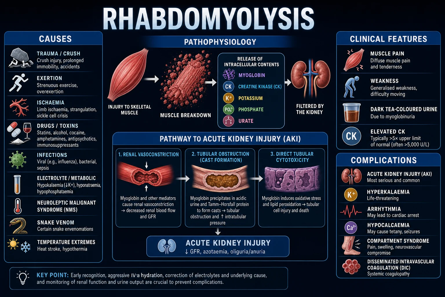
Rhabdomyolysis is the syndrome of skeletal-muscle (myocyte) injury that releases intracellular contents into the circulation. The cascade has four linked stages — each is fair game in the viva: [1]
- Myocyte membrane disruption (the initiating event). Direct mechanical trauma (crush, prolonged compression), ATP depletion from sustained contraction (seizures, exertion, agitation), membrane instability (hypokalaemia, hypophosphataemia, hypothyroidism), or direct myotoxicity (statins, illicit drugs, viral invasion) disrupts the sarcolemma and the sarcoplasmic reticulum, destroying membrane integrity.
- Calcium influx and cell death. Once the sarcolemma fails, extracellular Ca2+ floods down its gradient into the myocyte. The intracellular Ca2+ rise activates calcium-dependent proteases (calpains) and phospholipase A2, degrades myofilaments, injures mitochondria, and locks the cell into sustained contraction (rigor) → irreversible necrosis.
- Release of intracellular contents into the circulation. Necrotic myocytes pour out myoglobin, creatine kinase (CK), potassium (K+), phosphate, urate, lactate, AST/ALT and LDH. The volume of release tracks the mass of injured muscle.
- Systemic consequences. Myoglobin nephrotoxicity → AKI (the leading cause of death); hyperkalaemia → arrhythmia; phosphate binds calcium → hypocalcaemia; organic acids + renal failure → metabolic acidosis; muscle swelling within unyielding fascial compartments → compartment syndrome; and released tissue factor → DIC. [1]
Myoglobin nephrotoxicity — the three mechanisms of AKI
The kidney is the innocent bystander: it filters myoglobin, and myoglobin injures the tubule. Three concurrent mechanisms produce acute tubular necrosis (ATN): [1]
1. Intratubular cast formation
Obstruction
- Myoglobin is freely filtered at the glomerulus and enters the tubular lumen
- At acidic urinary pH, myoglobin precipitates with Tamm-Horsfall protein (uromodulin) → large proteinaceous casts → obstructs the tubular lumen → back-pressure injury to the nephron
- Worsened by dehydration (concentrated urine → higher myoglobin concentration → faster precipitation)
- Prevention: urine alkalinisation (keep urine pH >6.5 so myoglobin stays soluble) + aggressive IV fluids (dilute the tubular myoglobin — target UO 200-300 mL/hr)
2. Renal vasoconstriction
Hypoperfusion
- Myoglobin scavenges nitric oxide (NO — the dominant renal vasodilator) → loss of NO-mediated afferent arteriolar dilation → vasoconstriction → fall in GFR
- Volume depletion (fluid sequestered into damaged muscle — "third spacing") compounds the drop in renal perfusion
- Myoglobin-driven oxidant injury to renal endothelium impairs autoregulation
- Prevention: IV fluids (restore intravascular volume and perfusion pressure); avoid NSAIDs, ACEi and contrast (all worsen renal vasoconstriction)
3. Direct tubular toxicity
ATN
- Myoglobin is endocytosed by tubular cells and broken down via haem oxygenase → releases free iron
- Free iron catalyses the Fenton reaction (Fe2+ + H2O2 → Fe3+ + OH- + OH.) → hydroxyl radicals → lipid peroxidation of the tubular cell membrane → cell death
- Result: acute tubular necrosis (ATN) — the histological lesion of rhabdomyolysis-induced AKI
- Prevention: urine alkalinisation reduces free-iron release at acidic pH; avoid excess oxygen; N-acetylcysteine is unproven
What is released — and why each matters
Released intracellular contents and their clinical consequence
| Released substance | Why it matters | Clinical consequence |
|---|---|---|
| Myoglobin | Filtered by the glomerulus → toxic to the tubule + precipitates as casts | AKI (the #1 cause of death); dark/tea-coloured urine |
| Creatine kinase (CK) | Marker of injury magnitude; itself inert | Diagnostic (CK >5x ULN or >1000 U/L); CK >5000 raises AKI risk |
| Potassium (K+) | Continuously leaks from necrotic muscle; refractory to standard shifts | Life-threatening hyperkalaemia → arrhythmia → arrest |
| Phosphate | Binds calcium → precipitates in damaged muscle and kidney | Hyperphosphataemia + hypocalcaemia; Ca-P deposition worsens AKI |
| Urate | Purine breakdown from muscle nucleic acids | Hyperuricaemia → uric-acid crystallisation in tubules (adds to AKI) |
| Lactate + organic acids | Anaerobic metabolism + cell death | High-anion-gap metabolic acidosis (worsens myoglobin toxicity + hyperkalaemia) |
| AST/ALT | Released from muscle (AST > ALT typically) | Misleading "liver dysfunction" — it is muscle, not liver |
| LDH | Non-specific cell-damage marker | Markedly elevated; supports diagnosis |
| Tissue factor / thromboplastin | Released from damaged muscle | DIC (consumption coagulopathy) in severe cases |
Causes
Common causes
In ICU
- Trauma/crush injury (#1)
- Prolonged immobility (falls in elderly, overdose, post-operative)
- Status epilepticus
- Severe exertion (marathon, military training)
- Malignant hyperthermia, NMS, serotonin syndrome
Drugs and toxins
Iatrogenic/recreational
- Statins (especially simvastatin at high dose, with interacting drugs — macrolides, azoles, fibrates)
- Illicit drugs: cocaine, MDMA/ecstasy, amphetamines
- Alcohol (direct myotoxicity + immobility)
- Carbon monoxide, snake venom, insect venom
Infections and metabolic
Systemic
- Viral: influenza A/B, coxsackievirus, EBV, HIV, COVID-19
- Bacterial: Legionella, Streptococcus, Clostridium (gas gangrene)
- Electrolyte: severe hypokalaemia, hypophosphataemia (impairs muscle membrane potential)
- Endocrine: hypothyroid myopathy, DKA
- Genetic: McArdle disease, mitochondrial myopathies
Management
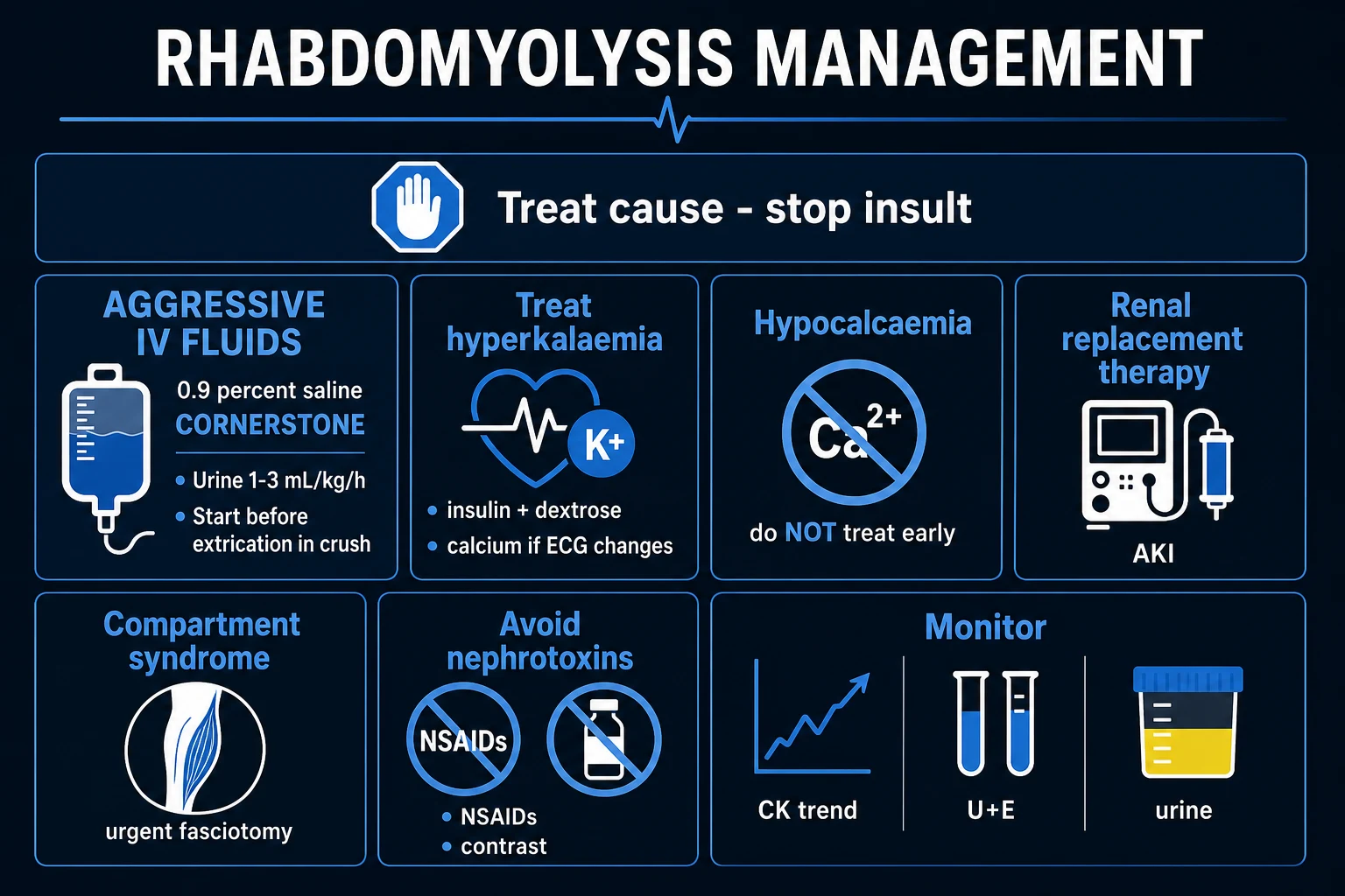
Rhabdomyolysis management protocol
Aggressive IV fluid resuscitation
Normal saline 10-15 mL/kg/h initially, titrate to urine output 1-2 mL/kg/h. Goal: flush myoglobin through kidneys before it precipitates in tubules. Volume: may need 6-12 L in first 24h. Monitor urine output (catheterise), CVP, and avoid fluid overload. If AKI develops despite fluids: consider RRT.
Treat hyperkalaemia (life-threatening)
Muscle breakdown releases intracellular K+. Check K+ every 2-4h initially. Treat per hyperkalaemia protocol: calcium gluconate (if ECG changes), insulin-dextrose, salbutamol, removal (diuretics/RRT). Hyperkalaemia may be refractory — RRT may be needed.
Do NOT treat hypocalcaemia unless symptomatic
Hypocalcaemia occurs because calcium binds to damaged muscle (phosphate release). If you give calcium: it precipitates as calcium-phosphate in tissues (nephrocalcinosis) → worsens AKI. ONLY treat if: symptomatic (tetany, seizures, QT prolongation with arrhythmia). Calcium will spontaneously correct as muscle heals and releases bound calcium.
Bicarbonate alkalinisation — controversial
Sodium bicarbonate to alkalinise urine (pH >6.5) may prevent myoglobin precipitation in renal tubules. THEORY: myoglobin is more soluble in alkaline urine. EVIDENCE: no clear benefit over saline alone. May cause: hypernatraemia, hypocalcaemia (worsens), fluid overload. NOT routinely recommended — use only if severe acidosis (pH <7.1) in addition to rhabdomyolysis.
Treat underlying cause
Stop offending drugs (statins). Treat infection (antibiotics). Correct electrolytes (K+, phosphate). Treat seizures. Cool if hyperthermia (NMS, malignant hyperthermia). Decompress if compartment syndrome. Remove from prolonged immobility.
Monitor for complications
Serial CK (peaks 24-72h, normalises over 1-2 weeks). Urine output hourly. Renal function (creatinine — may rise disproportionate to CK). Potassium every 2-4h. Calcium, phosphate, magnesium. Compartment pressures if limb swollen/tense. ECG (hyperkalaemia).
Renal replacement therapy
Indications: refractory hyperkalaemia, severe acidosis, fluid overload, symptomatic uraemia. CRRT preferred (continuous, avoids rapid solute shifts). Most AKI from rhabdomyolysis is reversible within 1-2 weeks.
Exam practice
SAQ — Crush injury with hyperkalaemia and myoglobinuric AKI
10 minutes · 10 marks
A 28-year-old man is extricated after 8 hours trapped under rubble following an earthquake. His right leg is swollen and tense. Initial bloods: CK 18 400 U/L, potassium 7.6 mmol/L (peaked T waves, QRS widening), creatinine 245 µmol/L, corrected calcium 1.62 mmol/L, phosphate 2.6 mmol/L, venous pH 7.16. Urine is tea-coloured and dipstick-positive for blood but with no red cells on microscopy.
SAQ — Statin-induced rhabdomyolysis and the medical aetiology
10 minutes · 10 marks
A 68-year-old woman is admitted with severe diffuse muscle pain and dark urine. She takes simvastatin 80 mg daily and was recently started on clarithromycin for community-acquired pneumonia. CK 9 600 U/L, creatinine 188 µmol/L, ALT 210 U/L, AST 280 U/L, K⁺ 5.9 mmol/L. Urine myoglobin positive.
Clinical pearls
Red flags
Diagnosis — the laboratory signature
Rhabdomyolysis is a clinical + biochemical diagnosis. No single test is definitive, but the constellation is unmistakable. In the ICU, suspect it in ANY patient with crush injury, prolonged immobilisation, status epilepticus, severe agitation/delirium, severe exertion, or unexplained AKI with dark urine. [1]
The diagnostic triad
- CK elevation — the gold-standard marker:
- Normal CK: <200 U/L (men), <170 U/L (women)
- Rhabdomyolysis: CK >5x ULN, or >1000 U/L (typically 10,000-100,000+ U/L in established cases)
- CK peaks at 24-72h, then falls ~40% per day if the insult has stopped
- A CK that fails to fall → ongoing muscle injury → re-evaluate the cause (missed compartment syndrome, continued seizure, continued toxin)
- Myoglobinuria — the bedside clue:
- Urine dipstick reads POSITIVE for "blood" (the dipstick detects the heme moiety — myoglobin contains heme)
- Urine microscopy shows NO (or very few) red blood cells — this "blood-positive, RBC-negative" discordance is the diagnostic clue
- Urine is classically dark, tea-coloured or cola-coloured
- Serum myoglobin rises early (within 1-2h) but has a short half-life (2-3h) and is rapidly cleared by the kidney → by the time CK is detected, serum myoglobin may already be normal → CK is the more reliable marker
- Biochemical derangement — the electrolyte signature:
- Hyperkalaemia (K+ continuously released from muscle — may be >7-8 and refractory)
- Hyperphosphataemia (phosphate is the most abundant intracellular anion)
- Hypocalcaemia (calcium precipitates in damaged muscle as Ca-P — do NOT routinely correct)
- Hyperuricaemia (purine release)
- Metabolic acidosis (high anion gap — lactate + organic acids + renal failure)
- Creatinine rising disproportionately (creatinine may climb faster than urea — the urea:creatinine ratio is characteristically low in rhabdomyolysis)
- AST > ALT elevation (both released from muscle; not liver injury) [1]
The "blood-positive, RBC-negative" urine — the single highest-yield exam pearl
The urinary dipstick pad detects heme, not haemoglobin specifically. Myoglobin carries a heme group → the dipstick reads strongly positive for "blood" (often 3+ or 4+). But microscopy shows no red cells (or only a few). This discordance — dipstick blood positive, microscopy RBC negative — is pathognomonic of myoglobinuria (or haemoglobinuria from haemolysis). Distinguish the two clinically: rhabdomyolysis has high CK + muscle pain; haemolysis has high LDH + low haptoglobin + fragmented red cells. [1]
Diagnostic markers — what to send and what it means
| Test | Finding in rhabdomyolysis | Significance |
|---|---|---|
| Creatine kinase (CK) | >5x ULN or >1000 U/L (often 10,000-100,000) | Diagnostic gold standard; tracks injury magnitude |
| Urine dipstick | Positive for "blood" (heme) | Myoglobin cross-reacts with the blood pad |
| Urine microscopy | No / few RBCs | Confirms the discordance → myoglobinuria |
| Serum myoglobin | Early rise then rapid fall (t1/2 2-3h) | May be normal by presentation — unreliable; use CK |
| Potassium | High (often >5.5, may be >7) | Continuously released; arrhythmia risk |
| Phosphate | High | Precipitates calcium → hypocalcaemia |
| Calcium (ionised) | Low (early) | Do NOT correct unless symptomatic |
| Urate | High | Adds to tubular crystalline injury |
| Venous gas | High-anion-gap metabolic acidosis | Worsens myoglobin toxicity + hyperkalaemia |
| Urea : creatinine ratio | Low (creatinine rises faster) | Suggestive (not diagnostic) of rhabdomyolysis vs prerenal |
| AST/ALT | Elevated (AST > ALT) | Muscle-derived — not liver |
| LDH | Markedly elevated | Supports muscle injury |
| Coagulation | PT/aPTT may rise + low platelets in severe cases | DIC from released tissue factor |
| CK-MB / troponin | CK-MB may be elevated (skeletal muscle has CK-MB) | Not necessarily MI — interpret with troponin and ECG |
The McMahon score — predicting AKI or death
The McMahon risk prediction score (McMahon, JAMA Intern Med 2013) stratifies the risk of kidney failure requiring RRT or in-hospital death in rhabdomyolysis, using age, sex, initial creatinine, initial CK, calcium, phosphate, bicarbonate, and the cause. It helps decide who needs the most aggressive fluid strategy and closest monitoring. [1]
McMahon score — risk strata
| McMahon score | Risk of death or RRT | Practical implication |
|---|---|---|
| ≤5 (low) | ~2-3% | Standard aggressive fluids; expect recovery |
| 6-10 (intermediate) | ~5-11% | Aggressive fluids + close biochemical monitoring |
| ≥11 (high) | ~20-25% | Highest-risk — maximise fluids, expect RRT, monitor for compartment syndrome and refractory hyperkalaemia |
Causes — the broad differential (expanded)
The original causes table above covers the major categories. The high-yield additions for the fellowship exam: [1]
Statin-induced rhabdomyolysis — the CYP3A4 / SLCO1B1 story
Statin myotoxicity is dose-dependent and multiplied by drug interactions. The cellular mechanism is depletion of mevalonate-pathway products → reduced CoQ10 (ubiquinone, an electron-transport-chain component) → mitochondrial ATP depletion → energy failure → Ca2+ overload → calpain-mediated cell death, compounded by reduced sarcolemmal cholesterol → membrane fragility. [1]
Lipophilic (higher myopathy risk)
Cross muscle membrane easily
- Simvastatin (highest risk, especially >40 mg)
- Atorvastatin
- Fluvastatin
- Lovastatin; cerivastatin (withdrawn in 2001 — caused fatal rhabdo)
Hydrophilic (lower risk)
Do not cross membrane as readily
- Rosuvastatin
- Pravastatin
- Preferred in patients with prior statin myopathy (after washout and supervised re-challenge)
Interactions that multiply risk
Mechanism
- Fibrates — gemfibrozil >> fenofibrate (both directly myotoxic; gemfibrozil also inhibits statin glucuronidation)
- Calcineurin inhibitors — ciclosporin/tacrolimus (inhibit OATP1B1 hepatic uptake → higher statin levels)
- Macrolides — erythromycin/clarithromycin (CYP3A4 inhibition)
- Azole antifungals — itraconazole/fluconazole (CYP3A4 inhibition)
- Amiodarone (CYP3A4 inhibition)
- Grapefruit juice (intestinal CYP3A4 inhibition)
- HIV protease inhibitors (CYP3A4 inhibition)
Patient risk factors
Host
- Age >80, female sex, low body mass
- Pre-existing CKD or hypothyroidism
- Heavy alcohol use
- Vigorous exercise, perioperative state
- SLCO1B1 c.521TC or CC genotype (4.5x and 16.9x increased simvastatin-myopathy risk)
Genetic and recurrent causes — when to refer
Suspect an underlying metabolic myopathy in any patient with recurrent rhabdomyolysis from childhood/young adulthood, exertional myalgia, or a family history: [1]
Glycogen storage
Carbohydrate metabolism
- McArdle disease (myophosphorylase deficiency) — exercise intolerance, "second-wind" phenomenon
- Pompe disease (acid alpha-glucosidase) — late-onset myopathy
Fatty-acid oxidation
Lipid metabolism
- CPT II (carnitine palmitoyltransferase II) deficiency — prolonged fasting/exercise → rhabdo
- VLCAD / MCAD deficiency
Mitochondrial
Energy failure
- MELAS, MERRF — multi-system, recurrent
Channelopathies / anaesthetic
Membrane
- Malignant hyperthermia (RYR1/CACNA1S) — volatile anaesthetics/suxamethonium → rigidity + hyperthermia + rhabdo
- Periodic paralysis (channelopathies) — can co-present with rhabdo
Crush syndrome — the mass-casualty scenario
Crush syndrome is the systemic manifestation of rhabdomyolysis after prolonged compression of a large muscle mass (earthquake, building collapse, mining accident, prolonged collapse on the floor). The defining risk is reperfusion on extrication — sudden death at the moment the crush is released. [1]
- Pre-extrication fluids. Start isotonic saline ± bicarbonate BEFORE releasing the crush to dilute and buffer the imminent K+/myoglobin/lactate surge. Aim ~1 L/h during extrication (adjust to age/comorbidity).
- Reperfusion injury. On release, blood returns to ischaemic-but-alive muscle → massive release of K+ (arrhythmia), myoglobin (AKI), lactate (acidosis), phosphate (hypocalcaemia). The cause of "death at extrication" is hyperkalaemic cardiac arrest.
- Continuous monitoring during extrication. ECG (peaked T → wide QRS → sine wave), blood pressure (may collapse from vasodilation and third-spacing), SpO2.
- Field amputation. Considered if a non-viable limb traps the patient and cannot be released in time.
- Post-extrication. Continue aggressive fluids (target UO 200-300 mL/hr), treat hyperkalaemia (calcium gluconate + insulin/dextrose + bicarbonate), check CK (often >10,000; may exceed 100,000), check all compartment pressures (crushed limbs develop compartment syndrome).
- Renal Disaster Relief Task Force. Vanholder et al. (Kidney Int 2001) documented the Marmara earthquake response — pre-planned dialysis logistics and field fluids reduced crush-related AKI mortality. Mass-casualty planning for dialysis is part of the answer.[6]
Management — expanded protocol
The cornerstone is aggressive IV fluid delivered early. The protocol below extends the flowchart above with the details an examiner expects. [1]
Why early, high-volume fluid is non-negotiable
- Dilutes tubular myoglobin (lower concentration → less cast precipitation)
- Restores intravascular volume (third-spacing into damaged muscle causes hypovolaemia → renal vasoconstriction)
- Maintains high urine flow to flush casts (target UO 200-300 mL/hr — NOT the routine 0.5 mL/kg/hr)
- Each hour of delay to first fluid is associated with a higher AKI rate — begin at first suspicion, before CK returns [1]
Fluid type
- Balanced crystalloid (Hartmann's / Plasma-Lyte 148 / Ringer's lactate) preferred over 0.9% saline — saline causes hyperchloraemic acidosis which (a) worsens myoglobin toxicity, (b) worsens hyperkalaemia, (c) lowers ionised calcium. Use saline only if deliberately alkalinising with added bicarbonate.
- Avoid starches (renal injury, coagulopathy) and albumin (no advantage, no evidence). [1]
Hyperkalaemia management — specific to rhabdomyolysis
Hyperkalaemia in rhabdomyolysis is continuously replenished by ongoing muscle necrosis, so standard shifts (insulin/dextrose, salbutamol) are only temporising — the K+ rebounds. Definitive removal is RRT or cessation of muscle injury (stop statin, treat infection, control seizure, decompress compartment). [1]
Hyperkalaemia in rhabdomyolysis — the staged approach
1. Stabilise the myocardium
Calcium gluconate 10 mL of 10% (10 mmol) IV over 2-5 min — does NOT lower K+ but raises the threshold for arrhythmia. Repeat if ECG changes persist. Onset within minutes, duration ~30-60 min.
2. Shift K+ intracellularly (temporising)
Insulin 10 units + 25 g dextrose (50 mL of 50%) IV over 15-30 min (monitor glucose — add a dextrose infusion to avoid hypoglycaemia). PLUS salbutamol 10-20 mg nebulised. PLUS sodium bicarbonate if the patient is acidotic (pH <7.1). Expect K+ to fall ~1 mmol/L, then rebound as more releases from muscle.
3. Remove K+ (definitive)
In the diuretic-responsive patient: loop diuretic (furosemide) to enhance urinary K+ excretion AND increase urine flow (synergy with the fluid strategy). In the oliguric/anuric or refractory patient: RRT (CRRT preferred — continuous, gentle, sustained K+ removal without rapid solute shifts). Potassium binders (patiromer, sodium zirconium cyclosilicate) are too slow for acute hyperkalaemia.
4. Treat the source
Stop ongoing muscle injury — discontinue statins/toxins, treat infection, control seizures, decompress compartment syndrome, correct primary hypokalaemia/hypophosphataemia (paradox: a primary low K+/PO4 can be the cause; correct cautiously). The hyperkalaemia will not resolve until muscle breakdown stops.
5. Continuous ECG monitoring
Peaked T waves → PR/QRS prolongation → loss of P wave → sine wave → ventricular fibrillation/asystole. Re-check K+ every 2-4h until stable.
Fluid resuscitation — worked calculation
Patient: 80 kg man, CK 45,000 U/L, K+ 7.2, creatinine 320 µmol/L, oliguric (UO 20 mL/hr). [1]
Goal: urine output 200-300 mL/hr (≈3-5 mL/kg/hr). [1]
- Step 1 — bolus. 1 L Hartmann's over 30 min to restore intravascular volume.
- Step 2 — infusion. 500 mL/hr × first 6h (=3 L), then reassess.
- Step 3 — reassess at 6h.
- UO improving (>100 mL/hr): continue 300-500 mL/hr.
- UO not improving: add sodium bicarbonate 100 mmol in 1 L 5% dextrose at 250 mL/hr ALONGSIDE crystalloid (only if pH <7.1).
- UO still poor despite fluids ± bicarbonate: prepare for RRT (a trial of furosemide is acceptable only if the patient is clearly euvolaemic).
- Step 4 — total first-24h volume. Typically 10-15 L — monitor for overload (daily weight, POCUS IVC/lungs, CVP). In elderly/cardiac patients, target the lower end and consider earlier CRRT for fluid removal.
- Step 5 — biochemistry. CK q12h (expect ~40%/day fall if insult stopped), K+ q4-6h, creatinine daily, UO hourly, calcium/ionised calcium daily, phosphate/magnesium daily.
- Step 6 — de-escalation. CK <5000 + stable UO + normal K+ → reduce to maintenance. [1]
Urine alkalinisation and mannitol — what the evidence actually shows
| Adjunct | Rationale | Evidence | Practical position |
|---|---|---|---|
| Sodium bicarbonate | Alkaline urine (pH >6.5) keeps myoglobin soluble → less cast precipitation; also corrects acidosis (which worsens myoglobin toxicity and hyperkalaemia) | Mixed/weak — retrospective series and small studies; no convincing RCT superiority over saline alone. Bicarbonate can cause hypernatraemia, hypocalcaemia, fluid overload | Give only if the patient is acidotic (pH <7.1) — not routinely. Do NOT give to non-acidotic patients |
| Mannitol | Osmotic diuretic → increases urine flow, may scavenge free radicals, may reduce compartment pressure | No proven benefit over fluids alone; risk of volume depletion (if inadequately resuscitated), hyperosmolarity, AKI from mannitol itself | Not routinely recommended. Consider only if proven volume-replete AND urine flow still low despite fluids — cap cumulative osmolar gap <55 mOsm/L |
| Loop diuretics (furosemide) | Increase urine flow, promote K+ excretion | No prevention benefit; risk of worsening volume depletion if given too early | Only after adequate volume resuscitation, to "push" urine output or treat overload |
| N-acetylcysteine | Free-radical scavenger — may blunt iron-mediated tubular injury | No evidence in rhabdomyolysis | Not recommended |
Renal replacement therapy — when and what
RRT in rhabdomyolysis is a bridge to renal recovery — the tubular lesion (ATN) is reversible over 2-4 weeks in the vast majority. Use the standard AEIOU indications plus a rhabdomyolysis-specific lens: [1]
- (A)cidosis — pH <7.1 refractory to bicarbonate
- (E)lectrolyte — refractory hyperkalaemia (the commonest rhabdomyolysis indication; K+ continuously released)
- (I)ngestion — of a dialysable toxin co-existing (salicylate, metformin, lithium)
- (O)verload — refractory to diuretics (especially in elderly/cardiac)
- (U)raemia — symptomatic (pericarditis, encephalopathy) or urea >30-35 mmol/L [1]
Modality: CRRT (CVVHDF ~20-25 mL/kg/h) is preferred — continuous K+ removal, haemodynamic stability, avoids rapid solute shifts that can provoke arrhythmia. Sustained low-efficiency dialysis (SLED) or intermittent HD are acceptable in haemodynamically stable patients. Do not start RRT early/preventatively — STARRT-AKI and AKIKI showed no benefit and more adverse events from accelerated initiation.[3]
Compartment syndrome — the limb-threatening co-traveller
Rhabdomyolysis and compartment syndrome are mutually reinforcing: injured muscle swells within an unyielding fascial compartment → pressure rises → perfusion falls → more ischaemia → more rhabdomyolysis → vicious cycle. Always examine compartments in rhabdomyolysis from crush/trauma/immobilisation. [1]
- The 5 Ps: Pain (disproportionate, on passive stretch — the earliest reliable sign), Pallor, Paraesthesia, Pulselessness (a LATE, ominous sign — nerve/muscle already dead), Pressure (a tense, woody compartment on palpation).
- Measure the pressure (Stryker needle or arterial-line transducer): absolute >30 mmHg OR delta pressure (diastolic BP − compartment pressure) <30 mmHg → fasciotomy.
- Time window: fasciotomy within 6 hours of onset → full recovery; beyond 6-8h → irreversible nerve/muscle damage → amputation risk.
- Check compartments every 2-4h — calf (deep/superficial posterior, anterior, lateral), forearm (volar/dorsal), thigh, gluteal. [1]
Differential diagnosis of an elevated CK
Not every high CK is rhabdomyolysis, and not every dark urine is myoglobinuria. [1]
Elevated CK — what else could it be?
| Condition | Distinguishing features |
|---|---|
| Rhabdomyolysis | Acute rise >5x ULN, myoglobinuria (dipstick blood +, RBC −), dark urine, electrolyte signature |
| Myocardial infarction | Troponin rise + ECG changes + ischaemic symptoms; CK-MB disproportionately high vs total CK |
| Polymyositis / dermatomyositis | Chronic, proximal weakness, elevated CK for weeks-months, autoimmune features; muscle biopsy confirms |
| Muscular dystrophy | Chronic, progressive, family history, onset in childhood/young adult |
| Neuroleptic malignant syndrome | Antipsychotic exposure, lead-pipe rigidity, hyperthermia, autonomic instability |
| Malignant hyperthermia | Anaesthetic trigger (volatile/suxamethonium), rapid hyperthermia + rigidity |
| Hypothyroid myopathy | Insidious, proximal weakness, high TSH, resolves with thyroxine |
| Haemoglobinuria (intravascular haemolysis) | Dark urine, dipstick blood +, RBC − — BUT low haptoglobin, high LDH, fragmented RBCs, normal CK |
| Porphyria (acute) | Abdominal pain + neurological signs; urine darkens on standing; CK normal unless seizures |
Monitoring schedule
Hour-by-hour to day-by-day monitoring in severe rhabdomyolysis
Continuous
ECG (hyperkalaemia surveillance), SpO2, urine output (hourly via catheter — target 200-300 mL/hr), blood pressure (third-spacing can cause hypotension)
Every 2-4h (first 24h)
Potassium, venous/arterial gas (pH, bicarbonate, lactate), glucose (if insulin-dextrose running), conscious state
Every 6-12h
CK (expect ~40% fall per day once insult stops), ionised calcium, phosphate, magnesium
Daily
Urea, creatinine, albumin, full blood count, coagulation (DIC surveillance), weight, fluid balance, examination of all compartments in crush/trauma
Decision points
RRT if AEIOU; fasciotomy if compartment pressure criteria met; dialysis-line insertion early if trajectory is toward RRT; nephrology follow-up for AKI-CKD transition after discharge
Prognosis
Outcomes by severity
| Scenario | Outcome |
|---|---|
| CK <5000, no AKI | Excellent — full recovery in days-weeks |
| CK >15,000 with AKI | ~40-50% require RRT |
| AKI requiring RRT | Mortality 20-50% (driven by underlying cause — crush, sepsis worse) |
| Overall mortality | 5-10% (higher in elderly, multi-organ failure, sepsis) |
| Renal recovery | Rhabdomyolysis-induced ATN is reversible in most — renal function recovers over 2-4 weeks; 10-30% of severe AKI survivors progress to CKD → nephrology follow-up |
| CK trend | Falls ~40% per day once the insult stops; a rising/plateauing CK → ongoing injury → re-evaluate |
| Late hypercalcaemia (days 7-14) | Calcium mobilises out of healing muscle → rebound hypercalcaemia — unique to rhabdomyolysis; monitor and treat if symptomatic (fluids ± bisphosphonate) |
Key trials and evidence
Bosch 2009 — Rhabdomyolysis and acute kidney injury (PMID 19571284)
Source
NEJM review — the definitive clinical reference
Key principle 1
CK >5x ULN (or >1000 U/L) is diagnostic; myoglobinuria (dipstick blood positive, microscopy RBC negative) is the bedside clue
Key principle 2
Aggressive IV fluids targeting urine output 200-300 mL/hr is the #1 intervention to prevent AKI
Key principle 3
Do NOT routinely correct hypocalcaemia — it precipitates in damaged muscle and rebounds as hypercalcaemia during recovery
Key principle 4
Urine alkalinisation with bicarbonate — give only if the patient is acidotic, not routinely; mannitol unproven
Clinical bottom line
Aggressive fluids + treat hyperkalaemia + do not correct hypocalcaemia + RRT for severe AKI
McMahon 2013 — Risk prediction score for kidney failure or mortality in rhabdomyolysis (PMID 24000014)
Study design
Retrospective cohort — 2377 hospitalised adults with rhabdomyolysis (CK >5000 U/L)
Population
Two US tertiary hospitals
Intervention
Derived a risk score from age, sex, initial creatinine, CK, calcium, phosphate, bicarbonate, and cause
Primary outcome
Kidney failure requiring RRT or in-hospital death
Finding
Score ≤5: ~2-3% risk; 6-10: ~5-11%; ≥11: ~20-25%. Well-calibrated and internally validated
Clinical bottom line
Use the McMahon score to stratify who needs the most aggressive fluids and closest monitoring — high scores predict RRT/death
Petejova 2014 — AKI from rhabdomyolysis and RRT: critical review (PMID 25043142)
Source
Critical Care review — pathophysiology and RRT strategy
Key point 1
The three mechanisms of myoglobinuric AKI — cast formation, renal vasoconstriction, direct tubular (free-iron) toxicity
Key point 2
RRT is a bridge — rhabdomyolysis-induced ATN recovers in 2-4 weeks in most patients
Key point 3
CRRT preferred over intermittent HD for haemodynamic stability and continuous potassium removal
Clinical bottom line
Prevent AKI with fluids; if AKI develops, RRT (preferably CRRT) supports the patient to recovery
SEARCH Collaborative Group 2008 — SLCO1B1 variants and statin-induced myopathy (PMID 18650507)
Study design
Genome-wide association study within the SEARCH trial — 85 subjects with definite/incipient simvastatin myopathy vs 90 controls
Population
Patients on 80 mg simvastatin daily
Finding
A single non-coding variant in SLCO1B1 (encoding the OATP1B1 hepatic statin transporter) strongly associated with myopathy — c.521TC OR 4.5, c.521CC OR 16.9
Clinical bottom line
SLCO1B1 genotype predicts statin myopathy risk; supports dose reduction/switching in high-risk genotypes and explains the statin interaction profile (CNIs, macrolides, azoles, fibrates)
Vanholder 2001 — Renal Disaster Relief Task Force, Marmara earthquake (PMID 11168962)
Source
Kidney International field report
Event
August 1999 Marmara (Turkey) earthquake — 5302 dead, 463 treated for crush syndrome, 439 needed RRT
Finding
Pre-planned field fluid resuscitation, coordinated dialysis logistics, and international nephrology support reduced crush-related AKI mortality
Clinical bottom line
Mass-casualty crush events need pre-event planning: pre-extrication fluids, dialysis surge capacity, and Renal Disaster Relief Task Force mobilisation
Additional clinical pearls — beyond the core 14
Red flags — expanded
Exam one-liners — quick recall for the viva
- Diagnosis in one line: CK >5x ULN (or >1000 U/L) + dipstick blood-positive/RBC-negative urine + the electrolyte signature (high K+, high PO4, low Ca, acidosis).
- Number-one intervention: aggressive IV fluids → UO 200-300 mL/hr.
- Number-one cause of death: AKI from myoglobinuria (prevent with fluids).
- The one thing NOT to do: correct asymptomatic hypocalcaemia.
- The one adjunct NOT to give routinely: bicarbonate (only if pH <7.1) and mannitol (unproven).
- Crush syndrome: pre-extricate with fluids; expect hyperkalaemic arrest at release.
- RRT: bridge to recovery; CRRT preferred; start for AEIOU, not "early".
- Outcome: ATN recovers in 2-4 weeks in most; watch for late rebound hypercalcaemia. [1]
References
- [1]Bosch X, Poch E, Grau JM. Rhabdomyolysis and acute kidney injury N Engl J Med, 2009.PMID 19571284
- [2]Cervellin G, Comelli I, Lippi G. Rhabdomyolysis: historical background, clinical, diagnostic and therapeutic features Clin Chem Lab Med, 2010.PMID 20298139
- [3]Petejova N, Martinek A. Acute kidney injury due to rhabdomyolysis and renal replacement therapy: a critical review Crit Care, 2014.PMID 25043142
- [4]McMahon GM, Zeng X, Waikar SS. A risk prediction score for kidney failure or mortality in rhabdomyolysis JAMA Intern Med, 2013.PMID 24000014
- [5]SEARCH Collaborative Group, Link E, Parish S, et al. SLCO1B1 variants and statin-induced myopathy--a genomewide study N Engl J Med, 2008.PMID 18650507
- [6]Vanholder R, Sever MS, De Smet M, et al. Intervention of the Renal Disaster Relief Task Force in the 1999 Marmara, Turkey earthquake Kidney Int, 2001.PMID 11168962