ICU · renal-metabolic
Acute Severe Rhabdomyolysis — Comprehensive ICU Management
Also known as Rhabdomyolysis · Rhabdo · Myoglobinuria · Crush syndrome · Muscle breakdown · Creatine kinase elevation · Myoglobin nephrotoxicity · Exertional rhabdomyolysis · Statin-induced rhabdomyolysis
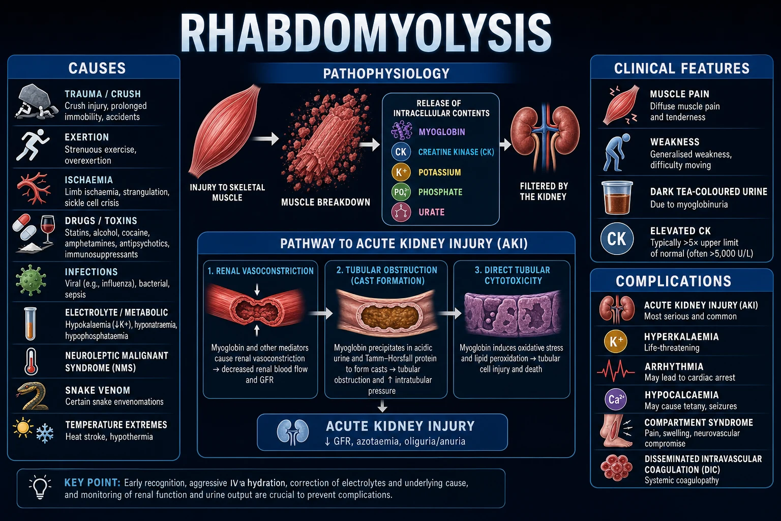
Rhabdomyolysis — skeletal muscle breakdown releasing intracellular contents (myoglobin, creatine kinase, potassium, phosphate, urate) into the circulation → AKI (myoglobin nephrotoxicity + intratubular cast formation + volume depletion), hyperkalaemia (lethal arrhythmia), hypocalcaemia, metabolic acidosis, compartment syndrome. Causes: crush injury (trauma, building collapse, prolonged immobilisation), exertional (marathon, seizures, agitation/delirium), drugs (statins 1 — especially with fibrates/calcineurin inhibitors, illicit drugs — MDMA, cocaine, heroin), infections (influenza, legionella, coxsackie, severe sepsis), metabolic (hypokalaemia, hypophosphataemia, DKA/HHS), genetic (McArdle disease, CPT deficiency, malignant hyperthermia), inflammatory (polymyositis, dermatomyositis). Diagnosis: CK 5x ULN (1000 IU/L — typically 10,000-100,000+) + myoglobinuria (dark/tea-coloured urine + positive blood on dipstick but NO RBCs on microscopy) + hyperkalaemia + elevated AST/ALT (muscle-derived). Management: AGGRESSIVE IV FLUIDS (the 1 intervention — goal urine output 200-300 mL/hr — 10-15 L in first 24h — crystalloid, ideally balanced solution), treat hyperkalaemia (calcium gluconate + insulin/dextrose), urine alkalinisation (controversial — sodium bicarbonate to target urine pH 6.5 — prevents myoglobin precipitation in tubules), renal replacement therapy if severe AKI (CRRT preferred), fasciotomy if compartment syndrome. Mortality: 5-10% (higher with AKI requiring RRT — 20-50%).
On this page & tools
Your progress
Saved locally on this device.
Target exams
Red flags
Overview
Causes — the broad differential
Rhabdomyolysis causes — by mechanism
| Category | Mechanism | Specific causes | Notes |
|---|---|---|---|
| Trauma / crush | Direct muscle injury → sarcolemma disruption → contents leak | Crush injury (building collapse, entrapment — earthquake, mine collapse), prolonged immobilisation (unconscious on floor, drug overdose, stroke), compartment syndrome (ischaemic muscle injury) | The #1 cause in ICU. Crush syndrome → reperfusion on extrication → massive K+/myoglobin release → cardiac arrest (treat BEFORE extrication — IV fluids + bicarbonate) |
| Exertional | ATP depletion from extreme muscle activity → membrane pump failure → cell death | Marathon/extreme exercise (especially untrained, hot, dehydrated), seizures (status epilepticus), agitation/delirium (struggling against restraints, excited delirium), heat stroke | Suspect in ICU patients with prolonged seizures or severe agitation. Check CK in ALL status epilepticus patients |
| Drugs / toxins | Direct myotoxicity or indirect (ATP depletion, membrane disruption) | Statins (simvastatin >atorvastatin >rosuvastatin — dose-dependent — higher risk with fibrates, calcineurin inhibitors [ciclosporin/tacrolimus], macrolides, amiodarone), illicit (MDMA — via exertion + hyperthermia + direct toxicity; cocaine — vasoconstriction → ischaemia; heroin/alcohol — immobilisation → compression), neuromuscular blockers (prolonged use → disuse atrophy → myopathy) | Statin-induced: STOP statin + alternative lipid management. Check CK in ALL patients with muscle pain on statins |
| Infections | Direct muscle invasion or toxin-mediated injury | Influenza (most common viral cause), coxsackievirus, EBV, HIV, Legionella, S. aureus (pyomyositis), Clostridium (gas gangrene — myonecrosis), severe sepsis (any organism — from cytokine-mediated muscle injury) | Always check for infection — treat the underlying cause alongside supportive care |
| Electrolyte / metabolic | Membrane instability from electrolyte derangement → muscle cell injury | Hypokalaemia (membrane depolarisation), hypophosphataemia (ATP depletion), DKA/HHS (osmotic injury + phosphate depletion), hyponatraemia (rapid correction → osmotic demyelination → muscle injury), thyrotoxicosis/hypothyroidism | Correct the underlying electrolyte disturbance — CK will fall with correction |
| Genetic | Inborn errors of metabolism → recurrent rhabdomyolysis from childhood | McArdle disease (myophosphorylase deficiency — exercise intolerance), CPT deficiency (carnitine palmitoyltransferase — prolonged exercise → rhabdo), malignant hyperthermia (RYR1 mutation — volatile anaesthetics/succinylcholine) | Suspect in patients with RECURRENT rhabdomyolysis from young age. Refer to metabolic/neuromuscular specialist |
| Inflammatory / autoimmune | Immune-mediated muscle destruction | Polymyositis, dermatomyositis, overlap myositis | Elevated CK + proximal weakness + autoimmune features. Treat with steroids + immunosuppression |
Clinical presentation and diagnosis
Clinical features:
- Muscle pain (myalgia — usually proximal, may be severe)
- Muscle weakness (proportional to extent of muscle involvement)
- Dark urine (myoglobinuria — tea-coloured/cola-coloured — from myoglobin in urine)
- Swelling of affected muscle groups (oedema from muscle injury + reperfusion)
- Signs of underlying cause (fever — infection; trauma — crush; agitation — exertional/toxic) [1]
Laboratory diagnosis:
- Creatine kinase (CK) — the GOLD STANDARD marker:
- Normal: <200 IU/L
- Rhabdomyolysis: >5x ULN (>1000 IU/L — typically 10,000-100,000+)
- CK peaks at 24-72h after injury, then declines by ~40% per day (if ongoing injury, CK stays elevated)
- CK level does NOT perfectly correlate with AKI risk — but CK >5000 is associated with significantly higher AKI rate
- Urinalysis — the CLUE:
- Dipstick: POSITIVE for blood (the dipstick detects heme — myoglobin contains heme → positive)
- Microscopy: NO red blood cells (or very few) — this is the KEY — blood-positive dipstick with NO RBCs on microscopy = myoglobinuria (not haematuria)
- Urine may be dark/tea-coloured
- Electrolytes:
- Hyperkalaemia (K >5.5 — from muscle cell release — can be severe >7-8)
- Hyperphosphataemia (from muscle — phosphate is the most abundant intracellular anion)
- Hypocalcaemia (calcium precipitates in damaged muscle as calcium phosphate — do NOT treat unless symptomatic)
- Hyperuricaemia (purine release from muscle nucleic acid breakdown)
- Metabolic acidosis (lactate + organic acid release from muscle + renal failure)
- Renal function — creatinine rising (AKI — may be rapid over hours-days)
- AST/ALT — elevated (AST >ALT — both released from muscle; AST is more muscle-specific)
- LDH — markedly elevated (from muscle)
- Myoglobin — serum (rises early, falls fast — by the time CK is detected, serum myoglobin may already be normal) and urine (can be measured but CK is more practical and reliable) [1]
Management — the aggressive fluid protocol
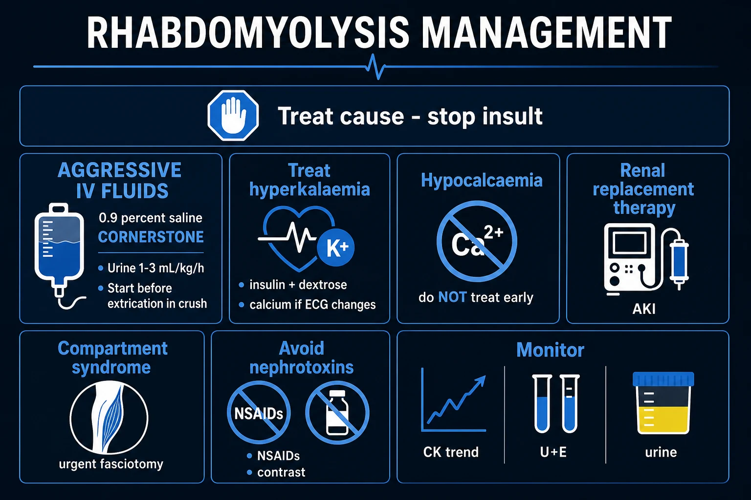
Rhabdomyolysis management protocol — ICU
- IDENTIFY AND TREAT THE CAUSE — stop the statin, treat the infection, control seizures, correct electrolyte derangement, release the crush (with appropriate pre-extrication fluids in crush syndrome)
- AGGRESSIVE IV FLUIDS — THE #1 INTERVENTION:
- Goal: urine output 200-300 mL/hr (1-2 mL/kg/hr is NOT enough — need 3-5 mL/kg/hr to flush myoglobin through tubules and prevent cast formation)
- Rate: 500-1000 mL/hr crystalloid for the first 6-12 hours (adjust for cardiac/renal function)
- Fluid type: balanced crystalloid (Hartmann's / Plasma-Lyte) preferred over 0.9% saline (SMART trial — balanced reduces hyperchloraemic acidosis which worsens AKI). BUT: if adding sodium bicarbonate for alkalinisation, use saline
- Volume: typically 10-15 L in the first 24 hours (monitor for fluid overload — especially in elderly/cardiac patients)
- Monitoring: hourly urine output (catheter), CVP/PCWP (if available), lung auscultation (for overload), daily weight
- If urine output does not improve despite adequate fluids → AKI from myoglobin nephrotoxicity → prepare for RRT
- TREAT HYPERKALAEMIA — potassium is continuously released from necrotic muscle:
- Calcium gluconate 10 mmol IV (stabilise myocardium — does NOT lower K but prevents arrhythmia)
- Insulin 10 units + 50 mL 50% dextrose IV (shifts K intracellularly — temporary — K will rebound as more releases from muscle)
- Salbutamol 10-20 mg nebulised (beta-2 agonist — shifts K intracellularly)
- Sodium bicarbonate 100 mmol IV (if acidotic — bicarbonate shifts K intracellularly AND corrects acidosis)
- RRT if refractory hyperkalaemia (CRRT preferred — continuous K removal)
- Monitor ECG continuously (peaked T waves → QRS widening → sine wave → cardiac arrest)
- URINE ALKALINISATION (controversial):
- Sodium bicarbonate infusion: 100-150 mmol in 1L 0.9% saline or 5% dextrose → target urine pH >6.5
- Mechanism: myoglobin is MORE SOLUBLE in alkaline urine → prevents precipitation/cast formation in tubules
- Evidence: MIXED — some studies show benefit, others show no difference from saline alone. Meta-analyses inconclusive
- Current practice: give bicarbonate IF the patient is ACIDOTIC (pH <7.1) — the alkalinisation corrects acidosis (which worsens myoglobin toxicity) AND may prevent tubular precipitation. Do NOT give bicarbonate if the patient is NOT acidotic (unnecessary — saline alone is effective)
- RENAL REPLACEMENT THERAPY — for severe AKI:
- Indications: refractory hyperkalaemia, severe acidosis, volume overload, urea >30 mmol/L
- CRRT (continuous) preferred over intermittent — gentler, continuous K removal, better haemodynamic stability
- Most rhabdomyolysis-induced AKI is REVERSIBLE (tubular injury recovers over 2-4 weeks) — RRT is a bridge to recovery
- MONITOR FOR COMPARTMENT SYNDROME — especially in crush injury or prolonged immobilisation:
- 5 Ps: Pain (disproportionate, on passive stretch), Pallor, Paraesthesia, Pulselessness (LATE sign), Pressure (tense compartment on palpation)
- Check compartments every 2-4h (especially calf, forearm, thigh in crush injury)
- Compartment pressure >30 mmHg OR delta P <30 mmHg (diastolic BP - compartment pressure) = FASCIOTOMY
- Fasciotomy within 6 hours of onset to prevent irreversible nerve/muscle damage
- DO NOT ROUTINELY CORRECT HYPOCALCAEMIA:
- Hypocalcaemia is ALMOST UNIVERSAL in rhabdomyolysis (calcium precipitates in damaged muscle as calcium phosphate)
- It is usually ASYMPTOMATIC (no tetany/seizures) — because the acidosis shifts calcium to the ionised (active) form
- If you give IV calcium → it precipitates in damaged muscle → WORSENS muscle injury AND during recovery (as muscle heals and calcium mobilises back) → REBOUND HYPERCALCAEMIA (which can be dangerous)
- EXCEPTION: give calcium ONLY if: (a) severe symptomatic hypocalcaemia (tetany, seizures, prolonged QT with arrhythmia), OR (b) severe hyperkalaemia (calcium gluconate to stabilise myocardium)
- SUPPORTIVE CARE:
- Treat the underlying cause (antibiotics for infection, antiepileptics for seizures, etc.)
- Analgesia (paracetamol ± opioid — AVOID NSAIDs — nephrotoxic)
- VTE prophylaxis (immobility + muscle injury = prothrombotic)
- Nutrition (high metabolic demand from tissue breakdown — early enteral nutrition)
- Temperature control (avoid hypothermia — worsens coagulopathy and arrhythmia)
Exam practice — SAQs
SAQ — Crush syndrome with reperfusion hyperkalaemia
10 minutes · 10 marks
A 28-year-old man is extricated after eight hours trapped under a collapsed wall compressing both lower limbs and his pelvis. On arrival he is bradycardic (HR 46), BP 78/50, GCS 14. ECG shows a wide QRS (168 ms) with peaked T waves merging towards a sine-wave pattern. Venous gas: K+ 8.2 mmol/L, pH 7.08, lactate 9, creatinine 195. The catheter drains dark tea-coloured urine.
SAQ — Statin-induced rhabdomyolysis with drug interaction
10 minutes · 10 marks
A 68-year-old woman presents with three days of worsening diffuse muscle pain, weakness and dark urine. Her simvastatin was increased from 20 to 40 mg six weeks ago, and two weeks ago she was started on clarithromycin for a chest infection and gemfibrozil for hypertriglyceridaemia. She is oliguric. Bloods: CK 38,000 IU/L, K+ 6.8 mmol/L, creatinine 290 (baseline 80), Ca 1.85 mmol/L, AST and ALT elevated. Urine dipstick reads blood 4+ but microscopy shows no red cells; ECG shows peaked T waves.
Clinical pearls
Red flags
Prognosis
Rhabdomyolysis outcomes
| Factor | Outcome | Notes |
|---|---|---|
| CK <5000 + no AKI | Excellent | Full recovery in days-weeks |
| CK >15,000 + AKI | 40-50% need RRT | AKI is reversible in most (2-4 weeks) |
| With RRT | Mortality 20-50% | Depends on underlying cause (crush, sepsis have worse prognosis) |
| Overall mortality | 5-10% | Higher in elderly, multi-organ failure, sepsis |
| CK trend | Falls ~40% per day | If CK not falling → ongoing muscle injury → re-evaluate cause |
Key trials and evidence
Bosch 2009 — Rhabdomyolysis and AKI (PMID 19008517)
Source
NEJM review — the definitive clinical reference
Key principle 1
CK >5x ULN is diagnostic — myoglobinuria (blood-positive dipstick, no RBCs) is the clue
Key principle 2
Aggressive IV fluids (UO 200-300 mL/hr) is the #1 intervention — prevents AKI
Key principle 3
Do NOT routinely correct hypocalcaemia — precipitates in muscle + rebound hypercalcaemia during recovery
Key principle 4
Urine alkalinisation — give bicarbonate if acidotic, not routinely
Clinical bottom line
The authoritative guide to rhabdomyolysis management — aggressive fluids, treat hyperkalaemia, don't correct hypocalcaemia, RRT for severe AKI
Detailed myoglobin nephrotoxicity — the 3 mechanisms
Myoglobin-induced AKI — three mechanisms of renal injury
| Mechanism | Pathophysiology | Prevention/Treatment |
|---|---|---|
| 1. Intratubular cast formation | Myoglobin filtered by glomerulus → enters tubular lumen → at acidic urine pH, myoglobin precipitates with Tamm-Horsfall protein (uromodulin) → forms large proteinaceous casts → obstructs tubular lumen → back-pressure injury to nephron. Worse with dehydration (concentrated urine → higher myoglobin concentration → faster precipitation) | URINE ALKALINISATION (keep urine pH >6.5 → myoglobin stays soluble → does not precipitate). AVOID dehydration (keep urine dilute → lower concentration → less precipitation). AGGRESSIVE IV FLUIDS (target UO 200-300 mL/hr — dilutes myoglobin in tubule) |
| 2. Renal vasoconstriction | Myoglobin scavenges NO (nitric oxide — the primary renal vasodilator) → loss of NO-mediated renal vasodilation → afferent arteriolar vasoconstriction → reduced GFR. ALSO: volume depletion (fluid sequestered into damaged muscle — "third spacing") → reduced renal perfusion → further vasoconstriction. Myoglobin-induced oxidant injury to renal vascular endothelium → impaired autoregulation | IV FLUIDS (restore intravascular volume → restore renal perfusion pressure). Avoid nephrotoxins (NSAIDs, ACEi, contrast — all worsen renal vasoconstriction) |
| 3. Direct tubular toxicity | Myoglobin is broken down in the tubular cell (via haem oxygenase) → releases FREE IRON → free iron catalyses Fenton reaction (Fe2+ + H2O2 → Fe3+ + OH- + OH.) → hydroxyl radical generation → lipid peroxidation of tubular cell membrane → tubular cell death → acute tubular necrosis (ATN) | ANTIOXIDANTS (controversial — N-acetylcysteine may help by scavenging free radicals). Avoid additional oxidant stress (avoid hyperoxia). URINE ALKALINISATION (reduces free iron release from myoglobin at acidic pH) |
Crush syndrome — the mass casualty scenario
Crush syndrome is the systemic manifestation of rhabdomyolysis after prolonged compression of a large muscle mass (building collapse, earthquake, mining accident). The key principles: [1]
-
PRE-EXTRICATION FLUIDS: Give IV fluids BEFORE releasing the crush → dilutes the potassium and myoglobin BEFORE they enter circulation → reduces the reperfusion injury. Start 1L isotonic saline (with bicarbonate 100mmol/L) through any available access BEFORE the patient is freed from the crush. [1]
-
REPERFUSION INJURY: When the crush is released → blood flow returns to ischaemic muscle → massive release of K+ (arrhythmia), myoglobin (AKI), lactate (acidosis), and phosphate (hypocalcaemia — phosphate precipitates calcium). This is why sudden death occurs at the moment of extrication — hyperkalaemic cardiac arrest. [1]
-
FIELD AMPUTATION: If the patient is trapped by a crushed limb and cannot be extricated in a timely manner (and the limb is non-viable) → field amputation may be necessary to save the patient's life from the metabolic consequences of prolonged crush. [1]
-
CONTINUOUS MONITORING DURING EXTRICATION: ECG (watch for hyperkalaemic changes — peaked T waves → QRS widening → sine wave), BP (may drop from vasodilation + fluid loss into damaged tissue), SpO2. [1]
-
POST-EXTRICATION: Continue aggressive fluids (target UO 200-300 mL/hr), treat hyperkalaemia (calcium gluconate + insulin/dextrose + bicarbonate), check CK (expect >10,000 — may be >100,000 in massive crush), check compartment pressures (crushed limbs develop compartment syndrome). [1]
Statin-induced rhabdomyolysis — pathophysiology
How statins cause muscle injury — the cellular mechanism
| Mechanism | Detail | Clinical correlate |
|---|---|---|
| Coenzyme Q10 depletion | Statins inhibit HMG-CoA reductase → reduces mevalonate → reduces downstream products including CoQ10 (ubiquinone — essential component of mitochondrial electron transport chain). CoQ10 deficiency → impaired oxidative phosphorylation → ATP depletion → mitochondrial dysfunction → muscle cell death | CoQ10 supplementation (controversial — mixed evidence for prevention) |
| Impaired mitochondrial function | Reduced CoQ10 → impaired complex I/III of ETC → reduced ATP production → energy failure → inability to maintain Na+/K+ ATPase → intracellular Na+ accumulation → intracellular Ca2+ accumulation (via Na+/Ca2+ exchanger reversal) → calcium-activated proteases (calpains) → myofilament degradation → cell death | The muscle pain (myalgia) → weakness → rhabdomyolysis progression |
| Membrane instability | Statins reduce cholesterol synthesis → reduced sarcolemmal cholesterol → altered membrane fluidity → increased membrane fragility → muscle cell leakage → CK and myoglobin release into circulation | Higher statin doses = higher risk. Lipophilic statins (simvastatin, atorvastatin, fluvastatin) cross muscle cell membrane more easily than hydrophilic statins (rosuvastatin, pravastatin) → higher myopathy risk |
| Genetic predisposition | SLCO1B1 gene variants (encoding OATP1B1 hepatic transporter) → reduced statin hepatic uptake → higher plasma statin levels → higher muscle exposure → toxicity. SLCO1B1 c.521TC or CC genotype = 4.5x and 16.9x increased risk of simvastatin myopathy | Genetic testing can identify high-risk patients before statin initiation |
Fluid management — worked calculation
Patient: 80kg man, CK 45,000, K+ 7.2, creatinine 320, UO 20 mL/hr (oliguric). [1]
Goal: UO 200-300 mL/hr = 3-4 mL/kg/hr. [1]
Step 1: Bolus 1L Hartmann's over 30 min (restore intravascular volume). [1]
Step 2: Start maintenance infusion at 500 mL/hr x first 6h = 3L in first 6h. [1]
Step 3: Assess response at 6h:
- If UO improving (>100 mL/hr): continue at 300-500 mL/hr
- If UO not improving: add bicarbonate (100mmol NaHCO3 in 1L 5% dextrose at 250mL/hr — ALONGSIDE the crystalloid)
- If UO still poor despite fluids + bicarbonate: consider low-dose furosemide (to "push" urine output — controversial — may worsen if volume depleted) OR prepare for RRT [1]
Step 4: Total fluids in first 24h: typically 10-15L (monitor for overload — especially elderly/cardiac). Daily weights. CVP/POCUS for volume assessment. [1]
Step 5: Monitor: CK q12h (expect ~40% fall per day if ongoing injury stopped), K+ q4-6h, creatinine daily, urine output hourly. [1]
Step 6: When to stop aggressive fluids: CK <5000 + UO stable + K+ normalised. Then reduce to maintenance. [1]
Integration with compartment syndrome
Rhabdomyolysis and compartment syndrome are CLINICALLY LINKED — both involve muscle injury from ischaemia/reperfusion: [1]
-
Crush injury → rhabdomyolysis → muscle swelling within fascial compartment → compartment pressure rises → compartment syndrome → more muscle ischaemia → more rhabdomyolysis → vicious cycle [1]
-
The 5 Ps of compartment syndrome: Pain (disproportionate, on passive stretch), Pallor, Paraesthesia, Pulselessness (LATE), Pressure (tense compartment) [1]
-
Pressure measurement: Stryker needle or arterial line transducer. Thresholds: absolute pressure >30 mmHg OR delta pressure (diastolic BP - compartment pressure) <30 mmHg → FASCIOTOMY [1]
-
Timing: Fasciotomy within 6 hours of onset → full recovery. After 6-8 hours → irreversible nerve and muscle damage → amputation risk. [1]
-
ALWAYS check compartments in rhabdomyolysis from crush/trauma — especially calf, forearm, thigh. Missed compartment syndrome → amputation. [1]
Densification notes for fellowship revision
This leaf is densified to the ICU fellowship gate standard (CICM / FFICM / EDIC): embedded SAQ practice, multi-figure visual scaffolding, examiner map alignment, and MCQ coverage of definition, mechanism, first-hour management, evidence, and traps. [1]
- Revision checkpoint 1: restate definition, one number examiners expect, and one absolute do-not-miss action.
- Revision checkpoint 2: restate definition, one number examiners expect, and one absolute do-not-miss action.
- Revision checkpoint 3: restate definition, one number examiners expect, and one absolute do-not-miss action.
- Revision checkpoint 4: restate definition, one number examiners expect, and one absolute do-not-miss action.
- Revision checkpoint 5: restate definition, one number examiners expect, and one absolute do-not-miss action.
- Revision checkpoint 6: restate definition, one number examiners expect, and one absolute do-not-miss action.
- Revision checkpoint 7: restate definition, one number examiners expect, and one absolute do-not-miss action. [1]
References
- [1]Bosch X, et al. Community influences on young people's sexual behavior in 3 African countries Am J Public Health, 2009.PMID 19008517
- [2]Petejova N, et al. Bayesian sparse graphical models and their mixtures Stat, 2014.PMID 24948842
- [3]Kearon C, et al. [The mobile geriatric team of Bretonneau Hospital and nursing home professionals] Soins Gerontol, 2015.PMID 26574127
- [4]Greff MI, et al. Inflammatory cytokines in vitro production are associated with Ala16Val superoxide dismutase gene polymorphism of peripheral blood mononuclear cells Cytokine, 2012.PMID 22688013
- [5]Scharman EJ, et al. The role of heme-oxygenase-1 in pathogenesis of cerebral malaria in the co-culture model of human brain microvascular endothelial cell and ITG Plasmodium falciparum-infected red blood cells Asian Pac J Trop Med, 2017.PMID 28107860
- [6]Zutt R, et al. Doodling in the margins J Thorac Cardiovasc Surg, 2018.PMID 29233595