ICU · Acid–base
Acid–Base Physiology and Blood-Gas Interpretation
Also known as Acid-base · Blood gas interpretation · Metabolic acidosis · Metabolic alkalosis · Stewart approach · Strong ion difference · Anion gap
Acid–base homeostasis is the chemistry of the hydrogen ion, and its disturbances are among the commonest and most informative derangements in critical illness. This topic builds the examiner's framework on four ideas. First, the physiology — the bicarbonate buffer system described by Henderson and Hasselbalch, and the more complete Stewart view in which pH is set by the carbon dioxide tension, the strong ion difference (largely sodium minus chloride) and the total concentration of weak acids (albumin and phosphate). Second, a systematic method for reading a blood gas — the pH, the pattern (metabolic versus respiratory), the appropriateness of compensation, the anion gap and the delta-delta, and the osmolar gap when a toxin is suspected. Third, the clinical disorders — the high anion-gap and normal anion-gap acidoses (lactic acidosis, ketoacidosis, the toxic alcohols, and renal tubular acidosis), the metabolic alkaloses, and the respiratory acid–base disturbances. Fourth, the evidence that has reshaped management — the SMART and SALT-ED trials showing balanced crystalloids modestly outperform saline, BICAR-ICU showing bicarbonate confers no overall benefit in severe acidaemia (with a signal in the acute-kidney-injury subgroup), and the principles of treating the cause rather than the number. Built entirely on these verified landmark trials.
On this page & tools
Your progress
Saved locally on this device.
Target exams
Red flags

Overview & definition
Acid–base homeostasis is the regulation of the hydrogen-ion concentration of the extracellular fluid, held within a narrow arterial range of pH 7.35 to 7.45 — corresponding to a free hydrogen-ion concentration of about 35 to 45 nanomoles per litre.[1] The healthy organism defends this range on three timescales: the chemical buffers (bicarbonate, phosphate, proteins, haemoglobin) act within seconds; the lung adjusts carbon dioxide excretion within minutes; and the kidney regulates bicarbonate reabsorption and acid excretion over hours to days. Acid–base disorders arise when one of these is deranged, and the pattern of the derangement is among the most diagnostic pieces of information available at the bedside of a critically ill patient.[1]
A metabolic disorder is a primary change in the bicarbonate concentration; a respiratory disorder is a primary change in the carbon dioxide tension. Each primary change provokes a compensatory response in the other system that is predictable in direction and — for the metabolic disorders, at least — predictable in magnitude. The skill of blood-gas interpretation is to identify the primary disorder, judge whether the compensation is appropriate, and detect the mixed disorders that inappropriate compensation reveals.[1][2]
Two conceptual frameworks coexist. The traditional Henderson–Hasselbalch approach describes pH as a function of the ratio of bicarbonate to dissolved carbon dioxide, and remains the practical, bedside framework for most clinical interpretation. The Stewart approach re-expresses the same chemistry in terms of three independent variables — the carbon dioxide tension, the strong ion difference, and the total weak-acid concentration — and gives a fuller account of why, for example, a chloride-rich fluid causes acidosis and albumin depletion masks an anion gap. Both describe the same physiology; the examiner expects fluency in the traditional method and an understanding of the Stewart insights.[3]
Pathophysiology: buffers, Henderson–Hasselbalch, and the whole blood
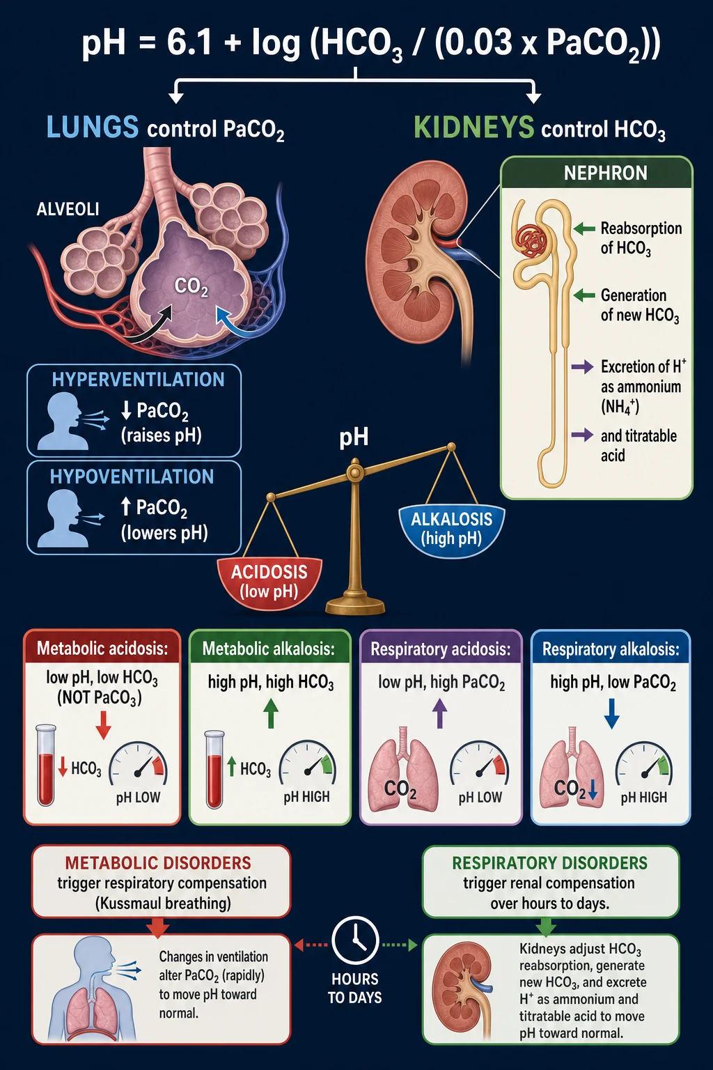
The dominant extracellular buffer is the bicarbonate–carbonic acid system, governed by the Henderson–Hasselbalch equation: [1]
pH = 6.1 + log₁₀( [HCO₃⁻] / (0.03 × PaCO₂) ) [1]
where 6.1 is the pK of the system and 0.03 is the solubility coefficient of carbon dioxide. The equation makes the central insight of acid–base physiology plain: pH is set by the ratio of bicarbonate to carbon dioxide, not by either alone. A fall in bicarbonate (a metabolic acidosis) and a rise in carbon dioxide (a respiratory acidosis) both lower the ratio and therefore the pH; a rise in bicarbonate or a fall in carbon dioxide both raise it.[1]
The bicarbonate system is effective not because bicarbonate is a strong buffer (its pK of 6.1 is distant from the physiological pH of 7.4) but because both of its components are independently regulated — the kidney controls bicarbonate and the lung controls carbon dioxide, so the system is "open" and can be driven to defend pH with enormous capacity. The non-bicarbonate buffers (haemoglobin, proteins, phosphate) matter most inside the cell and in the acute, unsteady state, and the base excess — the amount of acid or base required to return a litre of whole blood to pH 7.40 at standard conditions — is the laboratory's attempt to quantify the metabolic (non-respiratory) component in isolation.[2][1]
A negative base excess (a base deficit) signifies a metabolic acidosis; a positive value a metabolic alkalosis. The base excess, the bicarbonate and the standard bicarbonate all express the same metabolic information, and the candidate need not labour all three — but the base excess is the most robust to an acute respiratory disturbance.[1]
The Stewart approach: strong ions and the weak acids
The Stewart analysis re-derives pH from three independent variables, in place of bicarbonate (which Stewart treats as dependent).[3]
- The partial pressure of carbon dioxide (PaCO₂) — the respiratory variable, as in the traditional model.
- The strong ion difference (SID) — the difference between the fully dissociated cations and anions in plasma, dominated in practice by sodium minus chloride (about 40 mmol/L). Because the strong ions cannot combine with hydrogen, the SID acts like an electrical charge gap that water dissociation must fill; a small SID (a fall in sodium or, more commonly, a rise in chloride — as with saline) drives the dissociation of water towards more free hydrogen and an acidosis, while a large SID (chloride loss from vomiting, or diuretic-induced) produces an alkalosis.
- The total concentration of weak acids (Aₜₒₜ) — predominantly albumin and, to a lesser extent, phosphate. A fall in Aₜₒₜ (hypoalbuminaemia, ubiquitous in critical illness) is itself an alkalinising process, which is why a low albumin masks an underlying high anion gap. [1]
The Stewart framework earns its place at the bedside by explaining what the traditional model can only describe: why large-volume saline causes a hyperchloraemic acidosis (it lowers the SID by adding chloride without bicarbonate), why hypoalbuminaemia hides an anion-gap acidosis (it lowers Aₜₒₜ, an alkalinising effect that offsets the acid), and why a metabolic alkalosis accompanies chloride depletion. The unmeasured anions of critical illness (lactate, ketones, toxins) are, in Stewart terms, an addition of strong anions that shrinks the SID.[3]
Blood gas interpretation: a systematic method
A blood gas should be read in a fixed order, so that no step is omitted and mixed disorders are not missed.[1][1]
- The pH — is it acidaemia (pH below 7.35) or alkalaemia (above 7.45)? The pH names the net disturbance but not its cause.
- The primary disorder — compare the PaCO₂ and the bicarbonate (or base excess). A low bicarbonate with a low PaCO₂ is a primary metabolic acidosis; a high bicarbonate with a high PaCO₂ a primary metabolic alkalosis; a high PaCO₂ with a low pH a respiratory acidosis; a low PaCO₂ with a high pH a respiratory alkalosis.
- The appropriateness of compensation. For a metabolic acidosis, the expected PaCO₂ is given by Winter's formula: expected PaCO₂ = 1.5 × [HCO₃⁻] + 8 (± 2) mmHg. A measured PaCO₂ outside this range signals a mixed disorder — a concurrent respiratory acidosis or alkalosis. For chronic metabolic acidosis and for the alkaloses, other formulae apply; the principle is that compensation is predictable, and deviation from the prediction unmasks a second primary disorder.[1]
- The anion gap — Na⁺ − (Cl⁻ + HCO₃⁻), normally about 8 to 12 mmol/L. A high gap adds unmeasured anions (lactate, ketones, toxins, uraemia); a normal gap points to bicarbonate loss or chloride excess.
- The delta-delta (ΔAG / ΔHCO₃⁻). Compare the rise in the anion gap to the fall in bicarbonate. If they rise and fall roughly together (ratio 1 to 2), the disorder is a pure high anion-gap acidosis. A ratio below 1 means a second, normal-gap acidosis is present; a ratio above 2 means a concurrent metabolic alkalosis is elevating the bicarbonate. This step is what catches the mixed disorder that a single number conceals.[4]
- The osmolar gap when a toxin is suspected (below).
Metabolic acidosis and the anion gap
Metabolic acidosis — a primary fall in bicarbonate with a compensatory fall in PaCO₂ — is the commonest acid–base disorder in the intensive care unit, and its first subdivision is by the anion gap.[4]
The anion gap (Na⁺ − (Cl⁻ + HCO₃⁻)) estimates the concentration of unmeasured anions. In a healthy person it is 8 to 12 mmol/L, composed largely of albumin and phosphate. A raised anion gap means new unmeasured anions have appeared — lactate, ketoacids, the acid metabolites of toxins, uraemic acids, or drug anions — and each has displaced bicarbonate. A normal anion-gap (hyperchloraemic) acidosis means bicarbonate has been lost (diarrhoea) or chloride gained (saline, renal tubular acidosis) without the generation of new anions.[4]
Two corrections are essential before the gap is interpreted. Albumin is the largest contributor to the normal gap, so a low albumin lowers the expected gap and can hide an anion-gap acidosis: the corrected anion gap adds roughly 2.5 mmol/L for every 10 g/L that albumin falls below 40. And in mixed disorders the delta-delta (above) tells whether a second acid–base process accompanies the high gap. The anion gap is cheap, fast and powerful, but it is a calculated estimate with wide normal variability, and it does not replace a direct measurement (a lactate level, a toxicology screen) when a specific cause is suspected.[4]
High anion-gap metabolic acidosis: the causes
The mnemonic for the high anion-gap metabolic acidoses (HAGMA) is GOLD MARK (the modern, more complete successor to MUDPILES): [1]
- G — Glycols (ethylene glycol and its toxic metabolites; propylene glycol, the solvent in some IV preparations such as high-dose lorazepam and in some etomidate formulations).
- O — Oxoproline (5-oxoproline accumulation, classically in chronic paracetamol use with glutathione depletion).
- L — L-lactate (tissue hypoperfusion, sepsis, and the Type B causes below).
- D — D-lactate (from gut bacterial overgrowth in short-bowel syndrome).
- M — Methanol (metabolised to formic acid).
- A — Aspirin (salicylate late-stage).
- R — Renal failure (accumulation of uraemic organic acids).
- K — Ketoacidosis (diabetic, alcoholic, and starvation).[5][6]
The clinician's job at the bedside is to identify which — guided by the lactate, the glucose and ketones, the renal function, the drug history and, when more than one gap is raised, the osmolar gap (which is high in the toxic alcohols and normal in the others). [1]
Lactic acidosis
Lactic acidosis is the commonest cause of a high anion-gap acidosis in critical illness, and lactate is both a marker and, in excess, a contributor to the acidaemia.[5]
Lactic acidosis is classified by mechanism into Type A — tissue hypoperfusion and hypoxia, the high lactate of shock, severe hypoxaemia, mesenteric ischaemia and seizures, where anaerobic metabolism is the mechanism — and Type B, in which lactate accumulates without overt hypoperfusion: B1 from underlying disease (sepsis, malignancy, hepatic failure, diabetic ketoacidosis, seizures), B2 from drugs and toxins (metformin, the anti-retrovirals, propylene glycol, cyanide, the biguanides), and B3 from inborn errors of metabolism. The distinction is conceptual more than absolute — sepsis, the commonest ICU cause, has elements of both.[5]
A serum lactate above 5 mmol/L is severe, and a rising lactate (or a failure to clear) in septic shock is an independent predictor of mortality. The treatment of lactic acidosis is the treatment of its cause — restore perfusion and oxygen delivery, achieve source control, stop the offending drug — because the acid will clear when the underlying process resolves. The role of bicarbonate is addressed below; in summary it is not routinely indicated, and in the Type B causes (notably metformin-associated lactic acidosis) renal replacement therapy removes both the lactate and the causative drug.[5]
Diabetic ketoacidosis and hyperosmolar hyperglycaemic state
Diabetic ketoacidosis (DKA) is a high anion-gap acidosis driven by ketoacid (β-hydroxybutyrate and acetoacetate) production in absolute insulin deficiency, and its management is governed by the long-standing American Diabetes Association consensus.[8]
The DKA patient is volume-depleted (the osmotic diuresis of hyperglycaemia), potassium-depleted (total-body, despite a normal or high serum potassium driven by the acidosis and insulin lack), and acidotic. The ADA-consensus management rests on four pillars:[8]
- Fluid resuscitation — isotonic saline first (0.9 per cent, 15 to 20 mL/kg in the first hour), then adjusted to the corrected sodium; this alone lowers the glucose and, by improving perfusion, begins to clear the acid.
- Insulin — a continuous infusion of regular insulin at 0.1 units/kg/h (or a 0.1 unit/kg bolus then 0.1 units/kg/h), to suppress ketogenesis; the target is a fall in glucose of 3 to 4 mmol/L per hour and the closure of the anion gap, not merely normoglycaemia.
- Potassium — insulin and the correction of the acidosis drive potassium into cells, and the commonest preventable death in DKA treatment is hypokalaemia; potassium is replaced early, before insulin if the serum potassium is low, and held only if it is frankly high or the patient is anuric.
- Identify and treat the precipitant — infection (the commonest), omission of insulin, infarction, new-onset type 1 diabetes. [1]
Bicarbonate is not routinely given in DKA. The ADA consensus does not recommend it for pH above 6.9, because it does not improve outcome and may delay the clearance of ketones; the very lowest pH values (below 6.9) are a separate, contentious question, but the acid itself corrects as the ketogenesis is suppressed. The hyperosmolar hyperglycaemic state (HHS) shares the hyperglycaemia but lacks significant ketoacidosis; its management is dominated by more aggressive fluid resuscitation and a lower insulin dose.[8]
Toxic alcohol poisoning: the dual gap
The toxic alcohols — ethylene glycol and methanol (and to a lesser extent diethylene glycol and propylene glycol) — are themselves of low toxicity, but their metabolites are devastating: ethylene glycol yields glycolic and oxalic acids (a severe high anion-gap acidosis, acute kidney injury, and calcium oxalate crystalluria), and methanol yields formic acid (a severe high anion-gap acidosis and retinal toxicity — the characteristic visual disturbance).[6]
The diagnostic signature is the dual gap — a high anion-gap acidosis together with a high osmolar gap — that appears after the parent alcohol has been absorbed but is being metabolised. The osmolar gap is the difference between the measured serum osmolality and the calculated osmolality (2 × Na⁺ + glucose + urea); a gap above roughly 20 mOsm/kg suggests an unmeasured osmotically active solute, of which the toxic alcohols are the dangerous example, though the gap is imperfect and a normal gap does not exclude late presentation (once the parent has all been metabolised to anions).[6][7]
The treatment is time-critical, because the injury is done by the metabolites: inhibit alcohol dehydrogenase with fomepizole (15 mg/kg load then maintenance) or, where fomepizole is unavailable, ethanol, to halt the metabolism of the parent alcohol; and haemodialysis to remove both the parent alcohol and the formed metabolites in the severely acidotic, the renal-failure, or the very-high-concentration patient. The acidosis and the gaps fall as the toxin is cleared, and folate (methanol) and thiamine and pyridoxine (ethylene glycol) divert metabolism towards less harmful products.[6]
Normal anion-gap metabolic acidosis and renal tubular acidosis
A normal anion-gap (hyperchloraemic) acidosis means bicarbonate has been lost or chloride gained, with no new anions. The bedside discriminator is the urinary anion gap (Na⁺ + K⁺ − Cl⁻) or, more directly, the urine chloride, which separates gastrointestinal loss (a low urine chloride, the kidney appropriately excreting acid) from a renal cause (a high urine chloride, the kidney failing to acidify).[1]
- Gastrointestinal bicarbonate loss — diarrhoea, pancreatic or biliary drainage, fistulae, and the loss from ureteroenterostomies. The kidney compensates and the urine chloride is low.
- Renal tubular acidosis (RTA). Type 1 (distal) — an inability to excrete hydrogen into the distal nephron, with a high urine pH (above 5.5), a severe acidosis, nephrocalcinosis and nephrolithiasis. Type 2 (proximal) — impaired bicarbonate reabsorption, with a urine pH that can be high early but falls once the filtered load drops below the reduced threshold, and proximal tubular dysfunction (Fanconi syndrome). Type 4 — hypoaldosteronism or tubular resistance, with a hyperkalaemic acidosis that distinguishes it from the others (types 1 and 2 are hypokalaemic).[2]
- Iatrogenic — large-volume saline (a hyperchloraemic acidosis by Stewart's strong-ion mechanism), and the carbonic anhydrase inhibitor acetazolamide (a proximal, bicarbonaturic acidosis).[3]
Metabolic alkalosis: chloride-responsive and resistant
Metabolic alkalosis — a primary rise in bicarbonate — is the mirror image of the acidosis, and the first question is whether it is chloride-responsive.[2]
- Chloride-responsive (urine chloride below 10 to 20 mmol/L) — the common forms. Gastric acid loss (protracted vomiting or nasogastric suction), diuretic therapy (contraction alkalosis), and the post-hypercapnic state (the kidney that retained bicarbonate to compensate for chronic CO₂ retention now over-shoots when the hypercapnia is corrected). These share a low urine chloride and correct with chloride and volume (normal saline and potassium).
- Chloride-resistant (urine chloride above 20 mmol/L) — mineralocorticoid excess in its various forms: primary hyperaldosteronism, Cushing's syndrome, exogenous steroids, and the inherited Bartter and Gitelman syndromes. These are hypokalaemic and do not correct with saline alone; they require treatment of the underlying mineralocorticoid excess and potassium-sparing diuretics.[2]
The metabolic alkalosis matters in the ICU chiefly because it is most often iatrogenic (diuretics, gastric and citrate-from-transfusion losses) and because, in the ventilated patient, an alkalaemia suppresses the respiratory drive and delays weaning, while a leftward shift of the oxyhaemoglobin dissociation curve impairs tissue oxygen unloading.[1]
Respiratory acid–base disorders and compensation
A respiratory acidosis is a primary rise in PaCO₂ from alveolar hypoventilation (opiate or sedative overdose, neuromuscular weakness, airway obstruction, the exhausted asthmatic, the underventilated ventilated patient); a respiratory alkalosis is a primary fall in PaCO₂ from hyperventilation (pain, anxiety, the early septic or salicylate patient, hypoxaemia, and iatrogenic over-ventilation).[1]
The compensation differs from the metabolic disorders because it is biphasic: the acute compensation is a chemical buffering within minutes, while the chronic compensation, mediated by renal bicarbonate retention, takes two to three days to complete. For a respiratory acidosis, the pH falls by roughly 0.08 per 10 mmHg rise in PaCO₂ acutely but only 0.03 chronically (the kidney having retained bicarbonate to blunt the fall); for a respiratory alkalosis, the pH rises by roughly 0.08 per 10 mmHg fall acutely and 0.03 chronically. A deviation from these expected changes unmasks a mixed respiratory and metabolic disorder.[1][2]
The treatment of a respiratory acid–base disorder is the treatment of the ventilation: reverse the opiate, support the weak or exhausted patient, adjust the ventilator — and, for the chronic CO₂ retainer being mechanically ventilated, correct the hypercapnia slowly to avoid the post-hypercapnic alkalosis.[1]
Management and treatment: bicarbonate, balanced crystalloids and renal replacement
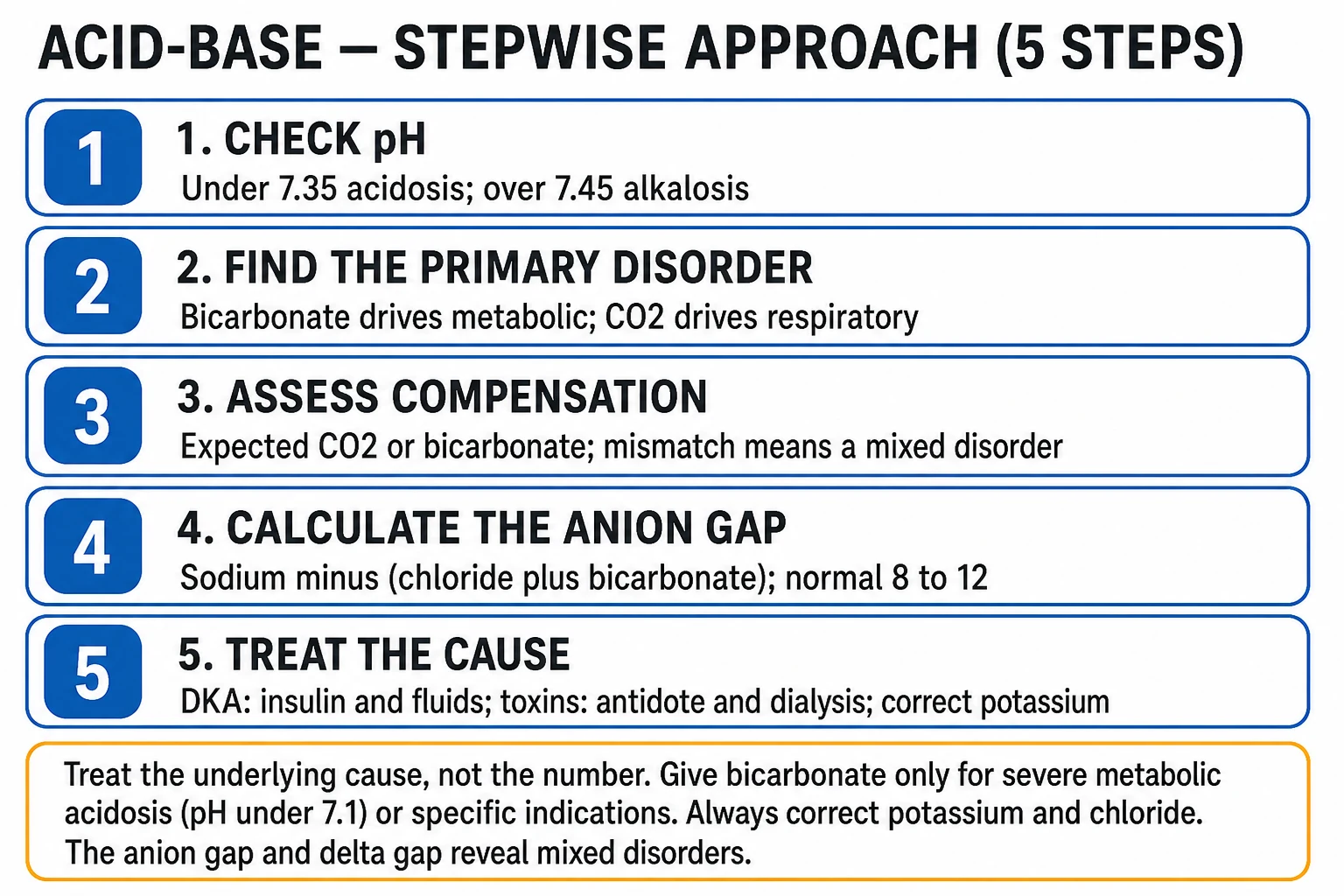
The over-arching principle of acid–base management is to treat the cause, not the number — the acid or base of a diabetic ketoacidosis, a lactic acidosis, or a renal-failure acidosis corrects when the underlying process is reversed. The specific interventions that have been tested in trials concern sodium bicarbonate and intravenous fluid composition.[9]
The two fluid and buffer trials
Sodium bicarbonate for severe metabolic acidaemia was tested in the BICAR-ICU trial (389 patients, Lancet 2018), which enrolled patients with a pH at or below 7.20. It found no significant difference in the primary composite outcome of death by day 28 and organ failure at day 7 (71 per cent control versus 66 per cent bicarbonate, P = 0.24), though a prespecified stratum with acute kidney injury showed a signal of benefit (day-28 survival 37 per cent versus 54 per cent, P = 0.028). Bicarbonate caused more metabolic alkalosis, hypernatraemia and hypocalcaemia, with no life-threatening complications. The synthesis is that bicarbonate is not routinely indicated for severe metabolic acidaemia outside the peri-arrest, the hyperkalaemic with ECG change, and the pre-dialysis settings; its possible role in the acidotic patient with acute kidney injury is the one nuance BICAR-ICU left open.[9]
The composition of intravenous fluid is an acid–base intervention in its own right, because chloride excess lowers the strong ion difference and causes a hyperchloraemic acidosis. The SMART trial (15 802 critically ill adults, NEJM 2018) found that balanced crystalloids (lactated Ringer's or Plasma-Lyte) modestly but significantly reduced a composite of death, new renal-replacement therapy and persistent renal dysfunction at 30 days compared with saline (14.3 per cent versus 15.4 per cent, OR 0.91, P = 0.04), and SALT-ED (13 347 non-critically ill adults) showed the same signal in the secondary kidney outcome (4.7 per cent versus 5.6 per cent, OR 0.82, P = 0.01). The practical implication is that balanced crystalloids are preferred over saline for most crystalloid resuscitation, both to avoid the hyperchloraemic acidosis and for a measurable renal benefit.[10][11]
Renal replacement therapy is the definitive treatment for the severe or refractory acidosis — it removes the acid anions and the causative toxins (lactate, methanol, ethylene glycol, salicylate, metformin) and corrects the acidosis without the volume, sodium and CO₂ load of bicarbonate. It is the treatment of choice for metformin-associated lactic acidosis, for the severe toxic-alcohol patient, and for the acidosis of acute kidney injury that has not responded to fluid and the correction of perfusion.[1]
Monitoring acid–base at the bedside
Acid–base monitoring in the ICU rests on the arterial blood gas — the pH, the PaCO₂, the bicarbonate or base excess, and the lactate — taken repeatedly to track the trend rather than a single value, and interpreted always with the electrolytes (for the anion gap) and the albumin (for the gap correction).[1][1]
- Frequency. A deteriorating or actively treated patient needs arterial gases at intervals short enough to track the response (often hourly); a stable patient, on a planned wean, less often. The trend is more informative than any single value.
- The venous gas. A central or peripheral venous gas gives a useful pH, bicarbonate and lactate when an arterial sample is not needed, with the caveat that the venous PCO₂ runs higher and the pH lower than arterial; it screens well but does not substitute for the arterial gas when the respiratory status matters.
- Point-of-care lactate. Lactate clearance (the fall over the first hours of resuscitation) is a marker of the adequacy of resuscitation in septic shock; a failure to clear is a poor prognostic sign and a prompt to look for ongoing hypoperfusion, a source, or an alternative cause.[5]
- The mixed disorders. In the complex patient, the delta-delta and the osmolar gap should be rechecked as the resuscitation proceeds, because an evolving second disorder (a dilutional or hyperchloraemic acidosis layered on a lactic one) is common and easily missed.[4]
Prognosis and common pitfalls
The prognosis of an acid–base disorder is the prognosis of its cause, but the severity and the trend of the acidaemia carry independent information: a severe metabolic acidaemia (pH below 7.1 to 7.2) is associated with increased mortality, and a failure of the lactate or the base deficit to clear portends a worse outcome independent of the diagnosis.[5]
The common pitfalls in interpretation are the ones the systematic method is designed to prevent:[1][4]
- Failing to correct the anion gap for albumin, so that a high-gap acidosis is missed in the hypoalbuminaemic ICU patient.
- Failing to apply the delta-delta, so that a concurrent metabolic alkalosis (elevating the bicarbonate) or a second normal-gap acidosis is overlooked.
- Misjudging the compensation — declaring a respiratory disorder "compensating" for a metabolic one without checking it against the expected value, and so missing a mixed disorder.
- Treating the number rather than the cause — giving bicarbonate for an acidaemia whose treatment is fluid, insulin, source control or dialysis.
- Forgetting that the toxic alcohols produce a dual gap — and that a normal osmolar gap does not exclude a late presentation once the parent has been metabolised. [1]
SAQ — The dual gap: toxic alcohol poisoning
10 minutes · 10 marks
A 39-year-old man is brought to the emergency department confused and ataxic, having been found unconscious next to an empty container of antifreeze. He is tachypnoeic. Arterial blood gas: pH 7.18, PaCO₂ 18 mmHg, HCO₃⁻ 7 mmol/L, base excess −19. Sodium 142, chloride 102, glucose 6.4, urea 5.1, albumin 38 g/L, measured osmolality 340 mOsm/kg. Urine microscopy shows envelope-shaped crystals. He has no visual symptoms.
SAQ — Mixed metabolic alkalosis delaying ventilator weaning
10 minutes · 10 marks
A 64-year-old woman, ventilated for ten days after emergency bowel surgery complicated by sepsis and acute kidney injury, is failing a spontaneous breathing trial. She has received furosemide 80 mg/day for volume overload and has a functioning nasogastric tube on free drainage. Arterial blood gas (FiO₂ 0.3): pH 7.55, PaCO₂ 49 mmHg, HCO₃⁻ 42 mmol/L, PaO₂ 88 mmHg. Sodium 148, potassium 2.9, chloride 88. Urine chloride is 12 mmol/L. The team asks whether to give acetazolamide.
Red flags
[1] [1]Densification notes for fellowship revision
This leaf is densified to the ICU fellowship gate standard (CICM / FFICM / EDIC): embedded SAQ practice, multi-figure visual scaffolding, examiner map alignment, and MCQ coverage of definition, mechanism, first-hour management, evidence, and traps. [1]
- Revision checkpoint 1: restate definition, one number examiners expect, and one absolute do-not-miss action.
- Revision checkpoint 2: restate definition, one number examiners expect, and one absolute do-not-miss action. [1]
References
- [1]Adrogué HJ, Madias NE. Management of life-threatening acid-base disorders. First of two parts N Engl J Med, 1998.PMID 9414329
- [2]Adrogué HJ, Madias NE. Management of life-threatening acid-base disorders. Second of two parts N Engl J Med, 1998.PMID 9420343
- [3]Emmett M. Stewart Versus Traditional Approach to Acid-Base Disorders Anesth Analg, 2016.PMID 27636585
- [4]Kraut JA, Madias NE. Serum anion gap: its uses and limitations in clinical medicine Clin J Am Soc Nephrol, 2007.PMID 17699401
- [5]Kraut JA, Madias NE. Lactic acidosis N Engl J Med, 2014.PMID 25494270
- [6]Kraut JA, Kurtz I. Toxic alcohol ingestions: clinical features, diagnosis, and management Clin J Am Soc Nephrol, 2008.PMID 18045860
- [7]Lynd LD, Richardson KJ, Purssell RA, et al. An evaluation of the osmole gap as a screening test for toxic alcohol poisoning BMC Emerg Med, 2008.PMID 18442409
- [8]Kitabchi AE, Umpierrez GE, Miles JM, Fisher JN. Hyperglycemic crises in adult patients with diabetes Diabetes Care, 2009.PMID 19564476
- [9]Jaber S, Paugam C, Futier E, et al.; BICAR-ICU Study Group. Sodium bicarbonate therapy for patients with severe metabolic acidaemia in the intensive care unit (BICAR-ICU): a multicentre, open-label, randomised controlled, phase 3 trial Lancet, 2018.PMID 29910040
- [10]Semler MW, Self WH, Wanderer JP, et al.; SMART Investigators and the Pragmatic Critical Care Research Group. Balanced Crystalloids versus Saline in Critically Ill Adults N Engl J Med, 2018.PMID 29485925
- [11]Self WH, Semler MW, Wanderer JP, et al.; SALT-ED Investigators. Balanced Crystalloids versus Saline in Noncritically Ill Adults N Engl J Med, 2018.PMID 29485926