ICU · Renal
Acute kidney injury and renal replacement therapy
Also known as Acute kidney injury (AKI) · AKI · Renal replacement therapy (RRT) · CRRT · Continuous venovenous haemofiltration (CVVH) · Haemodialysis · KDIGO classification · AKIKI trial
AKI is an abrupt reduction in kidney function (KDIGO: creatinine increase =26.5 umol/L in 48h, or =1.5x baseline, or urine output <0.5 mL/kg/hr for 6h). Classified Stage 1-3 by severity. Causes: pre-renal (most common, hypoperfusion), intrinsic (ATN, AIN, GN), post-renal (obstruction). Management: treat the cause, optimise haemodynamics, avoid nephrotoxins. RRT indications: AEIOU (Acidosis, Electrolyte, Ingestion, Overload, Uraemia). Timing: STARRT-AKI (2020): no benefit of early RRT. CRRT preferred in haemodynamically unstable.
On this page & tools
Your progress
Saved locally on this device.
Target exams
Red flags
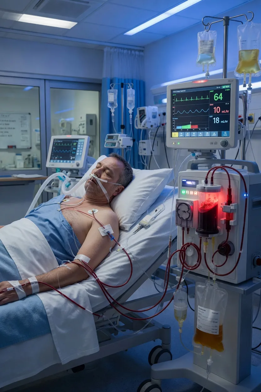
KDIGO definition and classification
The KDIGO 2012 classification defines AKI by creatinine and/or urine output criteria:[1]
KDIGO AKI staging (click each)
Stage 3 — Failure
Creatinine >=3x baseline OR >=354 umol/L OR RRT initiated. UO <0.3 mL/kg/hr for >=24h OR anuria for 12h. Severe injury — may require RRT.
Causes
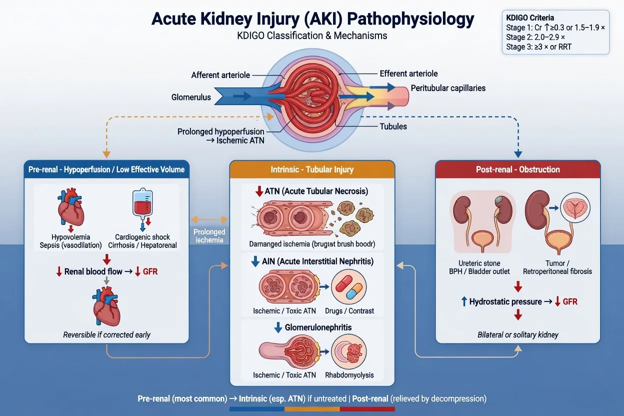
Pre-renal (most common)
Hypoperfusion — responsive to fluids
- Hypovolaemia (haemorrhage, dehydration, burns)
- Sepsis (vasodilation → effective hypovolaemia)
- Cardiogenic shock (low cardiac output)
- NSAIDs (afferent arteriolar constriction)
- ACE inhibitors/ARBs (efferent arteriolar dilation)
- Hepatorenal syndrome
Intrinsic
Kidney damage — structural
- ATN (most common intrinsic — ischaemic, nephrotoxic)
- AIN (allergic — penicillins, PPIs, NSAIDs)
- Glomerulonephritis
- Vascular (renal vein thrombosis, atheroembolism)
- Rhabdomyolysis (myoglobin toxicity)
- Tumour lysis syndrome (uric acid nephropathy)
Post-renal
Obstruction — exclude early
- BPH / prostate cancer
- Calculi (bilateral or single kidney)
- Retroperitoneal fibrosis
- Pelvic tumour (cervical, colorectal)
- Blocked urinary catheter (!)
- Bladder scan + ultrasound to exclude
Investigations
| Test | Finding | Significance |
|---|---|---|
| Urinalysis | Muddy brown casts → ATN; RBC casts → GN; WBC casts → AIN | Differentiates intrinsic causes |
| FENa (fractional excretion of Na) | <1% → pre-renal; >2% → intrinsic (ATN) | Less reliable with diuretics (use FEUrea) |
| FEUrea | <35% → pre-renal | Use if patient on diuretics |
| Urine Na | <20 mmol/L → pre-renal; >40 → ATN | Affected by diuretics and sepsis |
| CK | Elevated → rhabdomyolysis | Check if trauma, prolonged immobility, statins |
| Urate, phosphate, K | All elevated → tumour lysis syndrome | Especially after chemotherapy |
| Bedside bladder scan/US | Obstruction → post-renal | Exclude EARLY — easy reversible cause |
| Biomarkers (NGAL, KIM-1) | Elevated before creatinine rises | Research/emerging — not yet routine in ICU[8] |
AKI biomarkers
Creatinine and urine output are late and non-specific markers — creatinine only rises after ~50% GFR loss, lagging the actual injury by 24-48 h. Cell-stress biomarkers detect AKI at the subclinical stage (tubular stress before creatinine rises), enabling earlier recognition, prognostication, and avoidance of nephrotoxins.[8]
NGAL (neutrophil gelatinase-associated lipocalin)
Most studied — early tubular injury
- Small 25 kDa protein upregulated in distal tubule within 2-6 h of injury
- Plasma or urine; rises BEFORE creatinine (e.g. after CPB, contrast, sepsis)
- Useful for differential diagnosis: pre-renal vs intrinsic AKI (high in ATN)
- Limited specificity — also elevated in sepsis, inflammation, CKD, malignancy
- Predicts AKI severity, need for RRT, and mortality
Cystatin C
Freely filtered GFR marker
- Cysteine protease inhibitor produced at constant rate by all nucleated cells
- Freely filtered, fully reabsorbed and catabolised by proximal tubule
- Detects GFR fall EARLIER than creatinine (less muscle-mass bias)
- Better predictor of AKI than creatinine at 24 h before clinical diagnosis
- NOT a damage marker — a GFR marker (complements NGAL/KIM-1)
[TIMP-2]·[IGFBP7] (NephroCheck)
Cell-cycle arrest markers
- Two proteins released by tubular cells entering G1 cell-cycle arrest (stress response)
- FDA-cleared bedside urinary test; results in ~20 min
- Cut-off >0.3 = high risk of KDIGO Stage 2-3 AKI within 12 h (Sapphire study)
- Best NEGATIVE predictive value — a low value effectively rules out imminent moderate-severe AKI
- Useful to identify patients who should NOT receive contrast or nephrotoxins
KIM-1 (kidney injury molecule-1)
Proximal tubule-specific
- Type I transmembrane protein; undetectable in normal kidney
- Upregulated specifically in injured proximal tubule (ATN, AIN)
- High specificity for intrinsic (parenchymal) vs pre-renal AKI
- Correlates with severity and recovery potential
L-FABP / proenkephalin
Emerging
- L-FABP: marker of oxidative tubular stress
- Proenkephalin: stable surrogate of GFR independent of muscle mass
- Both under investigation; not yet routine in ICU practice
Management: pre-RRT
AKI management principles (KDIGO)
Identify and treat the cause
Sepsis → antibiotics + source control. Hypovolaemia → fluid resuscitation. Obstruction → catheter/nephrostomy. Nephrotoxin → STOP the drug. Rhabdomyolysis → aggressive fluid + bicarbonate.
Optimise haemodynamics
Ensure adequate MAP (>=65, higher if chronic HTN). Use noradrenaline for vasodilatory shock. Avoid excessive fluid — assess responsiveness. Target cardiac output that provides adequate renal perfusion.
Stop ALL nephrotoxins
NSAIDs (constrict afferent arteriole). ACEi/ARBs (dilate efferent arteriole). Aminoglycosides. Vancomycin (trough monitoring). Iodinated contrast (use lowest dose, pre-hydrate). Statins (rhabdomyolysis risk). Metformin (lactic acidosis risk).
Manage fluid balance
Assess volume status clinically + bedside US (IVC). If hypovolaemic → give balanced crystalloid boluses. If euvolaemic → maintain even balance. If hypervolaemic → fluid restriction + consider diuretics (do NOT use diuretics to "treat" AKI — only for fluid overload).
Monitor electrolytes
Daily K, phosphate, magnesium. Hyperkalaemia (K+ >6.0): calcium gluconate (membrane stabilisation) → insulin/dextrose → salbutamol → RRT if refractory. Hyperphosphataemia: phosphate binders.
Review medications
Renal dose adjustment for ALL medications. Use electronic renal dosing calculator. Vancomycin: trough-based dosing. Aminoglycosides: extended-interval dosing with level monitoring.
Nutrition
Enteral nutrition preferred. Do NOT restrict protein in AKI (target 1.5 g/kg/day — KDIGO). Protein restriction does NOT prevent AKI progression but worsens outcomes.
Monitor for complications
Fluid overload (pulmonary oedema), hyperkalaemia, metabolic acidosis, uraemia (platelet dysfunction, pericarditis, encephalopathy), infection risk.
AKI in sepsis (septic AKI)
Septic AKI is the most common AKI in ICU — about half of all ICU AKI cases and occurs in up to 60% of septic shock. It carries the highest mortality of any AKI subtype. Septic AKI is NOT simply "pre-renal" from hypoperfusion — it is a complex inflammatory, microcirculatory, and metabolic phenotype that can occur with preserved or even increased renal blood flow.[16][19]
Pathophysiology — why the kidney fails in sepsis
- Microvascular dysfunction: glomerular and peritubular capillary leak, leukocyte adhesion, endothelial swelling → heterogeneous cortical perfusion despite normal global renal blood flow.[19]
- Mitochondrial dysfunction / cell-cycle arrest: tubular cells "shut down" to conserve energy — a protective, not destructive, response. Apoptosis occurs only at extreme injury (analogous to hibernating myocardium in ischaemia).
- Inflammatory mediators: TNF-α, IL-6, HMGB1, and damage-associated molecular patterns (DAMPs) cause direct tubular toxicity.
- Macrocirculatory changes: vasodilation, venous pooling, low effective circulating volume, low mean arterial pressure.
- Concurrent nephrotoxic burden: vancomycin, aminoglycosides, contrast for imaging, hydroxyethyl starch (avoided).
Diagnosis
How septic AKI differs
- FENa often >2% even early — classic "pre-renal" urine indices are UNRELIABLE in sepsis
- Urinalysis often bland or shows granular casts; may mimic ATN
- Biomarkers (NGAL, [TIMP-2]·[IGFBP7]) elevated early — distinguish from pure pre-renal
- Occurs on top of vasoplegia, hyperlactataemia, multi-organ failure
- Urine microscopy score (Bagshaw) higher in septic ATN vs pre-renal picture
Management priorities
Sepsis bundle + kidney-specific
- **Source control + broad-spectrum antibiotics within 1 h** (Surviving Sepsis) — the single most important intervention
- Balanced crystalloid 30 mL/kg for hypotension or lactate ≥4 (but BEWARE fluid overload — see below)
- **Noradrenaline first-line** to MAP ≥65 mmHg. Vasopressin 0.03 U/min add-on.
- Do NOT delay vasopressors for "complete" fluid resuscitation — early vasopressors may PROTECT the kidney by restoring renal perfusion pressure
- Stop ALL nephrotoxins. Renal-dose adjust antibiotics (vancomycin AUC-guided, beta-lactams extended infusion)
- Avoid hydroxyethyl starch (CHEST/6S trials — increased RRT need). Use albumin only if needed for shock
- De-resuscitate once shock resolves — late positive fluid balance independently predicts AKI non-recovery and mortality
Contrast-associated AKI (CA-AKI)
Previously "contrast-induced nephropathy (CIN)". Defined as creatinine rise ≥26.5 umol/L (0.3 mg/dL) or ≥1.5x baseline within 48-72 h of iodinated contrast, with no other cause.[9][17]
Risk stratification and prevention
Identify high-risk patients
eGFR <30 (highest risk), diabetes, age >75, CKD, AKI at time of contrast, heart failure, hypovolaemia, multiple myeloma, concurrent nephrotoxins, high-osmolar or large contrast volume (>100 mL).
Pre-procedure (elective)
Hold nephrotoxins 24-48 h (NSAIDs, metformin, aminoglycosides). Hold diuretics if possible. Use low- or iso-osmolar non-ionic contrast at the lowest dose that gives diagnostic images. Consider alternative imaging (ultrasound, MRI without contrast).
Hydration — the only proven prevention
Isotonic saline 1 mL/kg/hr for 6-12 h before and after (or 3 mL/kg/hr for 1 h pre + 4-6 h post for day-case). Sodium bicarbonate is NOT superior to saline (PRESERVE trial, n=5177, NEJM 2018 — no benefit of bicarbonate or NAC over saline).<Cite id="17" />
N-acetylcysteine — no longer recommended
PRESERVE trial showed NO benefit of NAC over placebo. Do NOT use it as the sole preventative measure. Avoid relying on it.
In ICU/emergency
If AKI already present and contrast is essential (e.g. CT pulmonary angiogram for PE), do NOT withhold contrast solely for fear of CA-AKI — modern iso-osmolar agents and low-volume protocols carry minimal incremental risk. Document the risk-benefit discussion. Do NOT delay life-saving imaging.
Post-procedure monitoring
Check creatinine at 48-72 h. Most CA-AKI peaks at day 3 and recovers within 7-10 days. Persistent rise or oliguria → nephrology input, assess for RRT indication (rare).
PRESERVE
NEJM 2018
5177 pts with CKD undergoing angiography — 2×2 factorial: isotonic saline vs sodium bicarbonate AND NAC vs placebo
Key finding
No difference in death, need for dialysis, or persistent renal impairment at 90 days across all four groups. NAC and bicarbonate both ineffective.
Practice change
Use isotonic saline alone — drop routine NAC and bicarbonate
Renal replacement therapy
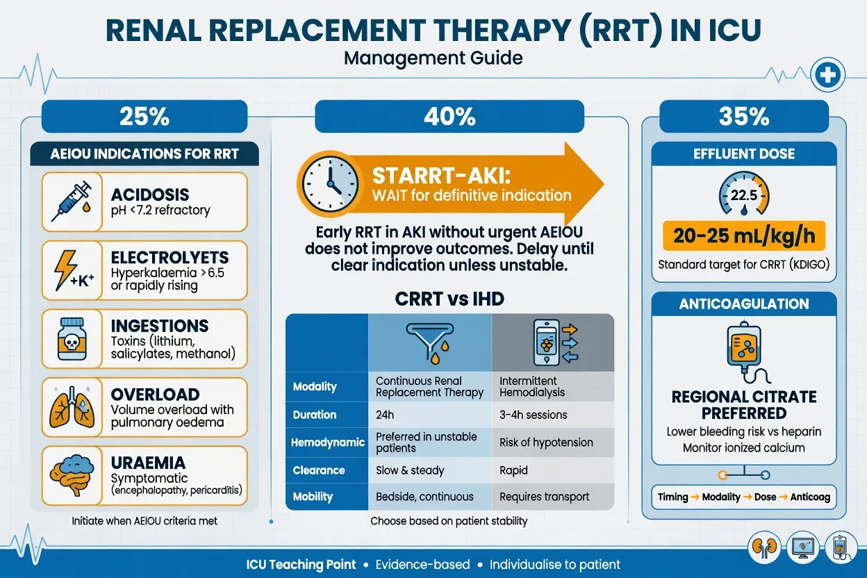
Indications — "AEIOU"
[1]Timing of RRT initiation
The three major RCTs:[2][3][4]
AKIKI
NEJM 2016
620 pts AKI Stage 3 (KDIGO) — early RRT vs delayed (until indication)
Key finding
No difference in 60-day mortality (48.5% early vs 49.7% delayed). Delayed group: 51% never needed RRT.
Practice change
Waiting for an indication is acceptable — do NOT start RRT solely for AKI Stage 3
ELAIN
JAMA 2016
231 pts AKI Stage 2 (with NGAL elevation) — early RRT vs delayed
Key finding
Trend toward lower 90-day mortality (39.3% vs 54.7%, p=0.11). Fewer complications with early.
Practice change
Suggested early RRT for Stage 2 with biomarker elevation — but underpowered
STARRT-AKI
NEJM 2020
3019 pts AKI Stage 2/3 — early RRT (within 12h) vs delayed (until indication)
Key finding
No difference in 90-day mortality (43.9% vs 43.7%). Early group had MORE adverse events (hypotension, bleeding, catheter infection).
Practice change
Definitive evidence: do NOT start early RRT — wait for an indication (AEIOU)
BICAR-ICU
Lancet 2018
389 pts with severe metabolic acidaemia (pH ≤7.20, HCO3 ≤20) — 8.4% sodium bicarbonate vs control
Key finding
No overall mortality benefit. Pre-specified subgroup with AKIN Stage 2 AKI had LOWER RRT need (35% vs 51%, p=0.03). Hypernatraemia common adverse effect.
Practice change
Consider bicarbonate in severe acidaemia with moderate-severe AKI to defer RRT — but no overall survival benefit
RENAL
NEJM 2009
1464 pts on CRRT — post-dilution CVVHDF 25 vs 40 mL/kg/hr effluent dose
Key finding
No difference in 90-day mortality (44.7% vs 44.7%). Higher dose did not improve outcomes.
Practice change
Standard CRRT dose = 25 mL/kg/hr (deliver 20-25); higher doses not beneficial
ATN (VA/NIH)
NEJM 2008
1124 pts — high-intensity (IHD/SLED 6×/wk or CVVHDF 35 mL/kg/hr) vs low-intensity (3×/wk or 20 mL/kg/hr)
Key finding
No difference in 60-day mortality (53.6% vs 51.5%) or RRT independence at day 28.
Practice change
Confirmed no benefit of higher RRT intensity across modalities
STARRT-AKI fluid substudy
Crit Care 2022
Secondary analysis of STARRT-AKI (n=2727) — cumulative fluid balance at day 3 vs RRT and mortality outcomes
Key finding
Higher cumulative fluid balance independently associated with adverse outcomes including mortality and dialysis dependence at 90 days, regardless of RRT timing strategy.
Practice change
Aggressive fluid de-resuscitation in AKI is as important as RRT timing
Bottom line: Do NOT initiate RRT solely for AKI Stage 3 without a clinical indication. Wait for one of the AEIOU criteria. Early RRT increases complications without survival benefit. [1]
RRT modalities
CRRT (CVVH/CVVHDF)
Continuous — for unstable
- Preferred in haemodynamically unstable patients
- Smooth solute clearance (less osmotic shift)
- Better fluid balance control
- Less risk of cerebral oedema (no rapid solute shift)
- Dose: 20-25 mL/kg/hr effluent rate
- Anticoagulation: citrate preferred (regional, lower bleeding risk)
- Disadvantage: requires continuous anticoagulation, immobilised
IHD (Intermittent)
For stable patients
- Preferred for haemodynamically stable patients
- Faster solute clearance (3-4 hour session)
- Less anticoagulation needed
- Patient can mobilise between sessions
- Higher risk of hypotension during session
- Rapid solute shift → disequilibrium syndrome risk
SLED
Hybrid — slow extended
- Slow Low-Efficiency Dialysis (6-12 hour sessions)
- Combines advantages of CRRT and IHD
- Less haemodynamic instability than IHD
- Less resource-intensive than CRRT
- Good option for resource-limited ICUs
CRRT settings
| Parameter | Target | Notes |
|---|---|---|
| Dose (effluent rate) | 20-25 mL/kg/hr | AKIKI/RENAL trials: no benefit of higher dose. Do NOT count downtime — deliver the prescribed dose.[6] |
| Blood flow | 150-200 mL/min | Adjust to maintain circuit life and patient stability |
| Dialysate/replacement fluid | Bicarbonate-based | Preferred over lactate-based (less metabolic issues) |
| Anticoagulation | Regional citrate | Preferred — lower bleeding risk. Monitor total/ionised Ca ratio. Contraindicated in severe liver dysfunction. Alternative: unfractionated heparin.[5] |
| Fluid removal (net ultrafiltration) | Individualised | Aim for even-to-negative balance in fluid-overloaded patients. Monitor haemodynamics during fluid removal. |
Complications of AKI
| Complication | Recognition | Management |
|---|---|---|
| Hyperkalaemia | K+ >6.0, peaked T waves, wide QRS | Calcium gluconate → insulin/dextrose → salbutamol → RRT if refractory |
| Metabolic acidosis | pH <7.2, low HCO3, high lactate | Treat the cause. Bicarbonate controversial (BICAR-ICU: no overall benefit). RRT if pH <7.15.[7] |
| Fluid overload | Pulmonary oedema, tissue oedema | Diuretics (furosemide) for fluid removal. Do NOT use diuretics to "treat" AKI. RRT if refractory. |
| Uraemia | Platelet dysfunction, pericarditis, encephalopathy | Initiate RRT. |
| Infection | Nosocomial infection, line sepsis | High infection risk due to uraemic immunosuppression. Aseptic technique for line insertion. |
| Bleeding | Uraemic platelet dysfunction | Desmopressin (DDAVP) for uraemic bleeding. Avoid unnecessary anticoagulation. |
Prognosis
AKI outcomes
- Prognostic factors: age, comorbidities (diabetes, CKD baseline), cause of AKI, severity (Stage 1/2/3), need for RRT, fluid overload, sepsis
- Recovery: most patients who survive recover renal function within 90 days; ~10% remain dialysis-dependent
- Long-term: AKI survivors are at increased risk of CKD progression, cardiovascular events, and recurrent AKI [1]
Exam practice
SAQ — AKI with refractory hyperkalaemia
10 minutes · 10 marks
A 65-year-old man in ICU with urosepsis has developed AKI. Creatinine has risen from 110 to 340 umol/L over 48 hours. Urine output 120 mL in the last 12 hours. K+ 6.8 mmol/L with peaked T waves and widened QRS on ECG. BP 95/60 on noradrenaline 0.2 mcg/kg/min. pH 7.28, HCO3 16.
SAQ — Septic AKI and fluid balance
10 minutes · 10 marks
A 72-year-old woman with community-acquired pneumonia is admitted to ICU with septic shock. BP 80/45, lactate 4.2 mmol/L, creatinine 95 umol/L (baseline 70). After 3 L of crystalloid over 6 h she requires noradrenaline 0.25 mcg/kg/min for MAP 70. On day 3 she is 6 L in positive balance, anuric, creatinine 280 umol/L, K+ 5.8 mmol/L, pH 7.25.
SAQ — Timing of RRT initiation in severe AKI
10 minutes · 10 marks
A 68-year-old man is admitted to ICU with pancreatitis and oliguric KDIGO Stage 3 AKI. Creatinine has risen from 90 to 410 umol/L over 4 days, urine output 180 mL/24h, K+ 5.6 mmol/L, pH 7.30, HCO3 17, and he is 3 L in positive balance with bibasal crackles that respond to furosemide. He is normotensive on low-dose noradrenaline 0.05 mcg/kg/min. The registrar asks whether to start CRRT now to 'stay ahead of the curve'.
SAQ — Contrast-associated AKI prevention
10 minutes · 10 marks
A 74-year-old woman with type 2 diabetes, baseline creatinine 180 umol/L (eGFR ~25 mL/min/1.73m²), heart failure, and on ramipril and furosemide requires an urgent CT pulmonary angiogram for suspected massive PE. She is currently oliguric (UO 0.3 mL/kg/hr) and the radiologist asks whether to administer IV iodinated contrast.
Clinical pearls
Red flags
References
- [1]KDIGO Acute Kidney Injury Work Group. KDIGO clinical practice guidelines for acute kidney injury Nephron Clin Pract, 2012.PMID 22890468
- [2]Gaudry S, Hajage D, Schortgen F, et al. Initiation Strategies for Renal-Replacement Therapy in the Intensive Care Unit N Engl J Med, 2016.PMID 27181456
- [3]Sch Schneider A, Bellomo R, Bagshaw SM, et al. Effect of Early vs Delayed Initiation of Renal Replacement Therapy on Mortality in Critically Ill Patients With Acute Kidney Injury: The ELAIN Randomized Clinical Trial JAMA, 2016.PMID 27209269
- [4]The STARRt-AKI Investigators. Timing of Initiation of Renal-Replacement Therapy in Acute Kidney Injury N Engl J Med, 2020.PMID 32668114
- [5]Ronco C, Bellomo R, Kellum JA. Continuous Renal Replacement Therapy: Who, When, Why, and How Chest, 2019.PMID 30266628
- [6]Schmidt GA, Kjaergaard J, Wetterslev J, et al. Renal replacement therapy intensity for acute kidney injury and recovery to dialysis independence: a systematic review and individual patient data meta-analysis Nephrol Dial Transplant, 2018.PMID 29186517
- [7]Hoste EA, Saraiva-Antunes R, De Corte A, et al. Management of acute metabolic acidosis in the ICU: sodium bicarbonate and renal replacement therapy Crit Care, 2021.PMID 34461963
- [8]Ostermann M, Zarbock A, Goldstein S, et al. Acute Kidney Injury: Biomarker-Guided Diagnosis and Management Medicina (Kaunas), 2022.PMID 35334515
- [9]Lenoir L, Ntsekas M, Brajadenta J, et al. [Prevention of contrast-induced nephropathy] Rev Med Liege, 2024.PMID 38869133
- [10]Tamma PD, Connelly S, Senchyna F, et al. Efficacy and safety of sulbactam-durlobactam versus colistin for the treatment of patients with serious infections caused by Acinetobacter baumannii-calcoaceticus complex: a multicentre, randomised, active-controlled, phase 3, non-inferiority clinical trial (ATTACK) Lancet Infect Dis, 2023.PMID 37182534
- [11]Jaber S, Paissant A, Pradel G, et al. Sodium bicarbonate therapy for patients with severe metabolic acidaemia in the intensive care unit (BICAR-ICU): a multicentre, open-label, randomised controlled, phase 3 trial Lancet, 2018.PMID 29910040
- [12]RENAL Replacement Therapy Study Investigators, Bellomo R, Cass A, et al. Intensity of continuous renal-replacement therapy in critically ill patients N Engl J Med, 2009.PMID 19846848
- [13]VA/NIH Acute Renal Failure Trial Network, Palevsky PM, Zhang JH, et al. Intensity of renal support in critically ill patients with acute kidney injury N Engl J Med, 2008.PMID 18492867
- [14]Kashani K, Al-Khafaji A, Ardiles T, et al. Discovery and validation of cell cycle arrest biomarkers in human acute kidney injury Crit Care, 2013.PMID 23388612
- [15]Bagshaw SM, Haase M, Haase-Fielitz A, et al. A prospective evaluation of urine microscopy in septic and non-septic acute kidney injury Nephrol Dial Transplant, 2012.PMID 21669886
- [16]Mehta RL, Bouchard J, Soroko SB, et al. Sepsis as a cause and consequence of acute kidney injury: Program to Improve Care in Acute Renal Disease Intensive Care Med, 2011.PMID 21152901
- [17]Weisbord SD, Gallagher M, Jneid H, et al. Outcomes after Angiography with Sodium Bicarbonate and Acetylcysteine N Engl J Med, 2018.PMID 29130810
- [18]Wald R, Bagshaw SM, Adhikari NKJ, et al. Fluid balance and renal replacement therapy initiation strategy: a secondary analysis of the STARRT-AKI trial Crit Care, 2022.PMID 36424662
- [19]Prowle JR, Ishikawa K, Ligons EV, et al. Measurement of renal blood flow by phase-contrast magnetic resonance imaging during septic acute kidney injury: a pilot investigation Crit Care Med, 2012.PMID 22487999