ICU · Renal / RRT
CRRT — Circuit, Prescription, Anticoagulation & Troubleshooting
Also known as Continuous renal replacement therapy · CRRT circuit · CRRT prescription · CRRT anticoagulation · Citrate anticoagulation · Regional citrate · Filtration fraction · Pre-dilution · Post-dilution · CRRT troubleshooting · Circuit clotting · Access pressure · Transmembrane pressure
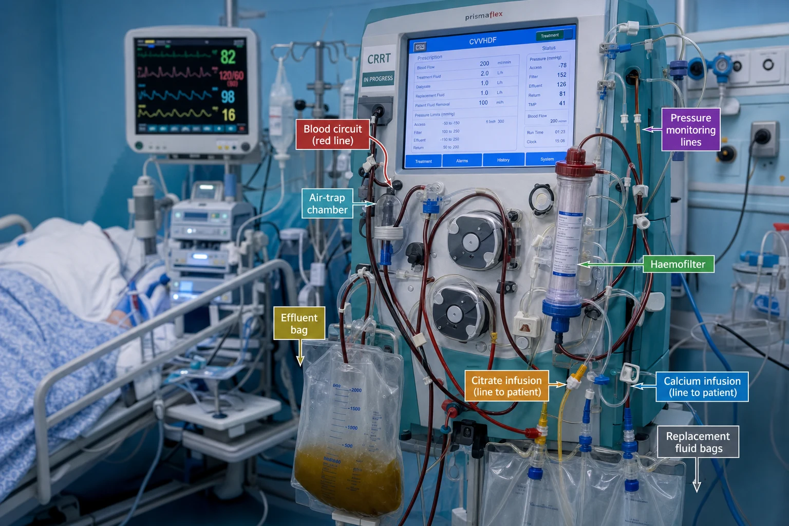
The CRRT is delivered through a blood circuit — the dual-lumen central access, the blood pump, the pre-dilution (the replacement fluid before the filter), the haemofilter (the hollow-fibre membrane), the post-dilution (the replacement fluid after the filter), the air-trap, and the return. The pressures (the access negative, the return positive, the filter transmembrane) monitor the circuit patency and the clotting. The prescription: the modality (the CVVH, the CVVHDF), the dose (the effluent 20 to 25 mL/kg/h), the blood flow, the fluid balance, the fluid composition, and the anticoagulation. The anticoagulation is the regional citrate (the preferred; the post-filter ionized calcium 0.25 to 0.4 mmol/L; the total-to-ionized ratio under 2.5; the risks the metabolic alkalosis, the acidosis in the liver failure, the hypercalcaemia) or the heparin or none. The troubleshooting: the circuit clotting (the inadequate anticoagulation, the high filtration fraction over 25 per cent, the low blood flow), the high access pressure (the catheter kink or the position), the air-in-line, and the electrolyte and the nutrient loss (replace the K, the phosphate, the Mg, the increased nutrition).
On this page & tools
Your progress
Saved locally on this device.
8 MCQs with explanations
Target exams
Overview & definition
The continuous renal replacement therapy (CRRT) is delivered through a blood circuit that runs continuously (the 24 hours) at the bedside. The circuit, the prescription, the anticoagulation, and the troubleshooting are the four operational domains. The goal is the efficient, the safe, the uninterrupted solute and fluid removal, with the haemodynamic stability and the minimal clotting.[1]
The circuit
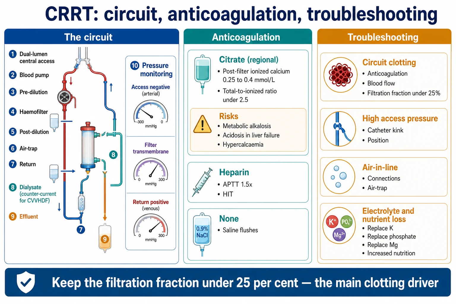
The blood circuit runs:[1]
- The dual-lumen central venous catheter (the access) — the right internal jugular or the femoral (the subclavian avoided for the stenosis risk).[1]
- The blood pump — the roller pump, the 150 to 200 mL/min.[1]
- The pre-dilution — the replacement fluid infused BEFORE the filter (the lower haematocrit, the less clotting, the less efficient).[1]
- The haemofilter — the hollow-fibre, the semi-permeable membrane (the 1.0 to 1.5 m² surface area).[1]
- The post-dilution — the replacement fluid infused AFTER the filter (the more efficient, the higher filtration fraction, the more clotting).[1]
- The air-trap and the return.[1]
- The dialysate (for the CVVHDF) — flows counter-current to the blood.[1]
- The effluent — the ultrafiltrate plus the dialysate (the waste).[1]
The pressure monitoring
- The access pressure (the negative) — the blood drawn from the patient; a high negative value suggests the catheter kinking or the inadequate flow.[1]
- The return pressure (the positive) — the blood returned to the patient; a high value suggests the return-line obstruction or the air.[1]
- The filter pressure (the transmembrane pressure) — the pre-filter minus the post-filter; a rising pressure drop suggests the filter clotting.[1]
- The effluent pressure.[1]
The prescription
- The modality — the CVVH, the CVVHDF (the commonest).[1]
- The dose (the effluent rate) — the 20 to 25 mL/kg/h (the ATN and the RENAL trials — the higher dose no benefit).[1]
- The blood flow — the 150 to 200 mL/min.[1]
- The dialysate flow (for the CVVHDF) — matches or exceeds the effluent target.[1]
- The pre- vs the post-dilution — the pre-dilution is the less efficient (the dilution of the blood before the filter) but the less clotting; the post-dilution is the more efficient but the higher filtration fraction and the clotting. The filtration fraction (the ultrafiltrate divided by the plasma flow) should be under 25 per cent.[1]
- The fluid balance (the net ultrafiltration) — the hourly fluid-removal target, titrated to the haemodynamic tolerance.[1]
- The fluid composition — the bicarbonate-buffered; the electrolyte (the K, the phosphate, the Mg) adjusted.[1]
- The anticoagulation.[1]
The anticoagulation
Regional citrate (the preferred)
- The citrate infused pre-filter chelates the calcium in the circuit (the local anticoagulation); the calcium infused systemically (to replace). The systemic anticoagulation is avoided (the bleeding risk reduced).[1]
- The monitoring: the post-filter ionized calcium 0.25 to 0.4 mmol/L (the anticoagulated circuit), the systemic ionized calcium normal, the total calcium (the total-to-ionized ratio under 2.5 to detect the citrate accumulation).[1]
- The risks: the metabolic alkalosis (the citrate metabolized to the bicarbonate — the commonest), the metabolic acidosis with the high anion gap (the citrate accumulation in the severe liver failure — the impaired metabolism), and the hypercalcaemia.[1]
- In the severe liver failure, switch to the heparin or the no-anticoagulation (the citrate accumulation).[1]
Heparin
- The bolus plus the infusion; the APTT monitoring (1.5 times the normal); the bleeding risk; the heparin-induced thrombocytopenia (the HIT).[1]
No anticoagulation
- For the high-bleeding-risk patient. The saline flushes (the pre-filter boluses every 30 minutes); the shorter circuit life; the higher blood flow to reduce the clotting.[1]
The troubleshooting
- The circuit clotting (the rising filter pressure drop, the darkening filter, the low effluent) → the inadequate anticoagulation, the high filtration fraction (over 25 per cent), the low blood flow, the catheter position. Check the anticoagulation, the pre/post-dilution ratio, the blood flow, and the access.[1]
- The high access pressure (the high negative) → the catheter kinking, the against-the-wall position, the inadequate flow. Reposition the patient, the flush the line, the check the access.[1]
- The high return pressure → the kinked return line, the air, the venous obstruction.[1]
- The air-in-line alarm → the air in the circuit. Check the connections, the air-trap, the re-prime.[1]
- The recurrent clotting → the anticoagulation review, the catheter position, the blood-flow rate, the filtration fraction (under 25 per cent), the haematocrit.[1]
- The hypothermia (the extracorporeal heat loss) → the blood warmer.[1]
- The electrolyte depletion (the K, the phosphate, the Mg) → the replacement (the CRRT clears them).[1]
- The nutrient loss (the amino acids, the water-soluble vitamins, the glucose absorption from the dialysate) → the supplementation and the increased nutrition.[1]
Red flags
The anticoagulation decision tree — the framework
The choice of the anticoagulation is the single biggest modifiable determinant of the filter life after the blood flow and the filtration fraction. The decision balances three forces: the bleeding risk of the patient, the clotting risk of the circuit, and the metabolic capacity of the patient (chiefly the liver, for the citrate metabolism).[5]
The practical algorithm: [1]
- Start with the regional citrate unless contraindicated — it gives the longest filter life at the lowest bleeding risk in the great majority of patients.[1][3]
- Switch to the unfractionated heparin when the citrate is contraindicated (the severe liver failure, the severe lactic acidosis, the persistent citrate accumulation).[5]
- Use the no-anticoagulation when the bleeding risk is extreme (the active bleeding, the recent neurosurgery, the coagulopathy) and only with the high blood flow and the pre-dilution.[16]
- Reserve the nadroparin, the epoprostenol, the nafamostat for the niche indications — the recurrent filter clotting despite the standard anticoagulation, the HIT, the hepatic failure where the citrate fails.[5][14]
The KDIGO summary, the French expert panel, and the Cochrane meta-analysis all converge: the regional citrate is the first-line for the CRRT anticoagulation in the great majority of patients.[7][3][18]
Unfractionated heparin (UFH) — the standard
The unfractionated heparin (UFH) was the standard anticoagulant for the CRRT for decades, and remains the workhorse where the citrate is contraindicated. It potentiates the antithrombin III to inhibit the thrombin (the factor IIa) and the factor Xa, producing a systemic anticoagulant effect.[5]
The dosing
- The bolus 30 to 50 units/kg, then the infusion 5 to 10 units/kg/h, titrated to the target.[5]
- The target ranges differ by the centre; the commonest is the aPTT 1.5 to 2.0 times the control (around 60 to 85 seconds).[1]
- An alternative, increasingly preferred, target is the anti-Xa level 0.25 to 0.35 IU/mL — it is unaffected by the acquired antithrombin deficiency (common in the septic ICU patient) and avoids the over-anticoagulation that the aPTT falsely flags as therapeutic.[5]
The monitoring
- The aPTT every 6 hours until stable, then every 12 hours. The aPTT is cheap and universal but is influenced by the factor VIII elevation (the acute-phase reactant — the false-low aPTT, the apparent heparin "resistance") and the antithrombin deficiency (the false-high, the bleeding without the protection).[5]
- The anti-Xa is more accurate in the critically ill, particularly in the sepsis and the liver disease, where the aPTT is unreliable. Monitor every 6 to 12 hours; aim for 0.25 to 0.35 IU/mL.[5]
- The platelet count every 2 to 3 days from day 4 to day 14 to screen for the heparin-induced thrombocytopenia (HIT).[9]
The complications
- The bleeding (the major drawback — the 4 to 10 per cent major bleeding in the historical series, the chief reason the citrate displaced it).[3]
- The heparin-induced thrombocytopenia (HIT) — the IgG against the platelet factor 4 (the PF4-heparin complex); the platelet fall of more than 50 per cent, or to under 100 × 10⁹/L, between days 5 and 14; the paradoxical thrombosis (the venous more than the arterial, the 30 to 50 per cent thrombosis risk in the first 30 days). The absolute contraindication to all heparins (the UFH, the LMWH) — switch to the citrate or the no-anticoagulation.[9]
- The antithrombin depletion in the sepsis and the cirrhosis — the heparin fails to anticoagulate despite the rising dose, because there is no antithrombin to potentiate; the heparin "resistance".[5]
Regional citrate anticoagulation (RCA) — the preferred deep dive
The regional citrate anticoagulation (RCA) is the preferred anticoagulation for the CRRT in the great majority of patients — it gives the longer filter life at the lower bleeding risk than the heparin, because the anticoagulation is confined to the circuit and is neutralised by the systemic calcium reinfusion.[1][4]
The mechanism — calcium chelation
- The trisodium citrate is infused pre-filter (into the access line). The citrate chelates the ionised calcium in the circuit blood, dropping the post-filter ionised calcium to 0.25 to 0.40 mmol/L — at this level the clotting cascade is arrested (the factor IXa, the Xa, the thrombin all require the ionised calcium as a cofactor).[4]
- The calcium-citrate complex is small enough to pass through most filters (it is lost in the effluent in the post-filter mode), so the systemic ionised calcium tends to fall and must be replaced by a separate systemic calcium infusion into the return line.[4]
- A portion of the citrate (about 20 to 30 per cent) reaches the patient and is metabolised to the bicarbonate (one citrate yields three bicarbonate equivalents) — chiefly in the liver, the skeletal muscle, and the renal cortex. This is the basis of the metabolic alkalosis and the basis of the contraindication in the hepatic failure.[6]
The two calcium targets — the bedrock of safe RCA
- The post-filter ionised calcium 0.25 to 0.40 mmol/L — measures the anticoagulation inside the circuit. Too high → the filter clots. Too low → no extra benefit and a risk of the hypocalcaemia if the systemic calcium infusion is insufficient.[4]
- The systemic (patient) ionised calcium 1.10 to 1.30 mmol/L — measures the calcium balance of the patient. Maintain by titrating the systemic calcium infusion; raise the rate if the systemic iCa falls.[4]
- Some modern protocols use the calcium-containing replacement/dialysate solutions (the Rhee 2021 approach) — the calcium in the fluid reduces or eliminates the need for the separate systemic calcium infusion, simplifying the circuit.[17]
The citrate accumulation monitor — the total-to-ionised calcium ratio
- The total-to-ionised calcium ratio (the CaT/CaI ratio) is the bedside marker of the citrate accumulation. The normal ratio is under 2.0. A ratio over 2.5 (with the rising total calcium and the falling or stable ionised calcium) signals the citrate accumulation — the citrate-bound calcium accumulates while the liver cannot metabolise it.[6]
- Check the ratio every 6 hours initially, every 12 hours once stable, and immediately if the metabolic acidosis worsens.[4][6]
The metabolic complications
- The metabolic alkalosis (the commonest derangement) — every citrate metabolised yields three bicarbonate molecules; over-metabolism (or a high citrate dose with a normal liver) pushes the bicarbonate up and the pH up. Manage by reducing the citrate dose, increasing the dialysate chloride (or the normal-saline replacement), or switching a portion of the dialysate to the 0.45 per cent saline.[6]
- The metabolic acidosis with the high anion gap — the citrate accumulation in the hepatic failure or the severe shock-lactic-acidosis. The citrate cannot be metabolised, so it accumulates as an unmeasured anion. A falling pH, a rising lactate, a rising CaT/CaI ratio over 2.5 — switch off the citrate and convert to the heparin or the no-anticoagulation.[6]
- The hypercalcaemia — the total calcium rises (the citrate-bound fraction) while the ionised calcium stays normal or low; the bio-available calcium is normal but the total is misleadingly high. Do not treat the high total calcium — treat the ionised.[6]
- The hypocalcaemia — the inadequate systemic calcium reinfusion; the tetany, the arrhythmia, the hypotension. Always check the ionised calcium after the circuit change (the new circuit may have a different calcium requirement).[6]
The absolute and relative contraindications
- The severe hepatic failure — the absolute contraindication. The cirrhosis with the INR over 2.0, the acute liver failure, the anicteric hepatic failure with the lactate over 4 mmol/L, the post-major-hepatectomy, and the post-liver-transplant with the graft dysfunction all cripple the citrate metabolism. The citrate accumulates within hours, the CaT/CaI ratio rises, and the metabolic acidosis deepens.[6]
- The severe lactic acidosis (the lactate over 5 mmol/L, particularly over 10) — relative contraindication; the lactate reflects the impaired tissue perfusion, and the citrate metabolism (which depends on the aerobic metabolism) is impaired. Many units will tolerate the citrate up to a lactate of 5 to 8 mmol/L if the CaT/CaI ratio stays under 2.5; beyond that, switch.[5]
- The severe shock with the escalating vasopressors — relative contraindication, for the same reason (the impaired hepatic perfusion cripples the citrate metabolism).[5]
Regional citrate anticoagulation vs unfractionated heparin — the comparison
Regional citrate anticoagulation (RCA) vs unfractionated heparin (UFH)
| Feature | Regional citrate (RCA) | Unfractionated heparin (UFH) |
|---|---|---|
| Filter life | Longer (40–70 h typical) | Shorter (20–40 h typical) |
| Bleeding risk | Low — regional only | Higher — systemic anticoagulation |
| Anticoagulation locale | Circuit only (post-filter iCa 0.25–0.40) | Systemic (aPTT 1.5–2.0×) |
| Monitoring | Post-filter iCa, systemic iCa, CaT/CaI ratio | aPTT or anti-Xa, platelets for HIT |
| Major metabolic effects | Alkalosis (over-metabolism); acidosis in liver failure | None metabolic |
| HIT risk | None — no heparin exposure | Yes — 1–3% after 5–14 days |
| Antithrombin dependence | None | Yes — fails in sepsis/cirrhosis |
| Contraindications | Severe liver failure; severe lactic acidosis | Active bleeding; HIT; coagulopathy |
| Drug cost | Higher (citrate + calcium + bags) | Low (cheap, off-patent) |
| Lab/staffing cost | Higher (frequent iCa) | Lower (aPTT cheaper) |
| Total cost-effectiveness | Better — longer filter life, fewer changes, less bleeding | Worse — more bleeding, more filters |
| Complexity | Higher — two infusions, two calcium targets | Lower — single infusion, single target |
| Evidence base | Preferred per KDIGO; longer filter life, less bleeding | Standard; largely displaced by citrate |
The bottom line: the citrate wins on the filter life and the bleeding, the heparin wins on the simplicity and the cost-per-bag. The sentinel trials (Hetzel, Monchi) and the Cochrane meta-analysis consistently show the citrate superior for the filter life and the bleeding; the cost-effectiveness analyses favour the citrate once the filter and the bleeding costs are counted.[1][2][3]
Low-molecular-weight heparin (nadroparin, dalteparin, enoxaparin)
The low-molecular-weight heparins (LMWH) — the nadroparin, the dalteparin, the enoxaparin — are sometimes used in place of the UFH. They have the more predictable pharmacokinetics (the longer half-life, the better bioavailability, the less platelet binding) and a lower HIT incidence than the UFH, but are harder to reverse (the protamine only partially neutralises them).[10]
The dosing
- The nadroparin — the bolus and the infusion protocol adapted to the CRRT; e.g. the initial bolus 30 to 50 IU/kg, then a continuous infusion at 5 to 10 IU/kg/h titrated to the anti-Xa 0.25 to 0.35 IU/mL.[10]
- The anti-Xa monitoring is essential (the aPTT does not reflect the LMWH effect reliably).[10]
- The LMWH is renally excreted — and since the patient is on the CRRT for the renal failure, the accumulation is unpredictable; the dose adjustment and the anti-Xa monitoring are mandatory.[10]
The role
- A reasonable option when the citrate is contraindicated and the UFH is undesirable (the borderline HIT suspicion, the convenience).[10]
- The nadroparin is widely used in the European and the Chinese ICUs; the anglophone literature is thinner.[10]
- A persistent limitation: the partial reversibility with the protamine (only about 60 per cent neutralised) makes the LMWH a less attractive option in the high-bleeding-risk patient.[10]
Prostacyclin / epoprostenol (PGI2)
The epoprostenol (the prostacyclin, PGI2) is a potent vasodilator and a platelet inhibitor. It is used as an alternative or an adjunct to the heparin in the CRRT — particularly in the HIT and the high-bleeding-risk patient who needs the circuit anticoagulation beyond the saline flushes.[11]
The dosing
- The infusion 2 to 10 ng/kg/min into the access line; titrate to the circuit pressure and the systemic haemodynamics.[11]
- No specific monitoring (the anti-Xa, the aPTT are unaffected) — the clinical signs (the bleeding, the hypotension) are the guide.[12]
The role and the adverse effects
- The main role is the HIT adjunct (the platelet-inhibition adds to the heparin-free regimen) and the high-bleeding-risk patient who needs the circuit anticoagulation beyond the saline flushes.[13]
- The main adverse effects — the hypotension (the vasodilatation, often dose-limiting in the vasopressor-dependent patient), the platelet dysfunction, the headache and the flushing.[11]
- The historical RCTs (the Langenecker 1994, the Kozek-Langenecker 2002, the Arcangeli 2010) compared the prostacyclin to the heparin; the prostacyclin had fewer bleeding events but a comparable or shorter filter life in some series.[11][12][13]
Nafamostat mesylate — the serine protease inhibitor
The nafamostat mesylate is a synthetic serine protease inhibitor (it inhibits the thrombin, the factor Xa, the trypsin, the complement, the kallikrein). It is widely used in the CRRT in Japan and Korea (less so in the West) and is a valuable option in the severe hepatic failure (it is metabolised in the liver AND cleared by the kidney, but to a lesser hepatic dependence than the citrate).[14][15]
The dosing
- The infusion 0.1 to 0.3 mg/kg/h into the access line; titrate to the activated clotting time (the ACT) or the aPTT.[14]
- The short half-life (around 5 to 8 minutes) — the anticoagulation is largely regional (the post-filter the effect is largely gone).[15]
The role and the adverse effects
- An option in the severe hepatic failure where the citrate is contraindicated and the heparin is undesirable (the bleeding risk, the HIT) — the recent meta-analyses (the Wang 2026, the Chen 2026) suggest a longer filter life than the heparin at a similar or lower bleeding rate.[14][15]
- The main adverse effects — the hyperkalaemia (the K-accumulation, particularly in the hepatic failure and the renal failure), the agranulocytosis (rare), the anaphylaxis (rare), and the pancreatitis (the enzyme elevation is common, the clinical pancreatitis is rare).[14]
- Limited availability in the anglophone countries — the supply and the cost often preclude its routine use; the evidence base is dominated by the Asian cohorts.[14]
No anticoagulation — the high-bleeding-risk patient
The no-anticoagulation strategy is reserved for the patient with the extreme bleeding risk — the active intracranial haemorrhage, the recent neurosurgery or the recent cardiac surgery (within 24 hours), the uncontrolled coagulopathy, the severe thrombocytopenia (the platelets under 20 × 10⁹/L).[16]
The technique
- The high blood flow (the 200 to 250 mL/min) — the fast flow reduces the stasis and the clotting.[1]
- The pre-dilution — the replacement fluid before the filter dilutes the haematocrit and the clotting factors, lowering the filtration fraction and the clotting.[1]
- The saline flushes — the 100 to 250 mL boluses every 30 minutes into the pre-filter line; the visual inspection of the filter, the air-trap, and the return line.[1]
- The acceptance of a shorter filter life (often under 24 hours) — the trade-off for the bleeding safety.[16]
The caveats
- The filter life is shorter — accept it, plan for the more frequent changes, and prescribe the dose 20 to 30 per cent above the target to compensate for the downtime.[16]
- The heparin-coated circuits and the saline flushes may extend the life modestly but do not eliminate the clotting.[16]
- The anticoagulant-free CRRT is gaining favour as the circuit technology improves (the better membranes, the better flow dynamics) — see the 2026 review by Dai.[16]
Pre-dilution vs post-dilution — the filtration fraction deep dive
The pre-dilution and the post-dilution are the two points at which the replacement fluid enters the circuit; the choice profoundly affects the solute clearance and the clotting.[1]
Pre-dilution
- The replacement fluid is infused BEFORE the filter (into the access line).[1]
- The blood entering the filter is diluted — the lower haematocrit, the lower protein concentration, the lower viscosity — and the filtration fraction falls.[1]
- The advantages: the less clotting (the lower haemoconcentration in the fibres), the longer filter life, the option to use the higher ultrafiltration rate without exceeding the filtration-fraction ceiling.[1]
- The disadvantages: the lower clearance — the solute concentration in the blood entering the filter is diluted, so the amount removed per litre of ultrafiltrate is lower (the efficiency loss of around 15 to 20 per cent for the small solutes).[1]
Post-dilution
- The replacement fluid is infused AFTER the filter (into the return line).[1]
- The blood entering the filter is undiluted — the higher haematocrit, the higher viscosity — and the filtration fraction rises.[1]
- The advantages: the higher clearance — the undiluted blood delivers more solute per litre of ultrafiltrate.[1]
- The disadvantages: the more clotting (the haemoconcentration in the filter, the protein deposition on the membrane) — the filter life is shorter.[1]
The combined (pre-post) dilution — the modern standard
- Most modern protocols use a combination — typically a 1:2 or 1:1 pre:post ratio — to capture the efficiency of the post-dilution and the safety of the pre-dilution.[1]
- The filtration fraction (the ultrafiltrate divided by the plasma water flow) should be kept under 25 per cent in the post-dilution; if it exceeds 25 per cent, add the pre-dilution (which reduces the post-filter haematocrit and the filtration fraction).[1]
Pre-dilution vs post-dilution
| Feature | Pre-dilution | Post-dilution |
|---|---|---|
| Infusion point | Before the filter (access line) | After the filter (return line) |
| Filter-in haematocrit | Lower (diluted) | Higher (undiluted) |
| Filtration fraction | Lower | Higher |
| Small-solute clearance | ~15–20% lower | Higher (the maximum efficiency) |
| Circuit life | Longer (less clotting) | Shorter (more clotting) |
| Use when | High bleeding/clotting risk, high Hct, low blood flow | Need maximum clearance, low clotting risk |
| Typical ratio in combined mode | 1 part pre | 2 parts post |
Heparin-induced thrombocytopenia (HIT) — the CRRT response
The heparin-induced thrombocytopenia (HIT) is the absolute contraindication to all heparins (the UFH, the LMWH, the nadroparin, the dalteparin, the enoxaparin) — the continued exposure risks the catastrophic thrombosis.[9]
The recognition
- The platelet fall of more than 50 per cent from the baseline, OR to under 100 × 10⁹/L, between days 5 and 14 of the heparin exposure (or sooner if the recent heparin in the last 30 days — the rapid-onset HIT).[9]
- The 4T score (the thrombocytopenia, the timing, the thrombosis, the other causes) — a score of 4 or more warrants the investigation and the presumptive switch.[9]
- The PF4 immunoassay and the serotonin release assay (the confirmatory) — the laboratory confirmation, but do not delay the switch in the high-probability case.[9]
The CRRT response
- Stop all heparins immediately — including the heparin flushes of the lines and the heparin-coated circuits.[9]
- Switch to the citrate (the first choice if no contraindication) or the no-anticoagulation (if the bleeding risk is also high).[5]
- Reserve the nafamostat or the epoprostenol for the niche case where the citrate fails and the bleeding risk is high.[14][11]
- The argatroban and the bivalirudin (the direct thrombin inhibitors) are systemic anticoagulants used in the HIT; they are difficult to titrate in the CRRT (the aPTT target) but are an option in the unit familiar with them.[9]
The circuit pressure interpretation — a deeper layer
The four pressures monitored on the CRRT machine are the access pressure, the pre-filter pressure, the post-filter pressure, the effluent pressure, and the return pressure. The trends (not the absolute values) drive the troubleshooting.[1]
- The access pressure (the negative) — the blood drawn FROM the patient; always negative. A high-negative (more negative than the baseline) → the catheter against the vessel wall, the kinking, the inadequate flow, the hypovolaemia. Reposition the patient, the flush the line, the check the catheter position.[1]
- The pre-filter pressure (the positive) — the blood pushed INTO the filter; always positive. A rising pre-filter pressure with the stable post-filter → the filter is clotting (the rising resistance). Check the anticoagulation, the filtration fraction.[1]
- The post-filter pressure (the positive, lower than the pre-filter) — the blood returning FROM the filter. A falling post-filter pressure with the rising pre-filter → the filter clotting (the pressure drop across the filter rises). The transmembrane pressure (the TMP) — the pressure across the membrane; the rising TMP signals the membrane fouling (the protein deposition) and the imminent filter failure.[1]
- The return pressure (the positive) — the blood returned TO the patient. A rising return pressure → the kinked return line, the air in the air-trap, the venous obstruction (the catheter against the wall).[1]
The flow steps — protocols at the bedside
Starting a regional citrate circuit — the first hour
- CONFIRM THE INDICATION AND THE ABSENCE OF CONTRAINDICATION — exclude the severe hepatic failure (the INR over 2.0, the acute liver failure) and the lactate over 5 mmol/L; if in doubt, start with the heparin and reassess.[5]
- SET THE PRESCRIPTION — the blood flow 150 to 200 mL/min; the dose 20 to 25 mL/kg/h effluent; the pre-dilution at a 1:2 ratio to the post.[1]
- START THE CITRATE AND THE CALCIUM — start the citrate at 22 to 30 mmol/h (per the protocol); start the systemic calcium infusion at 2 to 3 mmol/h into the return line.[4]
- CHECK THE POST-FILTER IONISED CALCIUM AT 30 MINUTES — titrate the citrate to the post-filter iCa 0.25 to 0.40 mmol/L (raise the citrate if too high; lower if too low).[4]
- CHECK THE SYSTEMIC IONISED CALCIUM AT 30 MINUTES — titrate the calcium infusion to the systemic iCa 1.10 to 1.30 mmol/L (raise the calcium infusion if the systemic iCa falls).[4]
- CHECK THE TOTAL CALCIUM, THE SYSTEMIC iCa, AND THE ARTERIAL BLOOD GAS AT 1 HOUR — calculate the CaT/CaI ratio; if over 2.0, watch closely; if over 2.5, switch off the citrate.[6]
- DOCUMENT THE PROTOCOL, THE TARGETS, AND THE MONITORING FREQUENCY — hand over to the nurse with the explicit escalation criteria (the pH, the iCa, the ratio).[5]
Responding to the circuit clotting — the systematic check
- CHECK THE ANTICOAGULATION FIRST — the post-filter iCa (the citrate, target 0.25 to 0.40), the aPTT/anti-Xa (the heparin), the ACT (the nafamostat). If under-anticoagulated, raise the dose.[5]
- CHECK THE FILTRATION FRACTION — if over 25 per cent, add the pre-dilution or reduce the ultrafiltration rate; the high filtration fraction is the commonest correctable cause.[1]
- CHECK THE BLOOD FLOW — if under 150 mL/min, raise it; the slow flow clots the filter through the stasis.[1]
- CHECK THE ACCESS — the high-negative access pressure, the catheter position, the kinking, the against-the-wall tip; reposition the patient, flush the line.[1]
- CHECK THE HAEMATOCRIT — the high Hct (over 0.40) raises the filtration fraction; consider the pre-dilution or the transfusion threshold review.[1]
- CHECK THE CONNECTIONS AND THE AIR-TRAP — the air promotes the clotting; the pre-filter air-trap is the commonest single site of the clot; aspirate and flush.[1]
- IF THE FILTER HAS CLOTTED, CHANGE IT — document the cause; prescribe the dose above the target to compensate for the downtime; review the anticoagulation strategy if the clotting recurs.[5]
Switching off the citrate for the accumulation — the safe conversion
- RECOGNISE THE ACCUMULATION — the rising CaT/CaI ratio (over 2.5), the worsening metabolic acidosis with the high anion gap, the rising total calcium with the stable or falling ionised calcium.[6]
- STOP THE CITRATE INFUSION — convert to the heparin (the bolus 30 to 50 units/kg, then 5 to 10 units/kg/h to the aPTT 1.5 to 2.0× or the anti-Xa 0.25 to 0.35 IU/mL) or the no-anticoagulation (if the bleeding risk is high).[5]
- MAINTAIN THE SYSTEMIC CALCIUM — keep the calcium infusion running; the systemic ionised calcium may fall as the citrate clears; titrate to the iCa 1.10 to 1.30 mmol/L.[4]
- CORRECT THE ACIDOSIS — the citrate clears within 4 to 6 hours of stopping; the bicarbonate may rise transiently as the accumulated citrate is metabolised; the acidosis usually resolves without the bicarbonate therapy.[6]
- IDENTIFY THE TRIGGER — the deteriorating hepatic function, the worsening shock, the new lactic acidosis; treat the underlying cause.[5]
- DO NOT RE-CHALLENGE THE CITRATE unless the hepatic function and the lactate have clearly recovered; the nafamostat is the alternative in the persistent hepatic failure.[14]
The trial cards — the evidence
Hetzel 2011 — regional citrate vs systemic heparin RCT
Citation
Hetzel GR, et al. Nephrol Dial Transplant 2011; 26: 232–239 (PMID 20876598)
Design
Single-centre prospective randomised controlled trial; 170 patients on CVVHDF
Population
Critically ill adults with AKI requiring CRRT
Intervention
Regional citrate anticoagulation (post-filter iCa 0.25–0.40 mmol/L) vs systemic UFH (aPTT 1.5×)
Primary outcome
Circuit life (hours of operation per filter)
Results
Citrate nearly doubled the median filter life (47 h vs 26 h, P<0.001) with a lower bleeding rate (5.9% vs 16.5%). No difference in mortality.
Bottom line
Regional citrate is superior to systemic heparin for the filter life and the bleeding in CRRT; supports citrate as the default anticoagulant.
Monchi 2004 — citrate vs heparin RCT
Citation
Monchi M, et al. Intensive Care Med 2004; 30: 260–265 (PMID 14600809)
Design
Single-centre prospective randomised trial; 71 patients on CVVH
Population
Critically ill adults with AKI requiring CRRT
Intervention
Calcium-free citrate-containing replacement fluid (post-filter iCa 0.30 mmol/L) vs systemic UFH (aPTT 1.5–2.0×)
Primary outcome
Filter life
Results
Citrate extended the median filter life from 26 h to 56 h (P<0.01); bleeding events reduced (0 vs 4). Mortality similar.
Bottom line
Citrate significantly prolongs filter life and reduces bleeding; the early sentinel RCT that drove the shift away from heparin.
Tsujimoto 2020 — Cochrane systematic review of pharmacological anticoagulation in CRRT
Citation
Tsujimoto H, et al. Cochrane Database Syst Rev 2020; 3: CD012428 (PMID 32164041)
Design
Systematic review and meta-analysis of pharmacological anticoagulation in CRRT
Population
Adults on CRRT across multiple RCTs
Intervention
Citrate vs heparin vs no anticoagulation vs other agents
Primary outcome
Filter life, bleeding, mortality
Results
Citrate prolonged filter life and reduced bleeding versus heparin (relative risk of bleeding around 0.4); no clear mortality difference. Quality of evidence low-to-moderate.
Bottom line
Citrate is the preferred anticoagulant for the filter life and the bleeding; the heparin and the alternatives remain the second-line options.
Wang 2026 — nafamostat vs heparin meta-analysis
Citation
Wang W, et al. Kidney Dis (Basel) 2026; 12: 1–12 (PMID 41909499)
Design
Systematic review and meta-analysis of nafamostat mesylate vs heparin in CRRT
Population
Adults on CRRT; predominantly Asian cohorts
Intervention
Nafamostat mesylate 0.1–0.3 mg/kg/h vs UFH
Primary outcome
Filter life and bleeding
Results
Nafamostat prolonged the filter life and reduced the bleeding versus heparin; signal of comparable or lower HIT and acid-base events.
Bottom line
Nafamostat is an effective alternative to heparin, particularly valuable in hepatic failure and bleeding-prone patients where citrate is contraindicated.
Arcangeli 2010 — heparin vs prostacyclin in CRRT
Citation
Arcangeli A, et al. Thromb Res 2010; 125: 315–318 (PMID 20185164)
Design
Randomised comparison of heparin vs prostacyclin in continuous hemodiafiltration
Population
Critically ill adults with acute renal failure on CRRT
Intervention
Systemic heparin (aPTT target) vs prostacyclin (epoprostenol) infusion
Primary outcome
Effects on the platelet count and the filter life
Results
Prostacyclin spared the platelet count and reduced bleeding events; filter life was comparable or modestly shorter.
Bottom line
Prostacyclin is a useful heparin-sparing/platelet-sparing option, particularly in the HIT and the high-bleeding-risk patient.
Exam practice
SAQ — Circuit prescription and modality for a polycythaemic septic patient
10 minutes · 10 marks
A 70-kg 49-year-old man with necrotising fasciitis and septic shock is about to commence CVVHDF for AKI. His haematocrit is 0.42, he is on noradrenaline 0.3 mcg/kg/min for MAP 68, and a right internal jugular haemodialysis catheter is in situ. The registrar asks how to set the prescription — modality, blood flow, dose, replacement-fluid distribution, and anticoagulation — to prevent the circuit clotting.
SAQ — Rising CaT/CaI ratio on regional citrate
10 minutes · 10 marks
A 55-year-old man on CVVHDF with regional citrate for septic AKI (admission lactate 3.8 mmol/L, no known liver disease) has run uneventfully for 18 hours. The 6-hourly blood gas returns: pH 7.24 (was 7.34), HCO3 16, anion gap 20, systemic ionised calcium 0.98 mmol/L, total calcium 2.70 mmol/L (was 2.40 mmol/L 6 hours ago). Post-filter ionised calcium remains 0.30 mmol/L.
Clinical pearls
Additional red flags
The exam answer — extended
[1]Summary — the one-paragraph consolidation
The CRRT anticoagulation is a four-strategy choice driven by the bleeding risk, the clotting risk, and the hepatic function. The regional citrate is the default (the longest filter life, the lowest bleeding, the CaT/CaI ratio the sentinel for the accumulation, the absolute contraindication the severe hepatic failure). The systemic heparin (the UFH with the aPTT or the anti-Xa, the LMWH with the anti-Xa) is the alternative when the citrate is contraindicated, with the HIT the absolute contraindication. The no-anticoagulation is the high-bleeding-risk strategy (the high blood flow, the pre-dilution, the shorter filter life, the prescribed dose above the target). The alternatives (the epoprostenol, the nafamostat) are the niche options for the HIT, the hepatic failure, and the recurrent clotting. The filtration fraction under 25 per cent is the main clotting driver across all strategies; the pre-dilution is the rescue. The trials (the Hetzel, the Monchi, the Cochrane) consistently favour the citrate; the modern practice is the citrate-first.[1][2][3][5]
References
- [1]Hetzel GR, Mirmailed N, Haxel D, et al Regional citrate versus systemic heparin for anticoagulation in critically ill patients on continuous venovenous haemofiltration: a prospective randomized multicentre trial Nephrol Dial Transplant, 2011.PMID 20876598
- [2]Monchi M, Berghmans D, Ledoux D, et al Citrate vs. heparin for anticoagulation in continuous venovenous hemofiltration: a prospective randomized study Intensive Care Med, 2004.PMID 14600809
- [3]Tsujimoto H, Tsujimoto Y, Nakata Y, et al Pharmacological interventions for preventing clotting of extracorporeal circuits during continuous renal replacement therapy Cochrane Database Syst Rev, 2020.PMID 32164041
- [4]Oudemans-van Straaten HM Citrate anticoagulation for continuous venovenous hemofiltration Crit Care Med, 2009.PMID 19114912
- [5]Legrand M, Darmon M, Joannidis M, Payen D Anticoagulation strategies in continuous renal replacement therapy Semin Dial, 2021.PMID 33684244
- [6]Bianchi NA, Altarelli M, Schneider AG Complications of Regional Citrate Anticoagulation for Continuous Renal Replacement Therapy: An Observational Study Blood Purif, 2020.PMID 32126564
- [7]Lameire N, Kellum JA, Group KDGAKIW Contrast-induced acute kidney injury and renal support for acute kidney injury: a KDIGO summary (Part 2) Crit Care, 2013.PMID 23394215
- [8]Wald R, Rabbat R, Bellomo R, et al Delivering optimal renal replacement therapy to critically ill patients with acute kidney injury Intensive Care Med, 2022.PMID 36066597
- [9]Warkentin TE Fondaparinux for Treatment of Heparin-Induced Thrombocytopenia: Too Good to Be True? J Am Coll Cardiol, 2017.PMID 29169471
- [10]Zhang W, Bai X Clinical experience with nadroparin in patients undergoing dialysis for renal impairment Hemodial Int, 2011.PMID 21722301
- [11]Arcangeli A, Antonelli M, Mancino G, et al Heparin versus prostacyclin in continuous hemodiafiltration for acute renal failure: effects on platelet function in the systemic circulation and across the filter Thromb Res, 2010.PMID 20185164
- [12]Kozek-Langenecker SA, Spiss CK, Gamsjager T, et al Anticoagulation with prostaglandins and unfractionated heparin during continuous venovenous haemofiltration: a randomized controlled trial Wien Klin Wochenschr, 2002.PMID 12060975
- [13]Langenecker SA, Felfernig M, Werba A, et al Anticoagulation with prostacyclin and heparin during continuous venovenous hemofiltration Crit Care Med, 1994.PMID 7956281
- [14]Wang W, Wang Y, Liu J, et al Comparison of Anticoagulant Effects of Nafamostat Mesilate and Heparin in Continuous Renal Replacement Therapy for Patients with High Bleeding Risk: A Meta-Analysis Systemic Review Kidney Dis (Basel), 2026.PMID 41909499
- [15]Chen Y, Liu Y, Chen S, et al Safety and effectiveness of nafamostat mesylate in continuous renal replacement therapy in patients with sepsis-associated acute kidney injury: a prospective randomized controlled trial Open Med (Wars), 2026.PMID 41737413
- [16]Dai T, Li J, Vinsonneau C, et al Evolution of continuous renal replacement therapy scenarios and the rise of anticoagulant-free continuous veno-venous hemodiafiltration Curr Opin Nephrol Hypertens, 2026.PMID 41263052
- [17]Rhee H, Jang KS, Shin MJ, et al Regional Citrate Anticoagulation for Continuous Kidney Replacement Therapy With Calcium-Containing Solutions: A Cohort Study Am J Kidney Dis, 2021.PMID 33798636
- [18]Vinsonneau C, Boffa JJ, Camus C, et al Renal replacement therapy in adult and pediatric intensive care : Recommendations by an expert panel from the French Intensive Care Society (SRLF) with the French Society of Anesthesia Intensive Care (SFAR) French Group for Pediatric Intensive Care Emergencies (GFRUP) the French Dialysis Society (SFD) Ann Intensive Care, 2015.PMID 26714808
- [19]Fabbri LP, Nucera M, Becchi P, et al Regional anticoagulation and antiaggregation for CVVH in critically ill patients: a prospective, randomized, controlled pilot study Acta Anaesthesiol Scand, 2010.PMID 19650808