ICU · renal
Continuous Renal Replacement Therapy (CRRT) — Comprehensive ICU Management
Also known as CRRT · CVVH · CVVHD · CVVHDF · Continuous venovenous haemofiltration · Regional citrate anticoagulation · AKIKI trial · STARRT-AKI trial
CRRT (continuous renal replacement therapy) — the preferred RRT modality for haemodynamically unstable ICU patients. Three modalities: CVVH (continuous venovenous haemofiltration — purely convective — solutes dragged by water flux across membrane — good middle molecule clearance), CVVHD (continuous venovenous haemodialysis — purely diffusive — solutes move down concentration gradient — less middle molecule clearance), CVVHDF (combination of both — most common). Circuit: double-lumen central venous catheter → blood pump → haemofilter → return to patient. Anticoagulation: REGIONAL CITRATE (preferred — citrate chelates calcium in circuit → prevents clotting → calcium returned to patient systemically → no systemic anticoagulation → lower bleeding risk — BUT requires calcium infusion + monitoring: circuit iCa target <0.35, systemic iCa target 1.1-1.3). Dose: effluent rate 20-25 mL/kg/hr (AKIKI: early vs late RRT — no benefit of early start; STARRT-AKI: accelerated vs standard — no benefit, possible harm with accelerated). Complications: clotting (circuit loss — most common — affects dose delivery), hypothermia (blood warming via extracorporeal circuit — use fluid warmer), electrolyte derangement (hypophosphataemia, hypokalaemia, hypomagnesaemia — lost in effluent — replace), drug clearance (vancomycin, beta-lactams cleared — need TDM + dose adjustment — DALI study: 75% of ICU patients are UNDERDOSED), bleeding (from systemic anticoagulation — citrate reduces this risk).
On this page & tools
Your progress
Saved locally on this device.
Target exams
Red flags
Overview
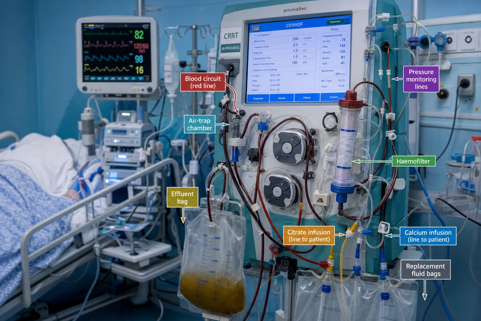
CRRT modalities — CVVH vs CVVHD vs CVVHDF [1]
CRRT modality comparison
| Feature | CVVH (haemofiltration) | CVVHD (haemodialysis) | CVVHDF (haemodiafiltration) |
|---|---|---|---|
| Solute removal | CONVECTIVE — water pulled across membrane by transmembrane pressure → solutes dragged along (solvent drag) | DIFFUSIVE — solutes move down concentration gradient from blood to dialysate | COMBINATION — both convective + diffusive |
| Middle molecule clearance | GOOD (β2-microglobulin, cytokines, vancomycin) | POOR (small molecules only — urea, creatinine) | GOOD (both small + middle) |
| Replacement fluid | YES — needed to replace ultrafiltrate (pre- or post-dilution) | NO — uses dialysate on the filtrate side | YES — both replacement fluid + dialysate |
| Blood flow rate | 150-200 mL/min | 150-200 mL/min | 150-200 mL/min |
| Effluent rate (dose) | Quf = Qrep + Qnet | Qeff = Qd | Qeff = Qrep + Qd + Qnet |
| Most common | — | — | ✅ MOST USED in ICU (best clearance) |
Regional citrate anticoagulation — the preferred method
Regional citrate anticoagulation setup and monitoring
- PRINCIPLE: Citrate (trisodium citrate) chelates IONISED CALCIUM in the extracorporeal circuit → calcium is required for the coagulation cascade → without ionised calcium, blood does NOT clot in the circuit. Citrate is infused PRE-filter (into the blood line before the haemofilter). A portion of the citrate-calcium complex is LOST in the effluent. The remaining citrate returns to the patient → metabolised by the liver (Krebs cycle) → releases calcium back into circulation. To compensate for the calcium lost in the effluent, a separate calcium infusion is given POST-filter (into the return line or via a separate central line)
- CITRATE INFUSION: Typically 4% trisodium citrate or Acid-Citrate-Dextrose (ACD-A) — infused into the pre-filter blood line at a rate proportional to blood flow (e.g., 25-30 mL/hr per 100 mL/min blood flow)
- CALCIUM INFUSION: Calcium chloride or calcium gluconate — infused into the post-filter return line or separate line — rate titrated to maintain SYSTEMIC ionised calcium 1.1-1.3 mmol/L
- MONITORING:
- Circuit ionised calcium (post-filter, pre-return): target <0.35 mmol/L → ensures anticoagulation is effective
- Systemic ionised calcium (patient's arterial line): target 1.1-1.3 mmol/L → ensures patient is normocalcaemic
- Total calcium / ionised calcium ratio: target <2.5 → if >2.5 = CITRATE ACCUMULATION (citrate chelating systemic calcium → metabolic acidosis from citrate anion) → reduce citrate rate or switch to heparin
- Check: circuit iCa q2-4h initially then q6-12h once stable. Systemic iCa q2-4h initially then q6-12h. Total Ca and ABG (for pH and bicarbonate) q12-24h
- CITRATE ACCUMULATION (the key complication):
- Occurs in HEPATIC FAILURE (citrate metabolised by liver — impaired metabolism → accumulation)
- Signs: rising total/ionised Ca ratio (>2.5), worsening METABOLIC ACIDOSIS (citrate is an unmeasured anion → high anion gap), hypocalcaemia (systemic)
- Management: REDUCE citrate rate, INCREASE calcium replacement, or SWITCH to heparin anticoagulation
- Severe hepatic failure (Child-Pugh C) is a RELATIVE contraindication to citrate — use heparin instead
- ADVANTAGES of citrate over heparin: (a) no systemic anticoagulation → lower bleeding risk, (b) longer circuit life (less clotting), (c) no heparin-induced thrombocytopenia risk, (d) no need for APTT monitoring
- DISADVANTAGES of citrate: (a) requires calcium infusion + close monitoring, (b) risk of citrate accumulation in hepatic failure, (c) metabolic alkalosis (citrate metabolised to bicarbonate), (d) sodium load (trisodium citrate contains sodium → hypernatraemia)
Clinical pearls
Red flags
Prognosis
CRRT outcomes
| Factor | Outcome | Notes |
|---|---|---|
| Overall mortality | 40-60% | Depends on underlying condition (sepsis, MOF) |
| Renal recovery | 60-70% | Most recover renal function (AKI is reversible) |
| Early vs late start (AKIKI/STARRT-AKI) | No difference | Do NOT start early — wait for urgent indication |
| Citrate vs heparin | Citrate: better circuit survival, less bleeding | Citrate preferred |
Key trials and evidence
AKIKI trial — Early vs delayed RRT (PMID 27568776)
Study design
Randomised — 620 patients with severe AKI (KDIGO stage 3)
Intervention
Early RRT (immediately upon randomisation) vs delayed (only if urgent indication: K+ >6, pH <7.15, overload, BUN >90)
Primary outcome
60-day mortality: 48% (early) vs 50% (delayed) — NO significant difference
Key finding
50% of the delayed group NEVER needed RRT — spontaneous recovery
Clinical bottom line
Do NOT start RRT early just for rising creatinine — wait for an urgent indication
STARRT-AKI trial — Accelerated vs standard RRT (PMID 32638054)
Study design
Randomised — 3,019 patients with AKI KDIGO stage 2-3 in 168 ICUs
Intervention
Accelerated (within 12h of stage 2-3) vs standard (only if urgent indication or persistent AKI >72h)
Primary outcome
90-day mortality: 43.9% (accelerated) vs 43.7% (standard) — NO benefit
Adverse events
Accelerated group had MORE adverse events (catheter infection, bleeding) — and MORE remained dialysis-dependent at 90 days
Clinical bottom line
Accelerated RRT may be HARMFUL — wait for urgent indication before starting RRT
CRRT circuit setup — components and configuration
A working knowledge of the extracorporeal circuit is examiner-favoured territory for the CICM Second Part viva. You must be able to draw the circuit and justify each component. The generic venovenous circuit is a closed loop: a double-lumen central venous catheter carries blood out of the patient, a blood pump generates flow across a semipermeable membrane (haemofilter), anticoagulant and/or replacement fluid is added at defined points, solutes and water leave as effluent, and blood returns to the patient. Every component is a potential point of failure and a potential exam question. [1]
The vascular access — double-lumen central venous catheter
CRRT requires a large-bore (12–14 Fr) dual-lumen catheter to support blood flows of 150–250 mL/min without excessive recirculation. The catheter has two lumina: a longer access (outflow) line with side holes that draws blood from a proximal vessel, and a shorter return (inflow) line that delivers blood downstream. The tip must sit in a high-flow central vein to dilute and disperse the returned blood and prevent recirculation. [1]
CRRT catheter site selection
| Site | Advantages | Disadvantages | Indication |
|---|---|---|---|
| Right IJV | Straight path to right atrium → best flow, least recirculation, lowest kinking, ultrasound-guided | Risk of pneumothorax (<1%), line infection (same as other central sites) | PREFERRED — first choice |
| Femoral | Safe insertion (no pneumothorax), patient can sit up less critical, good flow | Highest infection risk, DVT risk, patient immobility, higher recirculation if short | Second line; obese patients where IJV/clavicle access difficult |
| Left IJV | Available if right IJV thrombosed | Tortuous path through innominate vein → kinking → poor flow → frequent clotting | Avoid if possible |
| Subclavian | Comfortable, lowest infection | Stenosis risk — destroys future permanent dialysis access (AV fistula); pneumothorax; cannot easily compress | Avoid in patients likely to need long-term dialysis |
The tip should be confirmed (right IJV tip at cavo-atrial junction / upper right atrium on CXR; femoral tip in IVC) before starting CRRT. A malpositioned or kinked catheter produces low blood flow, repeated pressure alarms, access recirculation, and early circuit clotting — the single most fixable cause of recurrent filter loss. [1]
The blood pump — roller vs centrifugal
All modern CRRT machines use a peristaltic (roller) pump: rotating rollers compress a segment of flexible tubing, displacing blood forward. Roller pumps are cheap, simple, occlusive (deliver a fixed stroke volume per revolution), and generate the controlled blood flow (Qb 150–250 mL/min) and transmembrane pressure needed for CRRT. They are NOT used in ECMO, where centrifugal pumps are preferred because ECMO needs much higher flows (2–6 L/min) with less haemolysis. For CRRT the flow requirement is modest, so the roller pump's minor haemolysis is acceptable. [1]
Blood pump comparison — roller vs centrifugal
| Feature | Roller (peristaltic) pump | Centrifugal pump |
|---|---|---|
| Flow mechanism | Positive displacement — rollers compress tubing, fixed volume per revolution | Kinetic — impeller creates pressure gradient, flow is non-occlusive |
| Flow | Fixed (independent of downstream resistance) | Afterload-dependent (flow drops if line kinks) |
| Typical use | CRRT, standard IHD | ECMO, heart-lung machine |
| Haemolysis | Mild (tubing compression) | Less at high flow; more at low flow with high afterload |
| Air embolism risk | Low (closed) | Low |
| Caveat | Tubing spallation (particles shed over time) — change lines per protocol | Can generate negative pressure → suck air if access disconnects |
The pump also drives the replacement-fluid, dialysate, citrate, and calcium infusion lines in modern integrated machines (e.g., Prismaflex, multiFiltrate, Aquarius). Pump speeds for fluids are decoupled from blood flow and set independently to achieve the target effluent dose and anticoagulation. [1]
The haemofilter — membrane material, sieving, and cut-off
The haemofilter is the functional core of the circuit: a cartridge containing thousands of hollow fibres of a semipermeable synthetic membrane. Blood flows through the fibre lumens; water and small solutes cross the membrane wall driven by transmembrane pressure (filtration fraction) or diffuse down their concentration gradient (dialysis). The filtrate/effluent drains to a collection bag. [1]
Membrane material: modern CRRT filters use synthetic, biocompatible polymers — polyethersulfone (PES), polysulfone (PS), polyamide, polyacrylonitrile (AN69), or polymethylmethacrylate (PMMA). These replaced older cellulose-based (cuprophane) membranes, which activate complement and leukocytes (bioincompatible → worsen AKI). Synthetic membranes have large surface area (0.6–2.0 m²), high hydraulic permeability (allow ultrafiltration), and high biocompatibility. [1]
Replacement fluid — composition and buffer
Replacement fluid replaces the volume of ultrafiltrate removed (in CVVH and CVVHDF), preventing intravascular depletion. It is a crystalloid with electrolyte composition approximating plasma minus the uraemic toxins being cleared. Buffer: bicarbonate-buffered is preferred (lactate-buffered fluids are metabolised to bicarbonate but in shock/liver failure lactate accumulates and worsens acidosis). Bicarbonate comes as a two-compartment bag (mixed immediately before use because calcium phosphate precipitates in bicarbonate solution over time). [1]
Typical bicarbonate-buffered replacement fluid composition
| Component | Concentration (mmol/L) | Notes |
|---|---|---|
| Sodium | 140 | Slightly below plasma to mitigate citrate-induced hypernatraemia |
| Chloride | 108–115 | Lower than saline to allow correction of acidosis |
| Bicarbonate | 30–35 | Buffer — generates metabolic alkalosis if effluent rate low |
| Calcium | 0 (zero-Ca bags) or 1.5–1.75 | Zero-calcium bags MANDATORY with citrate anticoagulation |
| Magnesium | 0.5–1.0 | Often supplemented separately — lost in effluent |
| Potassium | 0, 2, or 4 | Choose based on patient's K+; many patients need K+ supplementation |
| Glucose | 0–11 | Some bags contain glucose → can contribute to hyperglycaemia |
| Phosphate | 0 | Add separately (glycerophosphate) — universally depleted on CRRT |
Critical rule with citrate: replacement fluid AND dialysate must be calcium-free when using regional citrate anticoagulation, otherwise exogenous calcium in the circuit defeats the citrate chelation. This is one of the commonest setup errors. [1]
Effluent drain
Effluent = the fluid that has crossed the membrane (ultrafiltrate + dialysate that has equilibrated). It is collected in a graduated bag (for volumetric control of net ultrafiltration / fluid removal) and discarded to drain. The effluent rate IS the CRRT dose (mL/kg/hr). Effluent is also a useful diagnostic fluid: it can be sent for vancomycin/beta-lactam levels (effluent concentration reflects cleared drug), and effluent biomarkers (NGAL, cell-cycle arrest markers) are research tools for predicting renal recovery. [1]
Regional citrate anticoagulation — detailed protocol and troubleshooting
Principle recap
Citrate (trisodium citrate or ACD-A) chelates ionised calcium (iCa) in the extracorporeal circuit → iCa is a cofactor for the coagulation cascade (factors IX, X, thrombin activation) → low iCa arrests clotting. Citrate is infused PRE-filter; a fraction of the citrate-calcium complex is lost in the effluent; the remainder returns to the patient where the liver metabolises citrate (Krebs cycle) → releasing calcium and generating bicarbonate. A separate calcium infusion (CaCl₂ or calcium gluconate) is given POST-filter (return line) to maintain systemic normocalcaemia. [1]
Step-by-step setup
Regional citrate anticoagulation — practical setup protocol
- CONFIRM CONTRAINDICATIONS first. Severe liver failure (Child-Pugh C, INR >2.0 from cirrhosis), severe lactic acidosis (lactate >5 mmol/L refractory to resuscitation), or profound shock with hepatic hypoperfusion → citrate will accumulate → USE HEPARIN instead. Mild-moderate liver dysfunction is NOT an absolute contraindication — start citrate with heightened monitoring.
- PRIME the circuit with heparinised or plain saline per machine protocol. Confirm calcium-free replacement fluid AND calcium-free dialysate are loaded. Load the citrate (4% trisodium citrate or ACD-A) and calcium (CaCl₂ 10% or calcium gluconate 10%) infusion lines.
- SET BLOOD FLOW (Qb). Start at 150 mL/min for a stable adult (100–120 mL/min if haemodynamically fragile, up to 200 mL/min once stable). Qb is the master variable — all other rates are referenced to it.
- SET CITRATE RATE. The citrate dose is calculated from blood flow: a common protocol targets a citrate-to-blood-flow ratio of ~3–3.5 mmol citrate per litre of blood. Practical formula: citrate rate (mL/hr) ≈ Qb (mL/min) × ratio factor. For 4% trisodium citrate: roughly 25–35 mL/hr per 100 mL/min blood flow. Example: Qb 150 mL/min → citrate 40–50 mL/hr. Modern machines (Prismaflex) auto-calculate this from a citrate-bolus dose.
- SET CALCIUM RETURN RATE. Start calcium infusion (CaCl₂ via return line) at ~60–90 mL/hr (or calcium gluconate ~2–3 mmol/hr). The calcium replaces what is lost in effluent AND titrates systemic iCa to target. Initial rate is an estimate — titrate to the first systemic iCa result.
- TARGETS (the three numbers you must know):
- Circuit (post-filter) iCa <0.35 mmol/L → confirms effective anticoagulation
- Systemic (patient) iCa 1.1–1.3 mmol/L → patient normocalcaemic
- Total Ca / iCa ratio <2.5 → no citrate accumulation
- MONITORING SCHEDULE: circuit iCa + systemic iCa at baseline, 1h, 2h, then q4–6h until stable, then q6–12h. Total calcium and ABG (pH, bicarbonate, base excess) q6–12h. Increase monitoring frequency if dose, blood flow, or citrate changed, or in liver dysfunction.
- NET FLUID BALANCE: the machine calculates net ultrafiltration (fluid removed from patient). Set target net removal per hour based on volume status. Citrate and calcium infusions contribute to fluid input — account for them in the balance.
Troubleshooting algorithm
The three measured values (circuit iCa, systemic iCa, total/iCa ratio) each map to a specific action. Memorise this table — it is viva gold. [1]
Citrate troubleshooting — the three-number algorithm
| Finding | Interpretation | Action |
|---|---|---|
| Circuit iCa >0.35 mmol/L (inadequate anticoagulation) | Not enough citrate reaching filter → blood will clot | INCREASE citrate rate by 10–20 mL/hr; recheck in 1h |
| Circuit iCa <0.35 (target met) | Anticoagulation effective | No change |
| Systemic iCa <1.1 mmol/L | Patient hypocalcaemic (insufficient calcium return OR citrate accumulation chelating systemic Ca) | INCREASE calcium return rate; ALSO check total/iCa ratio — if ratio >2.5 it is citrate accumulation, not simple hypocalcaemia → reduce citrate |
| Systemic iCa >1.3 mmol/L | Patient hypercalcaemic (excess calcium return) | DECREASE calcium return rate |
| Total/iCa ratio >2.5 | CITRATE ACCUMULATION — citrate chelating systemic calcium faster than liver metabolises it | REDUCE citrate rate by 30–50%; if persists or pH falling → SWITCH TO HEPARIN |
| Rising metabolic acidosis (pH down, HCO3 down, BE worsening) with normal lactate | Citrate accumulation (citrate is an unmeasured anion → high anion gap metabolic acidosis) | Reduce citrate or switch to heparin; check ratio confirms |
| Metabolic alkalosis (pH up, HCO3 up, BE high) | Excess citrate metabolised to bicarbonate (1 mol citrate → 3 mol bicarbonate) | Reduce citrate rate; increase effluent rate (removes more citrate before metabolism); consider ACD-A (less bicarbonate generation) |
| Rising serum sodium | Trisodium citrate sodium load (citrate 4% = ~420 mmol/L Na+) | Use lower-sodium replacement fluid; reduce citrate; consider calcium-citrate formulation |
Metabolic consequences of CRRT
CRRT is not metabolically neutral. Running blood and fluids through an extracorporeal loop for days alters temperature, electrolytes, acid-base, and glucose. Anticipating and replacing these losses is a daily (sometimes hourly) task. Examiners expect you to enumerate the metabolic consequences and their management. [1]
Heat loss — hypothermia
As blood traverses the extracorporeal circuit, heat is lost to the environment and to the room-temperature fluids. At a blood flow of 200 mL/min, a patient can lose 0.5–1.5°C/hr, producing hypothermia (core temperature 35–36°C or lower). Consequences: coagulopathy (hypothermia impairs platelet function and clotting enzyme activity — worsens bleeding), vasoconstriction (masks hypovolaemia), shivering (increases O₂ consumption, CO₂ production, work of breathing), and altered drug clearance (hypothermia reduces clearance of many drugs). This can be EXPLOITED therapeutically for targeted temperature management (TTM) post-cardiac arrest — CRRT with cooled fluids can hold a patient at 32–36°C without surface cooling — but in routine CRRT you must MAINTAIN NORMOTHERMIA with a blood/fluid warmer in the return line. Monitor core temperature continuously.[4]
Phosphate, magnesium, and potassium depletion
These three small, water-soluble ions are freely filtered across the membrane and lost in effluent. Phosphate is the most clinically significant: serum phosphate falls rapidly (often within 24–48h) to <0.6 mmol/L. Consequences: respiratory muscle weakness (diaphragm fatigue → failed ventilator weaning → reintubation), impaired cardiac contractility, rhabdomyolysis (paradoxically worsening AKI), haemolysis, and impaired leucocyte function (immune paresis). The RENAL trial found significantly MORE hypophosphataemia in the high-dose (40 mL/kg/hr) arm than the standard-dose arm (65% vs 54%), directly linking dose to phosphate loss.[6]
Magnesium is likewise depleted (arrhythmia risk, seizures, refractory hypokalaemia/hypocalcaemia — Mg is required to retain intracellular K+). Potassium is lost in effluent — many replacement fluids are K+-free or low-K+, so check K+ at least q6h initially and supplement via replacement fluid (K+ 4 mmol/L bag) or separate infusion. [1]
CRRT electrolyte depletion — replacement targets and protocols
| Electrolyte | Why depleted | Clinical effect | Target | Replacement |
|---|---|---|---|---|
| Phosphate | Small, freely filtered, lost in effluent | Respiratory failure, weaning failure, rhabdomyolysis, haemolysis | >0.8 mmol/L | Sodium/potassium glycerophosphate 10–20 mmol IV q12h OR add to replacement fluid; phosphate-bag protocols |
| Magnesium | Freely filtered | Arrhythmia, seizures, refractory hypoK/Ca | >0.8 mmol/L | MgSO₄ 10–20 mmol IV q12–24h or in replacement fluid |
| Potassium | Freely filtered; K+-free bags | Arrhythmia, weakness | 4.0–4.5 mmol/L | K+ in replacement fluid (4 mmol/L) or separate infusion; AVOID K+ <3.5 |
| Calcium | Lost as citrate-Ca complex in effluent | Hypocalcaemia, tetany, hypotension | Systemic iCa 1.1–1.3 (citrate) | Calcium return infusion (citrate protocols); Ca in fluid if no citrate |
Bicarbonate — gain or loss depending on buffer
The buffer in replacement fluid/dialysate determines the net acid-base effect. Bicarbonate-buffered fluids ADD bicarbonate to the patient → corrects metabolic acidosis of AKI (the usual goal). But if the effluent rate is LOW and hepatic metabolism normal, excess bicarbonate accumulates → metabolic alkalosis (also driven by citrate → bicarbonate conversion). Lactate-buffered fluids rely on hepatic conversion of lactate to bicarbonate — fine in normal liver, but in shock/liver failure the lactate accumulates and worsens acidosis. Hence bicarbonate-buffered fluids are preferred in ICU CRRT. Check bicarbonate/base excess on the daily (or q6h) ABG and titrate the buffer concentration (e.g., switch from 32 to 22 mmol/L bicarbonate bag if alkalotic). [1]
Glucose handling
Some replacement fluids and dialysates contain glucose (5–11 mmol/L) → continuous glucose infusion → can contribute to hyperglycaemia in the insulin-resistant critically ill. More importantly, glucose is a SMALL solute freely filtered — high serum glucose → high effluent glucose → 'glucose loss' in effluent (a marker the machine sometimes displays). Diabetic/hyperglycaemic patients need insulin titration; hypoglycaemia is rare unless insulin is over-dosed. Use glucose-free replacement fluid in brittle diabetics if hyperglycaemia is hard to control. Monitor blood glucose q1–4h initially. [1]
Metabolic consequences of CRRT — summary
| Consequence | Mechanism | Direction | Management |
|---|---|---|---|
| Hypothermia | Heat loss to circuit/fluids | ↓ temperature | Fluid/blood warmer; monitor core temp |
| Hypophosphataemia | Effluent loss | ↓ PO₄ | Glycerophosphate; check daily |
| Hypomagnesaemia | Effluent loss | ↓ Mg | MgSO₄ supplement |
| Hypokalaemia | Effluent loss | ↓ K | K+ in replacement fluid |
| Hypocalcaemia | Citrate chelation + effluent loss | ↓ iCa (systemic) | Calcium return infusion |
| Metabolic alkalosis | Citrate → bicarbonate; bicarbonate buffer | ↑ HCO₃ | Reduce citrate/buffer; raise effluent |
| Metabolic acidosis | Citrate accumulation (liver failure) | ↓ pH, ↑ anion gap | Reduce citrate → switch to heparin |
| Hypernatraemia | Trisodium citrate sodium load | ↑ Na | Lower-Na replacement fluid |
| Hyperglycaemia | Glucose in fluid + insulin resistance | ↑ glucose | Insulin; glucose-free fluid |
CRRT membrane characteristics
Sieving coefficient (SC)
The sieving coefficient quantifies how freely a solute crosses the membrane: SC = (effluent concentration) / (plasma concentration). An SC of 1.0 means the solute passes freely (concentration in effluent equals plasma); SC of 0 means complete retention. For standard CRRT membranes, SC is approximately 1.0 for solutes up to ~30 kDa (which covers urea, creatinine, electrolytes, glucose, β2-microglobulin, vancomycin, beta-lactams, most cytokines). Larger molecules (albumin 66 kDa, immunoglobulins) are retained (SC ≈ 0) — CRRT does NOT remove albumin or large proteins. This is why drug dosing on CRRT is predictable for small/medium water-soluble drugs (they behave like urea) but irrelevant for protein-bound or large drugs. [1]
Cut-off vs sieving coefficient
Cut-off = the molecular weight at which 90% of the solute is retained (10% passes). For standard CRRT membranes cut-off is ~30–40 kDa. High cut-off (HCO) membranes have cut-off ~45–60 kDa — they remove larger middle molecules (myoglobin 17 kDa in rhabdomyolysis, free haemoglobin, some cytokines, certain chemotherapeutics) but also lose albumin (66 kDa) and require albumin replacement. HCO membranes are used selectively: rhabdomyolysis with myoglobin clearance, sepsis (cytokine removal — controversial), drug removal in overdose of middle-molecular toxins. [1]
Adsorption
Beyond filtration and diffusion, some membranes adsorb proteins and drugs onto their surface (particularly the AN69/polyacrylonitrile and PMMA membranes). Adsorption removes substances WITHOUT them appearing in the effluent — so effluent drug levels underestimate clearance. This has two faces: [1]
- Beneficial: adsorption of cytokines (IL-6, TNF-α) and damage-associated molecular patterns — the rationale for 'blood purification' / 'cytokine adsorption' in septic shock or cytokine release syndrome (CAR-T). PMMA and AN69 membranes show the highest cytokine adsorption. This remains investigational — no mortality benefit in large RCTs.[9]
- Problematic: adsorption of drugs (antibiotics, antiepileptics) reduces their effective dose — contributes to the under-dosing problem. Adsorption is saturable and falls over the first 24h as binding sites fill. After a circuit change, a NEW membrane adsorbs again → drug levels transiently drop further. The DALI study and PK studies show this contributes to the 75% under-dosing rate.[2][8]
CRRT membrane characteristics — by polymer
| Membrane (polymer) | Cut-off | Biocompatibility | Adsorption | Notable use |
|---|---|---|---|---|
| Polyethersulfone (PES) | Standard (~30 kDa) | High | Low | Common CRRT filter; balanced clearance |
| Polysulfone (PS) | Standard (~30 kDa) | High | Low | Common CRRT filter; high flux |
| Polyamide | Standard | High | Low | Combined with PS in some filters |
| Polyacrylonitrile (AN69) | Standard / surface-treated | High | HIGH (cytokines, bradykinin) | Sepsis, cytokine adsorption; AVOID concurrent ACE-inhibitor (bradykinin → anaphylactoid reaction) |
| Polymethylmethacrylate (PMMA) | Standard | High | HIGH (cytokines, drugs) | Sepsis, cytokine removal; high drug adsorption |
| High cut-off (HCO) | ~45–60 kDa | High | Variable | Rhabdomyolysis (myoglobin), sepsis, chemotherapy clearance — needs albumin replacement |
Transitioning from CRRT to IHD
CRRT is the modality for haemodynamically unstable patients. As the patient recovers, the question becomes when and how to transition to intermittent haemodialysis (IHD) or stop dialysis entirely. The decision rests on three pillars: haemodynamic stability, renal recovery (urine output + falling creatinine), and absence of an ongoing dialysis indication. [1]
Criteria to transition CRRT → IHD
Transitioning from CRRT to IHD — criteria and process
- HAEMODYNAMIC STABILITY: patient off vasopressors (or on minimal, stable dose of a single agent) for >24h, MAP >65 mmHg without fluid boluses, no ongoing rapid fluid shifts needed.
- RECOVERING URINE OUTPUT: this is the single best predictor of renal recovery. Spontaneous urine output >500 mL/day without diuretics, OR >2 L/day with diuretics, predicts ability to wean.
- STABLE/IMPROVING BIOCHEMISTRY: creatinine falling or stable (not rising), K+ normal without replacement, bicarbonate normal, resolving acidosis, controlled phosphate/magnesium.
- NO ONGOING INDICATION: no refractory hyperkalaemia, no uncontrolled acidosis, no ongoing massive fluid removal needed, no uraemic complications.
- ADEQUATE VASCULAR ACCESS FOR IHD: the CRRT catheter can often be used for the first IHD session, but plan for an AV fistula if long-term dialysis anticipated.
- WEANING STRATEGY — two options:
- Reduce CRRT dose then stop: step dose down from 25 → 15 → 10 mL/kg/hr over 24–48h; if creatinine, K+, bicarbonate remain stable, stop CRRT and observe. This allows gradual transition.
- Direct switch to IHD: convert to alternate-day IHD sessions; the first session uses lower blood flow and shorter duration to avoid disequilibrium, especially after prolonged CRRT.
- MONITOR POST-STOP: check creatinine, K+, bicarbonate, volume status daily for 48–72h. If biochemical deterioration → restart RRT (the patient has not recovered).
- DIALYSIS DEPENDENCY RISK FACTORS: age, baseline CKD, severity/duration of AKI, number of failed organs, sepsis — counsel on possible long-term dialysis. STARRT-AKI showed accelerated-start patients had MORE residual dialysis dependence — underscoring that recovery is not guaranteed.[1]
CRRT vs IHD vs SLED — choosing and switching
RRT modality selection by clinical context
| Scenario | Modality | Rationale |
|---|---|---|
| Vasopressor-dependent shock | CRRT | No rapid solute/shift → haemodynamic stability |
| Brain injury (TBI, SAH, raised ICP) | CRRT | IHD causes osmotic shifts → ICP spikes; CRRT avoids this |
| Liver failure / hepatic encephalopathy | CRRT | IHD raises ICP and causes dysequilibrium; CRRT (with MARS if available) |
| Severe hyperkalaemia (K+ >7) — need rapid clearance | IHD | Diffusive K+ clearance faster; then switch to CRRT |
| Salicylate/lithium/metformin overdose (dialysable toxin) | IHD | Rapid diffusive clearance of toxin |
| Haemodynamically stable, chronic dialysis schedule | IHD | Standard, efficient, lets patient mobilise |
| Stable but doesn't tolerate 4h IHD | SLED | Slow extended (6–12h) — intermediate haemodynamic stability |
| Massive fluid overload needing slow removal | CRRT (or SLED) | Continuous slow ultrafiltration |
Key trials — CRRT dose and intensity
RENAL trial — CRRT dose 25 vs 40 mL/kg/hr (PMID 19846848)
Study design
Multicentre RCT — 1,508 critically ill adults with AKI needing RRT (Australia/NZ)
Intervention
Post-dilution CVVHDF at 25 mL/kg/hr (lower intensity) vs 40 mL/kg/hr (higher intensity)
Primary outcome
90-day mortality: 44.7% (higher) vs 44.7% (lower) — NO difference (OR 1.00, 95% CI 0.81–1.23, P=0.99)
Adverse events
Higher-intensity arm had SIGNIFICANTLY MORE hypophosphataemia (65% vs 54%, P<0.001)
Clinical bottom line
Delivered dose above 25 mL/kg/hr does NOT improve survival but DOES increase phosphate loss. Target effluent 20–25 mL/kg/hr (KDIGO standard)
ATN trial (VA/NIH) — Intensity of renal support in AKI (PMID 18492867)
Study design
Multicentre RCT — 1,124 critically ill patients with AKI + ≥1 non-renal organ failure
Intervention
Intensive (IHD/SLED 6×/week; CVVHDF 35 mL/kg/hr) vs less-intensive (IHD/SLED 3×/week; CVVHDF 20 mL/kg/hr)
Primary outcome
60-day mortality: 53.6% (intensive) vs 51.5% (less-intensive) — NO difference (P=0.47)
Key finding
No difference in duration of RRT, renal recovery, or non-renal organ recovery
Clinical bottom line
More intensive RRT does NOT improve outcomes — confirms the 20–25 mL/kg/hr standard. Two landmark trials (RENAL + ATN) together set the KDIGO dose
Sample exam question — worked answer
[1]Correct answer: C — Reduce the citrate rate by 30–50% and prepare to switch to heparin anticoagulation. [1]
Worked explanation (1500+ words)
This question tests the single most high-yield concept in CRRT management: the recognition and correct response to citrate accumulation. Let me work through it step by step, because the distractors are designed to exploit the three most common cognitive errors. [1]
Step 1 — Identify what each measured value tells you. The patient has three citrate-monitoring values plus an ABG. Work through each systematically. The circuit (post-filter) ionised calcium is 0.40 mmol/L — the target is <0.35 mmol/L, so 0.40 is marginally above target, meaning anticoagulation is slightly inadequate, but the filter is clearly not clotting catastrophically (it has run 48h). This is a minor deviation, not the headline finding. The systemic ionised calcium is 0.95 mmol/L — this is BELOW the target range of 1.1–1.3 mmol/L, so the patient is systemically hypocalcaemic. The total calcium is 2.50 mmol/L — which is normal-to-high. And the total/iCa ratio is 2.50/0.95 = 2.63, which is ABOVE the threshold of 2.5. Finally, the ABG shows a metabolic acidosis (pH 7.28, bicarbonate 18) with a NORMAL lactate (1.2 mmol/L). [1]
Step 2 — Recognise the pattern of citrate accumulation. The diagnosis rests on three converging pieces of evidence. First, the total/iCa ratio is >2.5 (2.63) — this is the cardinal biochemical marker of citrate accumulation; it means a disproportionate fraction of the patient's total calcium is bound to citrate (i.e., circulating as the citrate-calcium complex rather than as free ionised calcium). Second, the systemic ionised calcium is low (0.95) while the total calcium is normal/high — this dissociation (high total, low ionised) is pathognomonic of citrate chelation. Third, there is a new metabolic acidosis with a normal lactate — citrate is an unmeasured anion; when it accumulates faster than the liver can metabolise it (into bicarbonate), it contributes to a high-anion-gap metabolic acidosis. The normal lactate is critical here: it tells you the acidosis is NOT from tissue hypoperfusion or sepsis, and points instead to the citrate itself. [1]
Step 3 — Why is the liver failing to metabolise citrate? Citrate is metabolised in the mitochondria via the Krebs cycle, primarily in the liver (and to a lesser extent skeletal muscle and kidney). In this septic patient, the most likely explanation is hepatic hypoperfusion from shock (splanchnic vasoconstriction, mitochondrial dysfunction in sepsis) even in the absence of primary liver disease. Sepsis itself impairs hepatic mitochondrial function. The clinical lesson: citrate accumulation is not confined to overt liver failure — any patient with septic shock, particularly with rising lactate or impaired perfusion, is at risk, and monitoring the ratio is mandatory. [1]
Step 4 — Now evaluate each option against this diagnosis. [1]
Option A (increase calcium return to 110 mL/hr) is the trap for candidates who fixate on the low systemic iCa in isolation. Giving more calcium WILL raise the ionised calcium temporarily, but it does nothing about the accumulating citrate — the new calcium will simply be chelated by more citrate, the ratio will climb higher, and the metabolic acidosis will worsen. You are treating a number without treating the disease. More fundamentally, in citrate accumulation the problem is NOT a calcium deficit; it is an excess of citrate. Adding calcium is a band-aid that delays the correct intervention. This option is incorrect. [1]
Option B (increase citrate to 60 mL/hr) is wrong in the opposite direction — it would worsen accumulation. This distractor targets candidates who notice that the circuit iCa (0.40) is marginally above the 0.35 target and conclude that anticoagulation is inadequate. While 0.40 is indeed slightly above target, the overwhelming problem is systemic accumulation, and increasing citrate would push the ratio higher, worsen the acidosis, and risk precipitating severe hypocalcaemia and arrhythmia. The circuit iCa of 0.40 is acceptable in context; you would never escalate citrate in the face of a ratio >2.5. [1]
Option C (reduce citrate 30–50% and prepare to switch to heparin) is correct. Reducing the citrate rate immediately decreases the citrate load the failing liver must metabolise; the ratio will begin to fall. However, because the liver's capacity is impaired (this is WHY accumulation occurred), simply reducing the rate may be insufficient — the standard escalation is to switch to heparin anticoagulation (unfractionated heparin targeting APTT 1.5–2× baseline) or, if heparin is contraindicated (active bleeding), a no-anticoagulation protocol with high blood flow and pre-dilution. You continue the calcium return until the citrate is cleared (the calcium infusion compensates for the calcium lost in effluent while the citrate washes out). Monitor systemic iCa closely during the transition — it can transiently fall further as the accumulated citrate continues to chelate calcium. This option directly addresses the root cause. [1]
Option D (increase effluent to 35 mL/kg/hr) exploits a half-truth. A higher effluent rate DOES remove more citrate (some citrate-calcium complex is lost in effluent), and the RENAL trial demonstrated that higher doses are safe. However, in this scenario the dominant problem is hepatic under-metabolism of citrate, not inadequate clearance; pushing the dose up would also worsen phosphate loss (the RENAL trial's high-dose arm had significantly more hypophosphataemia), and it does not solve the accumulation. Dose escalation is a reasonable ADJUNCT but not the primary intervention, and it is not the 'most appropriate next step'. [1]
Option E (calcium gluconate bolus) is the distractor for the candidate panicking about the low iCa and arrhythmia risk. A calcium bolus is appropriate for SYMPTOMATIC hypocalcaemia (tetany, seizures, hypotension, prolonged QT with arrhythmia) but this patient's iCa of 0.95 is asymptomatic and the correct response is to address the cause. Bolusing calcium without reducing citrate will transiently raise iCa only for it to be re-chelated. Continue the infusion, do not bolus, and fix the citrate. [1]
Step 5 — Synthesise the management. The complete response is: (1) recognise citrate accumulation from the ratio >2.5 + systemic hypo-iCa with normal/high total Ca + high-anion-gap metabolic acidosis with normal lactate; (2) reduce the citrate rate by 30–50% immediately; (3) prepare to switch to heparin anticoagulation (this patient has sepsis with impaired hepatic metabolism — reduction alone is unlikely to suffice); (4) maintain or cautiously titrate the calcium return to keep systemic iCa ≥1.0 while the citrate washes out; (5) recheck systemic iCa, total calcium, and ABG in 1–2h; (6) investigate and treat the underlying hepatic hypoperfusion (optimise haemodynamics, ensure adequate cardiac output and splanchnic perfusion). The metabolic acidosis will correct as the citrate load falls. [1]
Step 6 — Why the other numbers matter for the viva. Be ready to discuss the mild hypernatraemia (146 mmol/L) — this is the sodium load from 4% trisodium citrate (~420 mmol/L sodium); it is expected and usually modest, managed with lower-sodium replacement fluid. Be ready to discuss the dose: 25 mL/kg/hr is the RENAL-trial-validated standard; the RENAL and ATN trials together proved that higher doses do not improve survival. Be ready to discuss the effluent as the dose surrogate, and that prescribed ≠ delivered (compensate for down-time). Be ready to discuss drug dosing: this patient on beta-lactams for pyelonephritis is at high risk of under-dosing (DALI: 75% of CRRT patients under-dosed) — use TDM and extended infusions. [1]
Bottom line for the exam: the total/ionised calcium ratio is the single most useful number in citrate CRRT. A ratio >2.5 in the setting of a new metabolic acidosis with normal lactate and a low systemic iCa but normal total calcium is citrate accumulation until proven otherwise. The correct response is to reduce citrate and switch to heparin — never simply to give more calcium. Memorise the three targets (circuit iCa <0.35, systemic iCa 1.1–1.3, ratio <2.5) and the action each abnormality dictates. [3][5][6][2]
Additional red flags
[1]Drug dosing on CRRT — practical summary
Antibiotic dosing on CRRT — common ICU agents
| Drug | Clearance on CRRT | Dosing principle | Monitoring |
|---|---|---|---|
| Vancomycin | Cleared (SC ~1.0) | Loading 25–30 mg/kg, then 15–25 mg/kg q24–48h (or continuous infusion) | Trough 15–20 mg/L before 4th dose |
| Piperacillin-tazobactam | Cleared | 4.5g q6–8h OR extended/continuous infusion (e.g., 16–18g/24h) | Beta-lactam level 4× MIC if available |
| Meropenem | Cleared | 1g q8–12h; extended infusion (3h) preferred | Beta-lactam level |
| Cefepime / ceftazidime | Cleared | 2g q8–12h | Beta-lactam level |
| Aminoglycosides | Cleared | Conventional weight-based, extended interval; risk of accumulation if residual function present | Trough |
| Linezolid | Partly cleared | Standard 600mg q12h | CBC (thrombocytopenia) |
| Levetiracetam | Cleared | Increased dose / more frequent (e.g., 1000mg q12h) | Anti-epileptic level |
The dose depends on the effluent rate (higher dose → more clearance), residual renal function, and the drug's volume of distribution and protein binding. Highly protein-bound drugs (e.g., ceftriaxone 90% bound) are NOT significantly cleared by CRRT (only the free fraction crosses). Always pair dosing with TDM where available, and re-evaluate after every filter change (new membrane adsorbs drug).[2][8]
Exam practice
SAQ — CRRT modality, citrate and timing
10 minutes · 10 marks
A 58-year-old man with septic shock and KDIGO stage 3 AKI is on noradrenaline 0.3 mcg/kg/min. K+ 6.6 mmol/L with ECG changes after medical therapy, pH 7.18, anuric, 5 L positive balance. Liver enzymes are normal.
References
- [1]Gaudry S, et al. Developing a policy to empower informal carers to administer subcutaneous medication in community palliative care; a feasibility project Int J Palliat Nurs, 2016.PMID 27568776
- [2]Roberts DM, et al. DALI: defining antibiotic levels in intensive care unit patients: are current β-lactam antibiotic doses sufficient for critically ill patients? Clin Infect Dis, 2014.PMID 24429437
- [3]Schneider AG, et al. Prostate cancer: Colorectal cancer increased by ADT? Nat Rev Urol, 2011.PMID 21287697
- [4]Ronco C, et al. Treatment satisfaction and efficacy of the rapid release formulation of sumatriptan 100 mg tablets utilising an early intervention paradigm in patients previously unsatisfied with sumatriptan Int J Clin Pract, 2008.PMID 19166436
- [5]Schneider AG, et al. Urea Transporter B and MicroRNA-200c Differ in Kidney Outer Versus Inner Medulla Following Dehydration Am J Med Sci, 2016.PMID 27650235
- [6]Bellomo R, et al. (RENAL Replacement Therapy Study Investigators) Intensity of continuous renal-replacement therapy in critically ill patients N Engl J Med, 2009.PMID 19846848
- [7]Palevsky PM, et al. (VA/NIH Acute Renal Failure Trial Network) Intensity of renal support in critically ill patients with acute kidney injury N Engl J Med, 2008.PMID 18492867
- [8]Schulman G, et al. Evaluation of Urinary KIM-1 for Prediction of Polymyxin B-Induced Nephrotoxicity Antimicrob Agents Chemother, 2017.PMID 28848003
- [9]Honore PM, et al. Regional Anesthesia to Scalp for Craniotomy: Innovation With Innervation J Neurosurg Anesthesiol, 2016.PMID 26083426