ICU · Electrolytes
Electrolyte Disorders in Critical Illness — Sodium, Potassium, Calcium, Magnesium and Phosphate
Also known as Dysnatraemia · Hyponatraemia · Hypernatraemia · Hyperkalaemia · Hypokalaemia · Electrolyte disturbances
Electrolyte disorders are among the commonest and most immediately dangerous derangements in critical illness, and their correction is governed less by the absolute concentration than by the speed and direction of change. This topic builds the examiner's framework around the two highest-yield cations — sodium and potassium — and a concise account of calcium, magnesium and phosphate. For sodium, the central ideas are that hyponatraemia is a disorder of water handling classified by osmolality and volume status, that the dangerous consequences are cerebral oedema (acute) and osmotic demyelination (over-correction), and that the rate of correction is the single most important decision. For potassium, the central ideas are that hyperkalaemia with ECG change is an emergency treated in a fixed sequence, and that hypokalaemia is a product of redistribution or total-body deficit. The evidence base is anchored on the Adrogué NEJM reviews of dysnatraemia, the Verbalis expert-panel hyponatraemia recommendations, the Kovesdy and Palmer potassium reviews, and the Ayuk hypomagnesaemia review, with the calcium and phosphate content resting on the standard ICU texts.
On this page & tools
Your progress
Saved locally on this device.
Target exams

Overview & definition
Electrolyte disorders are ubiquitous in critical illness, and several — a rapidly falling sodium, a rising potassium with ECG change — are immediately life-threatening. The unifying principle of their management is that the rate and direction of change matter more than the absolute value: a sodium corrected too fast causes osmotic demyelination, and a potassium lowered without addressing the cause rebounds.[1][2]
The topic is organised around the two cations that dominate the CICM exam and the bedside — sodium and potassium — with a concise account of calcium, magnesium and phosphate. Sodium disorders are disorders of water balance (and so of osmolality and brain cell volume); potassium disorders are disorders of a cation whose concentration is governed by intake, excretion and transcellular shift.[1]
Pathophysiology: sodium, water and the brain
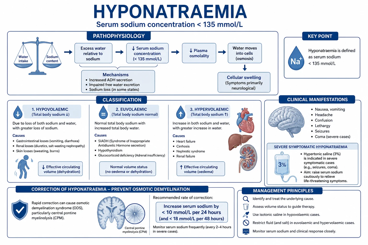
Serum sodium is the dominant determinant of serum osmolality, and serum osmolality is what the brain cells respond to. Water crosses cell membranes freely to equalise osmolality, so a fall in serum sodium (hypo-osmolality) drives water into brain cells, causing cerebral oedema, and a rise (hyperosmolality) draws water out, causing shrinkage.[1]
The brain defends itself against these volume changes. In acute hyponatraemia the brain has not yet adapted, so cerebral oedema develops quickly and the patient is at risk of herniation — this is the emergency. In chronic hyponatraemia the brain has extruded osmolytes to limit swelling, so it is symptomatic at much lower sodium concentrations than the acute form, but it is now vulnerable to a rapid rise in sodium: as the extracellular fluid becomes hyperosmotic, water is drawn out of the adapted brain faster than it can re-accumulate osmolytes, producing osmotic demyelination (the central pontine and extrapontine myelinolysis syndrome).[1][3]
This adaptation is the reason the rate of correction is the single most important decision in dysnatraemia: the acutely hyponatraemic brain needs rapid correction to relieve oedema, while the chronically hyponatraemic brain needs slow correction to avoid demyelination.[1]
Hyponatraemia: classification by osmolality and volume
Hyponatraemia is a serum sodium below 135 mmol/L. The first step is to measure serum osmolality, because a low sodium can be diluted by another osmole (glucose, in hyperglycaemia) and because not all hyponatraemia is hypo-osmolar.[1][3]
- Hypo-osmolar hyponatraemia (the common, dangerous form). Classified by volume status:
- Hypovolaemic — renal or gastrointestinal sodium loss (diuretics, diarrhoea, third-space losses); the urine sodium is low in extrarenal loss and high in renal loss.
- Euvolaemic — the syndrome of inappropriate antidiuresis (SIADH) is the archetype; also glucocorticoid deficiency, hypothyroidism, and primary polydipsia. SIADH shows an inappropriately concentrated urine (urine osmolality above 100, often above 300) with a normal or high urine sodium in a euvolaemic patient.
- Hypervolaemic — oedematous states (heart failure, cirrhosis, renal failure) in which effective circulating volume is low, driving vasopressin and water retention despite total-body sodium excess.
- Non-hypo-osmolar hyponatraemia — hyperglycaemic (the sodium falls by roughly 2.4 mmol/L for every 5.5 mmol/L rise in glucose; correct the glucose, not the sodium), and the rare pseudohyponatraemia of severe hyperlipidaemia or hyperproteinaemia.[1]
Severe hyponatraemia: the emergency and the correction limit
The severely symptomatic hyponatraemic patient — confusion, seizures, coma — has acute or rapidly evolving cerebral oedema and needs rapid correction with hypertonic saline (e.g. a 100 mL bolus of 3 per cent saline, repeated as needed) to raise the sodium quickly and relieve the brain swelling, with the airway and seizures managed in parallel.[1][3]
The danger that governs the rest is over-correction. Whether the patient is corrected rapidly for symptoms or slowly for chronicity, the sodium must not rise faster than the safe ceiling: roughly 8 to 10 mmol/L in any 24 hours (a stricter limit than older guidance), because exceeding this risks osmotic demyelination. Correction that threatens to exceed the ceiling is capped — with desmopressin to hold the water, free water, or (in extreme over-shoot) hypotonic fluids. The genuinely symptomatic patient is corrected fast to relieve oedema and then throttled back; the asymptomatic patient is corrected slowly throughout.[1][3]
Hypernatraemia (serum sodium above 145 mmol/L) is, almost always, a disorder of water deficit — insufficient intake or excessive loss, in a patient who cannot access water (the elderly, the intubated, the unconscious). The causes are loss of pure water (insensible, fever), hypotonic loss (diuresis, diarrhoea), or sodium gain (hypertonic saline, mineralocorticoid excess).[2]
The correction is to replace the free-water deficit (and ongoing losses), calculated from the sodium and body weight, given as enteral water or a hypotonic infusion, and again governed by the rate: a fall of no more than roughly 10 mmol/L per 24 hours in the chronic case, to avoid cerebral oedema from too-rapid correction. The genuinely dangerous hypernatraemia is the one generated iatrogenically by hypertonic saline or sodium bicarbonate, and it is prevented by vigilance and by matching water replacement to ongoing losses.[2]
Hyperkalaemia (serum potassium above 5.0 to 5.5 mmol/L) is the electrolyte emergency, and its danger is the cardiac arrhythmia.[4]
The ECG changes progress with severity — peaking of the T waves, then loss of P waves, widening of the QRS, and finally a sine-wave pattern and arrest — and any ECG change at a high potassium is treated as an emergency. The management is a fixed sequence:[4][5]
- Stabilise the myocardium — intravenous calcium gluconate (or chloride), which does not lower the potassium but restores the membrane threshold and protects the heart within minutes.
- Shift potassium into cells — insulin with dextrose (the most reliable), nebulised or intravenous salbutamol, and (where appropriate) sodium bicarbonate for the acidotic patient.
- Remove potassium from the body — gastrointestinal cation-exchange resins (patiromer, sodium zirconium, the older polystyrene sulfonate) for the more stable patient, and renal replacement therapy for the severe, refractory, or renal-failure case. [1]
The error to avoid is being reassured by a normal-looking ECG at a very high potassium: the heart can arrest without classic warning changes, so a markedly raised potassium is treated on the number.[5]
Hypokalaemia (serum potassium below 3.5 mmol/L) reflects either a transcellular shift (alkalaemia, insulin, beta-agonists — which move potassium into cells without changing the total body store) or a true total-body deficit from renal (diuretics, mineralocorticoid excess, recovery phase of acute tubular necrosis) or gastrointestinal loss.[5]
The consequences are muscular weakness (and, at very low levels, paralysis and respiratory failure), an arrhythmia tendency (especially in the patient on digoxin or with ischaemia), and potentiation of digoxin toxicity. The treatment is potassium replacement — enterally where possible and safe, intravenously (at a controlled rate, with cardiac monitoring, never as a rapid bolus) when severe or symptomatic — and the correction of the magnesium at the same time, because hypomagnesaemia perpetuates a refractory hypokalaemia by increasing renal potassium loss.[5][6]
Calcium, magnesium and phosphate
The divalent cations and phosphate are lower-yield than sodium and potassium but produce characteristic, examinable syndromes.[1][1]
- Calcium. Hypercalcaemia (of malignancy, hyperparathyroidism) presents with confusion, polyuria, constipation and nephrolithiasis, and is treated with aggressive normal-saline hydration followed by a bisphosphonate. Hypocalcaemia (after parathyroidectomy, in acute pancreatitis, with citrate chelation in massive transfusion or on citrate CRRT) causes tetany, a prolonged QT, and perioral and peripheral paraesthesiae; it is treated with intravenous calcium gluconate, monitoring the ionised (not total) calcium.
- Magnesium. Hypomagnesaemia (diuretics, proton-pump inhibitors, diarrhoea, alcoholism) causes a hypokalaemia and hypocalcaemia that will not correct until the magnesium is replaced, together with arrhythmias and seizures; it is repleted enterally for the mild and intravenously for the severe case.[6]
- Phosphate. Hypophosphatemia (re-feeding, sepsis, respiratory alkalosis, recovery) causes muscle weakness — including respiratory-muscle weakness complicating weaning — and is replaced enterally or intravenously. Hyperphosphataemia, in renal failure, is managed with phosphate binders and dialysis.
An integrated overview of ICU electrolyte disorders
Electrolytes are not independent variables at the bedside — they are coupled through shared renal and endocrine machinery, through the intracellular–extracellular distribution governed by the membrane potential, and through the fluids and drugs the intensivist delivers. The examiner's "integrated" question asks how a single insult (sepsis, diabetic ketoacidosis, a session of renal replacement therapy, a massive resuscitation) disturbs several electrolytes at once, and how correcting one perturbs the others. The framework below maps the coupling.[1][1]
The two governing axes are water–sodium (which sets the serum sodium through tonicity) and cell-shift–excretion (which sets the serum potassium through transcellular distribution and renal loss). The divalents sit underneath: magnesium governs potassium and calcium handling (a low magnesium refracts both), phosphate tracks nitrogen balance and cellular anabolism (the refeeding swing), and calcium is governed by parathyroid hormone, vitamin D, the kidneys, and — in the ICU — by albumin and chelation. Chloride is the silent determinant of the strong ion difference and thus of acid–base. Each of these axes is destabilised in characteristic ways by critical illness and by its treatment.[1]
The ICU electrolyte axes — what sets the serum value, and what critical illness does to it
| Electrolyte | Principal determinant | Transcellular shift? | The ICU accelerators of change | The dangerous end of the range |
|---|---|---|---|---|
| Sodium (Na⁺) | Total body water relative to Na⁺ (tonicity) | Minimal — governed by water balance | Hypertonic saline, diuretics, SIADH, polyuria, vomiting, DKA | Acute fall → cerebral oedema; chronic over-correction → ODS |
| Potassium (K⁺) | Intake, renal excretion, distribution | Yes — major (insulin, β₂-agonists, acid–base) | AKI, acidosis, rhabdomyolysis, CRRT, insulin/dextrose | Hyperkalaemia → arrest; hypo → TdP, paralysis |
| Calcium (Ca²⁺) | PTH/vitamin D/renal; albumin binding; chelation | Minimal (correct for albumin, act on ionised) | Parathyroidectomy, pancreatitis, citrate CRRT, malignancy | Hypo → tetany, QT; hyper → coma, stones |
| Magnesium (Mg²⁺) | Renal excretion (CaSR, ROMK); intake | Minor | Diuretics, PPIs, alcohol, diarrhoea, CRRT | Hypo → refractory K⁺/Ca²⁺ loss, torsades |
| Phosphate (PO₄³⁻) | Renal excretion, intake, intracellular shift | Yes — major (refeeding, glucose, insulin) | Refeeding, CRRT, sepsis, respiratory alkalosis | Hypo → respiratory and cardiac muscle failure |
| Chloride (Cl⁻) | Renal handling, fluid choice | Minimal | 0.9% saline (high Cl⁻), diuretics, GI loss | High → SID acidosis; low → SID alkalosis |
Sodium in depth: correction rates, the Adrogué–Madias formula, and osmotic demyelination
The empirically derived ceiling for chronic hyponatraemia correction is 8 mmol/L in any 24 hours (the European guideline and the Verbalis panel converge here), with an absolute ceiling of about 16–18 mmol/L over 48 hours; some authorities permit up to 10–12 mmol/L in 24 hours for the symptomatic patient but the trend is to the lower figure because osmotic demyelination is irreversible. The ceiling for hypernatraemia correction is the mirror image: no faster than 10 mmol/L per 24 hours (about 0.5 mmol/L per hour), to avoid cerebral oedema in an adapted brain.[7][8]
The expected rise in serum sodium per litre of a given infusate is given by the Adrogué–Madias formula: ΔNa = (Na_infusate − Na_serum) / (TBW + 1), where TBW = total body water (0.6 × weight in men, 0.5 in women, 0.5 in elderly men, 0.45 in elderly women).[9] For 3 per cent saline (Na 513 mmol/L) in a 70 kg man (TBW 42 L) with a serum Na of 110, one litre raises the sodium by (513 − 110)/(42 + 1) ≈ 9.4 mmol/L — close to the entire daily ceiling, which is why 3 per cent saline is given in 100 mL boluses, not litre loads, for symptomatic hyponatraemia. The formula is an estimate; the serum sodium is rechecked every two to four hours and the rate adjusted to the measured change.[9]
The symptomatic hyponatraemia protocol — relieve the oedema, then defend the ceiling
Recognise the emergency
Severe symptoms (seizure, coma, agitation) imply evolving cerebral oedema. Na⁺ is typically below 125 and the onset is acute (under 48 h) or chronic-but-decompensated.
Bolus 3% saline
100 mL of 3% (Na⁺ 513) over 10 minutes, repeated up to three times, targeting a rise of 4–6 mmol/L over the first few hours (enough to relieve oedema). Recheck Na⁺ between boluses.<Cite id="7" />
Recheck at 1, 2, 4 hours
The early rise is what relieves brain swelling. Aim for the 4–6 mmol/L rise in the first 4–6 h, then SLOW to the 8 mmol/L/24 h ceiling for the rest of the day.
Defend the ceiling
If Na⁺ threatens to rise more than 8 mmol/L/24 h: stop hypertonic saline, give desmopressin 1–2 μg IV/SC (to lock urine osmolality and stop a water diuresis), and add D5W or oral water. Recheck every 2–4 h.<Cite id="7" /><Cite id="8" />
Investigate the cause
Once stable, classify by osmolality and volume, measure urine Na⁺ and osmolality, review drugs (thiazides, SSRIs, carbamazepine), exclude adrenal insufficiency and hypothyroidism, and treat the underlying SIADH or loss.
The risk factors for osmotic demyelination are the key to understanding why the ceiling is so low: they are the states in which the brain has adapted maximally and cannot re-accumulate osmolytes quickly. The classic high-risk profile is serum sodium below 105–110 mmol/L, hypokalaemia, alcoholism, malnutrition, advanced liver disease, burn injury, and the post-transplant patient.[7] In a high-risk patient the correction ceiling is lowered further — to 4–6 mmol/L per 24 hours — and desmopressin (the "DDAVVP clamp") is used proactively to prevent an uncontrolled water diuresis once the stimulus to vasopressin resolves.[8]
Osmotic demyelination presents two to six days after the over-correction, with dysarthria, dysphagia, mutism, quadriparesis, and the classic (but not universal) "locked-in" syndrome; MRI shows T2 hyperintensity in the central pons (and extrapontine sites) but the imaging often lags the clinical presentation by days. There is no proven treatment once it occurs; management is supportive. The risk is the reason the ceiling is defended so aggressively.[7]
Acute vs chronic hyponatraemia — why the correction rate differs
| Feature | Acute (under 48 h) | Chronic (over 48 h) |
|---|---|---|
| Brain adaptation | None — osmolytes still in brain cells | Adapted — osmolytes extruded |
| Symptom threshold | Na⁺ below 125 (cerebral oedema) | Na⁺ often 110–115 (well tolerated) |
| Main danger | Herniation from cerebral oedema | Osmotic demyelination from over-correction |
| Correction target | Rapid rise (1–2 mmol/L/h for first few h) | Slow rise (≤ 0.5 mmol/L/h, ≤ 8 mmol/L/24 h) |
| Hypertonic saline | Indicated for symptoms | Reserved for seizures/coma only |
| Daily ceiling | Still 8 mmol/L/24 h total | 8 mmol/L/24 h; 4–6 if high ODS risk |
Potassium in depth: ECG signatures, the dangerous arrhythmia, and the refractory hypokalaemia
The ECG is the single most important branch point in hyperkalaemia, but it is also the most unreliable. The classical progression — peaked (tented) T waves → PR prolongation → loss of P waves → widening of the QRS → sine-wave → asystole/VF — is taught as orderly, but in practice it is patchy and the heart may arrest without warning changes, especially in the elderly, the uraemic, and the diabetic. A serum potassium above 6.5 mmol/L with any ECG change, or above 7.0 regardless of the ECG, is an emergency. The reverse holds: treat the number, not the trace.[4][5]
The calcium given first does not lower the potassium — it raises the threshold potential of the myocardial membrane and abolishes the depolarising effect of the hyperkalaemia within minutes, "buying time" for the insulin-dextrose and salbutamol to shift potassium into cells. Calcium gluconate (10 mL of 10 per cent) is preferred peripherally; calcium chloride (more Ca²⁺ per mL) is reserved for central access because it is vesicant if extravasated. The effect lasts 30–60 minutes; re-dose if the ECG changes recur.[4]
Hyperkalaemia vs hypokalaemia — the ECG signatures
| ECG feature | Hyperkalaemia (high K⁺) | Hypokalaemia (low K⁺) |
|---|---|---|
| T wave | Tall, peaked, "tented", narrow base | Flattened, then inverted |
| P wave | Diminished → absent | May be prominent |
| QRS | Widened → sine wave | Normal or widened (late) |
| ST segment | May mimic ischaemia | Depressed |
| QT interval | Shortened (or normal) | Prolonged — risk of torsades de pointes |
| U wave | Absent | Prominent U wave (pathognomonic when large) |
| The dangerous rhythm | VF / asystole / sine-wave → PEA | Torsades de pointes, especially with hypomagnesaemia |
| First treatment | Calcium gluconate (membrane stabilisation) | Potassium (with magnesium first) |
Hypokalaemia and torsades. A low potassium prolongs the action-potential duration (late repolarisation), generating the prominent U wave and the prolonged QT that predispose to early after-depolarisations and torsades de pointes (TdP). The risk is amplified by concurrent hypomagnesaemia (almost always present in the hypokalaemic patient and itself a TdP substrate), by hypocalcaemia (the prolonged QT of hypocalcaemia stacks on top), and by any drug that prolongs the QT (macrolides, fluoroquinolones, antipsychotics, ondansetron). A torsades in the ICU is a hypokalaemia and a hypomagnesaemia until proven otherwise.[5][6]
The refractory hypokalaemia is the syndrome in which potassium replacement fails to raise the serum potassium — and the explanation is almost always a coexisting hypomagnesaemia. Magnesium is the cofactor that closes the ROMK channel in the distal nephron; without it, potassium is secreted into the urine regardless of the body store. Replete the magnesium first (or alongside), or the potassium will continue to be wasted.[5][6]
Severe hyperkalaemia (K⁺ ≥ 6.5 with ECG change, or ≥ 7.0) — the fixed sequence
1. Stabilise the membrane
Calcium gluconate 10 mL of 10% IV over 2–5 min (calcium chloride 10 mL 10% via a central line if arrested). Onset minutes, lasts 30–60 min. Does NOT lower K⁺. Re-dose if ECG changes recur.
2. Shift K⁺ into cells
Insulin 10 units with 25–50 g dextrose IV (most reliable; onset 15–30 min, peak 1–2 h, lasts 4–6 h). Add nebulised salbutamol 10–20 mg (synergistic; beware tremor/tachycardia). Sodium bicarbonate if acidotic (slow effect; not effective alone).<Cite id="5" />
3. Remove K⁺ from the body
Patiromer / sodium zirconium cyclosilicate / polystyrene sulfonate (slower; hours to days). Dialysis for the severe, refractory, oliguric or profoundly hyperkalaemic patient (the fastest removal; K⁺ falls ~1 mmol/L in the first hour).
4. Identify and reverse the cause
Check for AKI, acidosis, rhabdomyolysis, tissue breakdown (tumour lysis), drugs (ACEi/ARB, K⁺-sparing diuretics, NSAIDs, trimethoprim, heparin), adrenal insufficiency. The medical shift is temporary; K⁺ rebounds when it wears off.
5. Continuous ECG and serial K⁺
Recheck K⁺ at 1, 2, 4 h. Monitor for hypoglycaemia after insulin-dextrose (especially in renal failure — the insulin half-life is prolonged).
Calcium in depth: the corrected calcium, the ionised fraction, and the ICU traps
About half of serum calcium is ionised (the physiologically active fraction), about 40 per cent is albumin-bound, and about 10 per cent is complexed to anions. The total calcium is misleading whenever albumin or chelation changes — which is most of the time in the ICU. Two corrections are essential:[1][14]
- The albumin correction (corrected total calcium): corrected Ca²⁺ = measured Ca²⁺ + 0.02 × (40 − albumin in g/L). The classical formula "add 0.02 mmol/L per 1 g/L albumin below 40" rescues the low total calcium of the hypoalbuminaemic patient. The correction is an estimate; the ionised calcium is the truth.
- The ionised calcium is the actionable measurement in critical illness, especially during citrate-anticoagulated CRRT (where citrate chelates calcium) and massive transfusion (where the citrate in stored blood does the same). Measure it directly and act on it. [1]
Hypercalcaemia vs hypocalcaemia in the ICU
| Feature | Hypercalcaemia (corrected/ionised high) | Hypocalcaemia (ionised low) |
|---|---|---|
| Common ICU causes | Malignancy (PTHrP, lytic metastases), hyperparathyroidism, immobilisation, vitamin-D intoxication, thiazides | Parathyroidectomy ("hungry bone"), acute pancreatitis, sepsis, rhabdomyolysis, citrate CRRT, massive transfusion, hypomagnesaemia |
| Symptoms | Confusion, polyuria, constipation, nephrolithiasis ("stones, bones, groans, psychic moans") | Perioral/peripheral paraesthesiae, tetany, Chvostek/Trousseau signs, carpopedal spasm, laryngospasm |
| ECG | Shortened QT, sometimes broad T | Prolonged QT — risk of ventricular arrhythmia |
| Treatment | Aggressive 0.9% saline (force diuresis), then bisphosphonate (zoledronate); calcitonin for rapid effect; dialysis if severe with AKI | IV calcium gluconate (10 mL 10%); correct the magnesium; treat the cause |
The two hypocalcaemia traps in the ICU are: (1) treating the low total calcium of hypoalbuminaemia that is not a true hypocalcaemia (the ionised is normal) — unnecessary calcium is arrhythmogenic; and (2) missing the citrate-induced hypocalcaemia on CRRT, where the total calcium may read near-normal because the calcium-citrate complex is measured, but the ionised calcium is critically low. Monitor the ionised calcium (and the total-to-ionised calcium ratio, which rises above about 2.25 in citrate accumulation/toxicity) on every citrate CRRT circuit.[1][13][14]
Magnesium in depth: the master cation of refractory electrolyte deficits
Magnesium is the fourth most abundant cation and a cofactor in over 300 enzymatic reactions, including the Na⁺/K⁺-ATPase that maintains the intracellular potassium and the ROMK channel that governs renal potassium excretion. Hypomagnesaemia is ubiquitous in critical illness (diuretics, proton-pump inhibitors, alcohol, diarrhoea, refeeding, CRRT losses) and it perpetuates two other deficits:[6]
- A refractory hypokalaemia — because magnesium is needed to close ROMK and prevent urinary potassium wasting. Replacing potassium without magnesium is futile.
- A refractory hypocalcaemia — because hypomagnesaemia both suppresses PTH secretion and blunts the end-organ response to PTH. The calcium will not correct until the magnesium is repleted. [1]
The severe or symptomatic patient (seizure, arrhythmia, torsades, or a complicated cardiac arrest) is repleted intravenously: 2 g (8 mmol) of magnesium sulfate over 15–30 minutes for the arrhythmia/torsades, repeated as guided by levels; the arrest/torsades setting permits a faster bolus. Repletion is slower in renal failure (magnesium is renally excreted; check the reflexes for the earliest sign of hypermagnesaemia — loss of the patellar reflex).[6]
Phosphate in depth: refeeding syndrome and CRRT losses
Phosphate is the intracellular anion par excellence — about 85 per cent is intracellular, where it is the backbone of ATP, of 2,3-DPG, and of the phospholipid membrane. A low serum phosphate reflects either a true total-body deficit (inadequate intake, renal wasting — including the proximal tubular recovery phase of ATN, where phosphate spills with the rest of the solute) or, far more commonly in the ICU, an acute intracellular shift.[12]
The two high-yield ICU syndromes are:[11][12]
- Refeeding syndrome. When a starved or chronically undernourished patient (the alcoholic, the anorexic, the chronic malabsorber, the prolonged NPO post-operative patient) is fed — especially with carbohydrate — insulin rises and drives phosphate (and potassium and magnesium) into the cells for ATP and phosphorylation. The serum phosphate can collapse within 12–72 hours, with respiratory and cardiac muscle weakness (diaphragm and myocardial failure, contributing to weaning failure and arrhythmia), haematological dysfunction (impaired leucocyte and platelet function), and rhabdomyolysis. Prevention is the entire game: identify the at-risk patient, start feeding at low caloric intake (5–20 kcal/kg/day), replete thiamine before the first feed, and monitor phosphate, potassium and magnesium with each escalation.[11]
- CRRT-associated hypophosphataemia. Continuous renal replacement therapy removes phosphate across the filter; on a high-dose or high-flow circuit a patient can lose 30–60 mmol/day, enough to drive the serum phosphate down to the weaning-failure range. Replacement phosphate (intravenous sodium or potassium phosphate, titrated to the serum level) is built into the CRRT prescription in most units, alongside phosphate in the replacement fluid where available.[12]
The clinical signature of severe hypophosphataemia (serum phosphate below 0.3–0.4 mmol/L) is muscle weakness affecting the diaphragm (the failing wean, the re-intubation) and the myocardium (a reversible cardiomyopathy), together with a haemolytic anaemia and impaired oxygen delivery (low 2,3-DPG shifts the dissociation curve left). It is reversible with replacement.[12]
Chloride and the strong ion difference (SID)
Chloride is the most abundant anion and the silent determinant of the metabolic component of acid–base, because it sets the strong ion difference (SID) — the difference between the fully dissociated cations and anions, dominated by Na⁺ − Cl⁻, normal about 40 mmol/L. The SID acts like an electrical charge gap that water dissociation must fill: a fall in the SID (rising chloride or falling sodium) drives acidosis; a rise in the SID (chloride loss) drives alkalosis.[17]
The clinical importance is that the choice of intravenous fluid changes the SID and thus the acid–base: 0.9% saline (Na⁺ 154, Cl⁻ 154) loads chloride without bicarbonate, shrinking the SID and producing the hyperchloraemic metabolic acidosis that is the hallmark of large-volume saline resuscitation; balanced crystalloids (Hartmann, Plasma-Lyte, Ringer's lactate) carry a lower chloride-to-sodium ratio and preserve the SID, avoiding the acidosis. The SMART and SALT-ED trials translated this into a measurable clinical benefit for balanced crystalloids (less AKI, less RRT); Yunos showed the same with chloride-restriction.[15][16]
The intravenous fluids and their effect on the SID and acid–base
| Fluid | Na⁺ (mmol/L) | Cl⁻ (mmol/L) | SID effect | Acid–base effect |
|---|---|---|---|---|
| 0.9% saline | 154 | 154 | Shrinks (loads Cl⁻) | Hyperchloraemic metabolic acidosis |
| Hartmann / lactated Ringer's | 131 | 111 | Preserves (metabolised lactate → bicarbonate) | Neutral |
| Plasma-Lyte | 140 | 98 | Preserves / widens | Neutral / mildly alkalinising |
| 5% dextrose (D5W) | 0 | 0 | Free water — dilutional | Dilutional effect on Na⁺ |
| 4% albumin | 140 | 128 | Near-neutral | Mildly acidifying in older formulations |
| 3% saline (hypertonic) | 513 | 513 | Shrinks (massive Cl⁻ load) | Hyperchloraemic acidosis at high volume |
The Stewart approach — the physical-chemical view of electrolytes and acid–base
The Stewart (strong ion) approach (Peter Stewart, 1983) re-derives the pH from three independent variables and treats bicarbonate as a derived quantity. The three independent variables are:[17]
- The PaCO₂ — the respiratory variable.
- The strong ion difference (SID) — dominated by Na⁺ − Cl⁻, normal about 40 mmol/L. A fall drives acidosis; a rise drives alkalosis.
- The total weak acid concentration (A_tot) — predominantly albumin and phosphate. A fall (hypoalbuminaemia) is alkalinising; a rise (hyperphosphataemia of renal failure) is acidifying. [1]
The Stewart framework earns its place at the ICU bedside by explaining what the traditional model can only label — why saline causes acidosis (it shrinks the SID), why hypoalbuminaemia masks a high-AG acidosis (it lowers A_tot, alkalinising), why the hyperphosphataemia of renal failure is acidifying (it raises A_tot), and why a metabolic alkalosis accompanies chloride depletion (it widens the SID). The "unmeasured anions" of critical illness (lactate, ketones, toxin metabolites) are quantified as the strong ion gap (SIG) — the Stewart equivalent of the anion gap, but corrected automatically for albumin and phosphate.[17]
Electrolyte derangements by clinical syndrome
The examiner's integrated question is almost always framed as a syndrome. The four highest-yield syndromes, with their characteristic electrolyte signatures, are:[18]
The syndrome signatures — what to expect and what to monitor
| Syndrome | Na⁺ | K⁺ | Ca²⁺/Mg²⁺/PO₄³⁻ | Cl⁻/SID | The single highest-risk derangement |
|---|---|---|---|---|---|
| DKA | Pseudo(hypo)natraemia of glucose — correct Na⁺ by ~2.4 per 5.5 mmol/L glucose rise; total body low | Total body low but serum often normal/high (acidosis, insulin deficiency) → falls rapidly with insulin | Mg²⁺ and PO₄³⁻ total-body low; PO₄³⁻ falls with insulin (refeeding-like) | Hyperchloraemic acidosis from saline (AG normalises, HCO₃⁻ stays low) | K⁺ — falls with insulin; replete early |
| AKI | Variable — Na⁺ usually normal; dilutional if overloaded | Rising (impaired excretion) | Hypocalcaemia (phosphate retention, vit-D deficiency); hyperphosphataemia | Hyperchloraemia if saline-loaded | K⁺ — the immediate killer |
| CRRT | Governed by the replacement-fluid Na⁺ | Low-normal (K⁺ removed across filter) — needs replacement | Hypocalcaemia (citrate chelation); hypomagnesaemia and hypophosphataemia (filter loss) | Governed by the replacement-fluid chloride | Ionised Ca²⁺ (citrate toxicity) and PO₄³⁻ |
| Sepsis | Variable; SIADH common; hypoNa from resuscitation dilution | Variable; redistribution; hypoK with GI loss; hyperK with AKI | Hypocalcaemia (sepsis-induced); Mg²⁺ often low | Low SID acidosis (lactate) + hyperchloraemic acidosis (saline) | Na⁺ (SIADH) + SID acidosis |
The DKA electrolyte swing — what happens, in order, when insulin starts
At presentation
Glucose high → translocational hyponatraemia (correct Na⁺ by +2.4 per 5.5 mmol/L glucose above 5.5). Total body K⁺, Mg²⁺, PO₄³⁻ all low (osmotic diuresis), but serum K⁺ often normal/high (acidosis shift, insulin deficiency).<Cite id="10" />
Insulin + fluids start
Insulin drives K⁺ into cells → serum K⁺ falls (often by 1–2 mmol/L over the first few hours). Replete K⁺ when serum K⁺ is under 5.0–5.5 — keep it 4–5.
6–12 hours
PO₄³⁻ falls (insulin-driven intracellular shift, the "refeeding" effect). Mg²⁺ falls. The anion gap closes as ketones clear.
12–24 hours
If resuscitated with 0.9% saline, a hyperchloraemic (normal-AG) metabolic acidosis appears — the AG has normalised but the HCO₃⁻ stays low and the chloride is high. This is NOT ongoing ketoacidosis; switch to balanced crystalloid.<Cite id="16" />
The monitoring
Glucose hourly; K⁺ every 2 h for the first 8 h; venous gas + AG every 2–4 h; Mg²⁺ and PO₄³⁻ at 6 and 12 h; replete as guided.
The CRRT electrolyte budget — what the filter takes and what to replace
Potassium
Removed across the filter at a rate set by the effluent dose and the serum K⁺. A high-dose CVVHDF can remove 30–60 mmol/day. Replacement K⁺ is added to the replacement fluid or given separately; target serum K⁺ 4–4.5.
Phosphate
Filter loss of 30–60 mmol/day → serum phosphate falls into the weaning-failure range within days. Replace IV sodium/potassium phosphate or use a phosphate-containing replacement fluid.<Cite id="12" />
Calcium (citrate circuits)
Citrate chelates Ca²⁺; the ionised Ca²⁺ falls (and the total-to-ionised ratio rises above ~2.25 in citrate accumulation). Monitor ionised Ca²⁺ systemically; the circuit Ca²⁺ is held low (0.25–0.35) by the citrate and restored by the calcium infusion.
Magnesium
Filter loss → hypomagnesaemia; replace to keep Mg²⁺ above 0.7 mmol/L (supports the K⁺).
Sodium and chloride
Governed by the replacement-fluid composition; the SID of the prescription sets the acid–base. A hypoNa replacement fluid causes iatrogenic hyponatraemia.
Management principles: the safe rate governs everything

The over-arching principle of electrolyte management is that the rate and direction of change are the decisions that harm or protect the brain and the heart.[1][2]
- In dysnatraemia, the ceiling of correction (about 8 to 10 mmol/L per 24 hours) is defended with desmopressin and free water if over-correction threatens, because osmotic demyelination is a devastating, largely irreversible complication of an iatrogenic rise.[1][3]
- In hyperkalaemia, the sequence is fixed (calcium, then shift, then removal), and the cause is sought and treated in parallel, because the medical measures are temporary and the potassium rebounds when insulin and salbutamol wear off.[4]
- In hypokalaemia, the magnesium is corrected first or alongside, and intravenous potassium is rate-limited and monitored.[5]
- Throughout, the trend is followed more closely than any single value, and the underlying cause (a drug, a loss, an endocrine state) is identified and reversed.[1]
Monitoring electrolytes at the bedside
Electrolyte monitoring rests on the point-of-care or laboratory panel (sodium, potassium, ionised calcium, magnesium, phosphate, urea, creatinine, and the osmolality), taken frequently enough to track the response to treatment, and interpreted with the ECG (for the potassium and calcium effects on the myocardium) and the clinical state (the conscious state for the sodium, the muscle strength for the potassium).[1][1]
- Frequency. A patient being actively corrected needs the relevant electrolyte at intervals short enough to track the rate (often two- to four-hourly for sodium, hourly with the ECG for severe hyperkalaemia).
- The urine. In hyponatraemia the urine osmolality and sodium separate SIADH (concentrated, sodium-rich urine) from the hypovolaemic and hypervolaemic causes, and they are rechecked during correction because a brisk water diuresis can over-correct the sodium.[1]
- The ionised calcium and magnesium. The total calcium is misleading in hypoalbuminaemia and during citrate CRRT; the ionised calcium, and the magnesium (which governs the potassium), are the actionable measurements.[6]
- The trend. A single value is a snapshot; the trajectory — and whether the correction is within its safe limits — is the information that drives the next decision.[1]
Prognosis and pitfalls
The prognosis of an electrolyte disorder is the prognosis of its cause, but two iatrogenic complications dominate the harm done by their treatment: osmotic demyelination from over-correction of chronic hyponatraemia, and rebound hyperkalaemia (or a fatal arrhythmia from under-treatment) in hyperkalaemia.[1][4]
The common pitfalls are:[1][5]
- Correcting chronic hyponatraemia faster than the 8-to-10 mmol/L-per-24-hour ceiling, or failing to cap an over-shoot with desmopressin.
- Treating pseudohyperkalaemia (a haemolysed sample) or hyperglycaemic hyponatraemia as primary electrolyte disorders rather than artefacts.
- Reassuring the clinician that a normal ECG excludes dangerous hyperkalaemia — the heart can arrest without classic changes.
- Failing to correct the magnesium in a refractory hypokalaemia, or the glucose in hyperglycaemic hyponatraemia.
- Relying on the total calcium in the hypoalbuminaemic or citrate-anticoagulated patient instead of the ionised value. [1]
Fellowship SAQs — electrolyte disorders
SAQ — Severe hyperkalaemia with ECG changes in acute kidney injury
10 minutes · 10 marks
A 72-year-old man with diabetic nephropathy and an acute kidney injury on day 3 of a severe pneumonia is on the cardiac monitor in ICU. The nurse calls you to review the trace, which shows widening of the QRS to 140 ms, peaking of the T waves and loss of the P waves. A venous gas taken five minutes ago returns a potassium of 7.3 mmol/L. He is drowsy but rousable, blood pressure 95/55, and has not had a ventricular arrhythmia. He has not yet passed urine today and is due to start aminoglycoside-based therapy.
SAQ — Severely symptomatic chronic hyponatraemia: correction, ceiling and the DDAVP clamp
10 minutes · 10 marks
A 58-year-old woman is brought to the emergency department after a generalised tonic-clonic seizure. She has been taking a thiazide diuretic for hypertension for six months and has had several days of malaise and drowsiness. On arrival she is confused and drowsy, and she has a further seizure in the resuscitation bay. Her serum sodium is 108 mmol/L, serum osmolality 248 mOsm/kg, urine osmolality 420 mOsm/kg, urine sodium 60 mmol/L. She is clinically euvolaemic. Glucose and renal and thyroid function are normal; a random cortisol is in the stress range.
Clinical pearls
Trials and evidence
Adrogué & Madias — Hyponatraemia and Hypernatraemia (NEJM 2000, PMID 10824078 & 10816188)
Source
New England Journal of Medicine, 2000 — the two foundational reviews of dysnatraemia
Concept
Hyponatraemia is a disorder of water (classified by osmolality and volume); hypernatraemia is a water deficit. The rate of correction governs outcome
Correction ceiling
Established the principle that chronic hyponatraemia must not be corrected faster than ~8–10 mmol/L/24 h, because osmotic demyelination follows over-correction
Enduring status
Still the most-cited single reference for the bedside approach to the dysnatraemias; the framework the examiners expect you to reproduce
Sterns — Disorders of plasma sodium (NEJM 2015, PMID 26063272)
Source
New England Journal of Medicine, 2015 — the modern authoritative review of sodium disorders
Key teaching
Refined the over-correction risk: the ceiling is 8 mmol/L/24 h, lowered to 4–6 in the high-ODS-risk patient. Emphasised the desmopressin (DDAVP) clamp to halt an over-correction
ODS risk factors
Serum Na⁺ < 105–110, hypokalaemia, alcoholism, malnutrition, advanced liver disease, burns, post-transplant — the states of maximal brain adaptation
Bottom line
The reference the modern examiner expects when you defend a tight correction ceiling and reach for desmopressin
European guideline on hyponatraemia (Spasovski et al, 2014, PMID 25499739)
Source
European Journal of Endocrinology, 2014 — the combined ESE/ESICM/ERA clinical practice guideline
Key recommendation
Symptomatic severe hyponatraemia: 3% saline 100 mL bolus over 10 min, repeated to a target 5 mmol/L rise in the first hour, then the 8 mmol/L/24 h ceiling. Asymptomatic chronic: fluid restriction, urea, vaptans
ODS prevention
Daily ceiling 8 mmol/L/24 h (10 in the first 24 h, 8 in the next); desmopressin + D5W if over-correction threatens
Bottom line
The pragmatic, modern, bolus-based protocol now used at most ICU bedsides for symptomatic hyponatraemia
KDIGO Controversies — Potassium homeostasis and dyskalaemia (Palmer & Clegg, 2020, PMID 31706619)
Source
Kidney International, 2020 — the KDIGO Controversies Conference conclusions on potassium
Key teaching
Hyperkalaemia is treated in a fixed sequence (membrane stabilisation → cellular shift → removal); hypokalaemia requires concurrent magnesium correction. The medical shift is temporary and the potassium rebounds
ECG
Peaked T waves, PR prolongation, loss of P waves, QRS widening, sine wave — but the heart can arrest without classic changes
Bottom line
The single best modern reference for the potassium management algorithm the examiners expect
Semler et al — SMART (NEJM 2018, PMID 29768150)
Design
Pragmatic, cluster-crossover, single-centre RCT — 15,802 critically ill adults across 5 ICUs
Intervention
Balanced crystalloids (lactated Ringer's or Plasma-Lyte) vs 0.9% saline for all IV fluid
Primary outcome
Composite of death, new RRT, or persistent renal dysfunction at 30 days — balanced crystalloids modestly but significantly reduced it (14.3% vs 15.4%, p = 0.04)
Electrolyte relevance
The benefit is attributable to the avoidance of the saline-induced hyperchloraemic (low-SID) acidosis. The clinical validation of Stewart's prediction
Bottom line
Prefer balanced crystalloids for resuscitation; reserve saline for the hyponatraemic or chloride-losing patient
Yunos et al — Chloride-liberal vs chloride-restrictive (JAMA 2012, PMID 22409271)
Design
Before-and-after, open-label, sequential trial in a single tertiary ICU
Intervention
Chloride-restrictive fluids (balanced crystalloids, colloids in saline-free vehicles) vs chloride-liberal (0.9% saline, 4% albumin, gelatin)
Key result
The chloride-restrictive period had a significant reduction in the incidence of AKI and the use of RRT
Electrolyte relevance
The first ICU-level evidence that chloride-rich fluids (low SID) cause measurable renal harm — the precursor to the SMART/SALT-ED trials
Mehanna et al — Refeeding syndrome (BMJ 2008, PMID 18829623)
Source
BMJ, 2008 — the accessible review that re-established refeeding syndrome as a preventable ICU hazard
Concept
Carbohydrate feeding of the starved patient raises insulin, which drives phosphate, potassium and magnesium into cells; the serum phosphate collapses with respiratory/cardiac muscle failure and arrhythmia
Prevention
Identify the at-risk patient, give thiamine before the first feed, start at 5–20 kcal/kg/day, monitor phosphate/potassium/magnesium with each escalation
Bottom line
Refeeding syndrome is preventable, and the prevention is in the diet prescription — not in the rescue phosphate
Additional red flags
Red flags
References
- [1]Adrogué HJ, Madias NE. Hyponatremia N Engl J Med, 2000.PMID 10824078
- [2]Adrogué HJ, Madias NE. Hypernatremia N Engl J Med, 2000.PMID 10816188
- [3]Verbalis JG, Goldsmith SR, Greenberg A, et al. Diagnosis, evaluation, and treatment of hyponatremia: expert panel recommendations Am J Med, 2013.PMID 24074529
- [4]Kovesdy CP. Management of hyperkalaemia in chronic kidney disease Nat Rev Nephrol, 2014.PMID 25223988
- [5]Palmer BF, Clegg DJ. Potassium homeostasis and management of dyskalemia in kidney diseases: conclusions from a Kidney Disease: Improving Global Outcomes (KDIGO) Controversies Conference Kidney Int, 2020.PMID 31706619
- [6]Ayuk J, Gittoes NJ. Treatment of hypomagnesemia Am J Kidney Dis, 2014.PMID 24100128
- [7]Sterns RH. EMPOWER--pathways for supporting the self-management of diabetes patients Stud Health Technol Inform, 2015.PMID 26063272
- [8]Spasovski G, Vanholder R, Allolio B, et al. The adenosine A2A receptor antagonist, istradefylline enhances the anti-parkinsonian activity of low doses of dopamine agonists in MPTP-treated common marmosets Eur J Pharmacol, 2015.PMID 25499739
- [9]Adrogué HJ, Madias NE. Cardiac tamponade--a rare complication in acute pancreatitis Z Gastroenterol, 1997.PMID 9231991
- [10]Hillier TA, Abbott RD, Barrett EJ. Beneficial effect of proprioceptive physical activities on balance control in elderly human subjects Neurosci Lett, 1999.PMID 10505621
- [11]Mehanna HM, Moledina J, Travis J. Prospective follow-up oral food challenge in food protein-induced enterocolitis syndrome Arch Dis Child, 2009.PMID 18829623
- [12]Geerse DA, Bindels AJ, Kuiper MA, Roos AN, Spronk PE, Schultz MJ. Long-term effect of latanoprost/timolol fixed combination in patients with glaucoma or ocular hypertension: a prospective, observational, noninterventional study BMC Ophthalmol, 2010.PMID 20825668
- [13]Stewart AF. Effects of identical context on visual pattern recognition by pigeons Learn Behav, 2005.PMID 15971496
- [14]Cooper MS, Gittoes NJ. Contact angle measurement - a reliable supportive method for screening water-resistance of ultraviolet-protecting products in vivo Int J Cosmet Sci, 2007.PMID 18489356
- [15]Yunos NM, Bellomo R, Hegarty C, Story D, Ho L, Bailey M. The busiest of all ribosomal assistants: elongation factor Tu Biochemistry, 2012.PMID 22409271
- [16]Semler MW, Self WH, Wanderer JP, et al. Balanced Crystalloids versus Saline in Critically Ill Adults N Engl J Med, 2018.PMID 29768150
- [17]Story DA, Morimatsu H, Bellomo R. A rationale for a stone on the heart--subepicardial lipoma Cardiovasc Pathol, 2006.PMID 16697936
- [18]Singer M, Deutschman CS, Seymour CW, et al. The Third International Consensus Definitions for Sepsis and Septic Shock (Sepsis-3) JAMA, 2016.PMID 26903338