ICU · Renal
Fluid overload management in ICU: comprehensive
Also known as Fluid overload · Positive fluid balance · De-resuscitation · ROSE model fluid therapy · Fluid accumulation syndrome · Global Increased Permeability Syndrome
Fluid overload is one of the most common and most dangerous iatrogenic complications in the ICU. A positive cumulative fluid balance is an independent predictor of mortality, AKI, ARDS, prolonged mechanical ventilation, and increased ICU length of stay. The ROSE model conceptualises fluid therapy in four phases — Rescue (salvage boluses for life-threatening shock), Optimisation (goal-directed titration using dynamic indices), Stabilisation (zero or slightly negative balance, organ support only), and De-escalation/Evacuation (active fluid removal or de-resuscitation with diuretics or CRRT). Key evidence: the SOAP study (Vincent 2006) demonstrated that positive fluid balance is an independent predictor of ICU mortality in sepsis. The FACTT trial (ARDSNet 2006) showed that a conservative fluid strategy in ALI/ARDS increased ventilator-free days and reduced ICU stay without increasing non-pulmonary organ failure. The CLASSIC trial (Meyhoff 2022, NEJM) found that a restrictive fluid strategy in septic shock was safe and showed signals toward benefit in pre-specified subgroups. Cordemans 2012 described Global Increased Permeability Syndrome (GIPS) — the 'third hit' of critical illness where capillary leak, endothelial dysfunction, and ongoing inflammation make fluid accumulation self-perpetuating. Management requires phase-appropriate fluid stewardship: give fluid only to responsive patients (passive leg raise, pulse pressure variation, IVC collapsibility), track cumulative balance daily (cumulative 10% body weight = significant overload), and de-resuscitate aggressively once shock resolves (furosemide IV bolus or infusion, thiazide for sequential nephron blockade, albumin 20% for oncotic pressure, CRRT for refractory overload). The FEAST trial (Maitland 2011) remains a cautionary landmark: fluid boluses INCREASED mortality in African children with severe infection, demonstrating that fluid is not always benign.
On this page & tools
Your progress
Saved locally on this device.
Target exams
Red flags
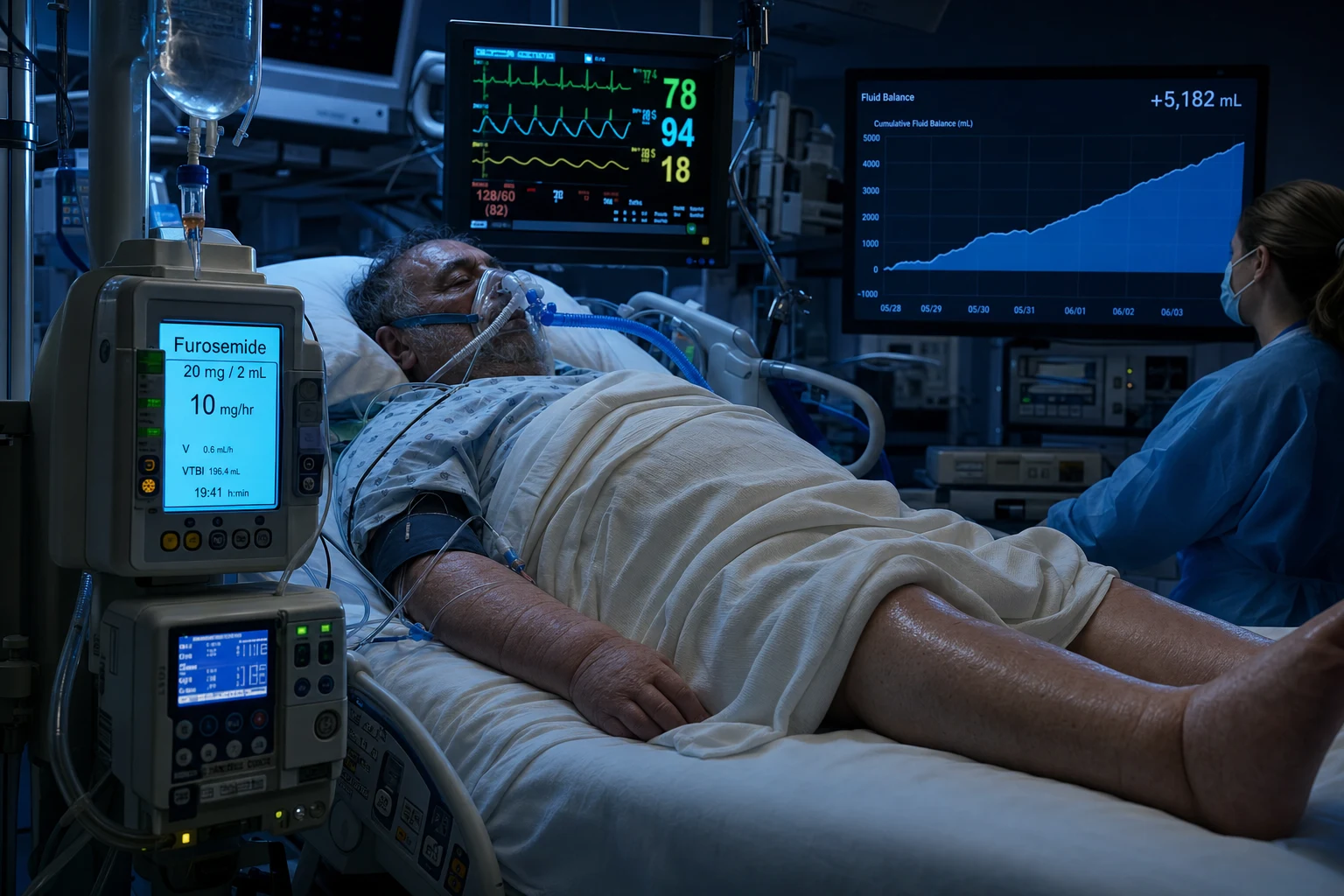
The ROSE model: four phases of fluid therapy
Fluid therapy in the critically ill is not a single act but a dynamic, time-dependent process. The ROSE concept (Rescue → Optimisation → Stabilisation → Evacuation) frames fluid management as a therapeutic arc: early aggressive resuscitation gives way to conservative management and then active fluid removal. The cardinal error is failing to transition between phases — continuing to give fluid when the patient is no longer in shock, or failing to de-resuscitate accumulated fluid during recovery. [1]
ROSE model: the four phases of fluid therapy
Phase 1 — Rescue (Salvage)
TIME: minutes to first few hours. GOAL: restore life-threatening perfusion deficit — correct hypotension, restore cardiac output, reverse shock. ACTION: rapid boluses of balanced crystalloid (250–500 mL over 15–30 min, up to 30 mL/kg in septic shock per SSC). Give to any patient with overt shock (hypotension, mottled skin, altered mentation, oliguria). Endpoints are macroscopic: MAP >65 mmHg, improving lactate, skin perfusion. This phase is life-saving — do NOT delay fluid for dynamic assessment when shock is obvious. The patient should be POSITIVE in fluid balance.
Phase 2 — Optimisation (Titration)
TIME: hours 2–24. GOAL: optimise tissue perfusion while avoiding overload. ACTION: give fluid ONLY to fluid-responsive patients, using DYNAMIC indices (passive leg raise, pulse pressure variation >13%, stroke volume variation, IVC collapsibility). Smaller titrated boluses (100–250 mL). Introduce vasopressors early to reduce total fluid volume. Use inotropes if low cardiac output. Consider the four pillars of fluid stewardship: (1) give fluid only if responsive, (2) use the right fluid (balanced crystalloid), (3) stop before overload, (4) remove excess fluid. Endpoints shift from macro to micro: ScvO2 >70%, lactate clearance >10%/h, urine output >0.5 mL/kg/h. Balance should still be positive but with diminishing magnitude.
Phase 3 — Stabilisation (Equilibrium)
TIME: day 2 onward (once shock resolved). GOAL: achieve ZERO or slightly negative fluid balance — neither gain nor lose. ACTION: STOP maintenance fluids (enteral nutrition provides most water needs). Eliminate all unnecessary fluid: drug dilution in minimal volume, concentrated medications, avoid fluid "creep" from flushes and carrier fluids. Monitor daily weights, fluid balance charts, IVC ultrasound. The patient should no longer require boluses. If haemodynamics are stable without fluid, you have reached this phase. Continue vasopressor weaning, nutrition, rehabilitation.
Phase 4 — De-escalation / Evacuation (De-resuscitation)
TIME: recovery phase — days to weeks. GOAL: achieve NEGATIVE fluid balance — actively remove accumulated fluid to return to admission (dry) weight. ACTION: furosemide IV bolus (20–40 mg, titrate) or continuous infusion (5–20 mg/h — more predictable, preferred for ICU). Add thiazide (metolazone 5–10 mg or chlorothiazide) for sequential nephron blockade if loop diuretic-resistant. Use albumin 20% to maintain oncotic pressure if hypoalbuminaemic (albumin <25 g/L). CRRT for refractory overload (especially with concomitant AKI). Target: net negative balance of 1–3 L/day, guided by haemodynamic tolerance, electrolytes, and daily weights. Monitor IVC, B-lines on lung ultrasound, oxygenation. Failure to de-resuscitate independently predicts mortality.
Why fluid overload kills: pathophysiology
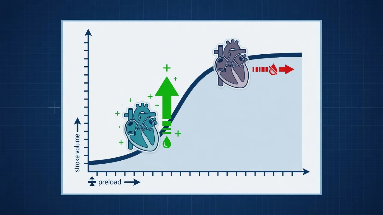
Tissue and organ oedema
The core mechanism
- Fluid leaks into interstitium → organ swelling → increased tissue pressure → impaired microvascular perfusion and oxygen diffusion
- PULMONARY: interstitial and alveolar oedema → reduced compliance, worsened V/Q mismatch, hypoxaemia, prolonged mechanical ventilation (FACTT trial showed conservative fluid strategy increased ventilator-free days)
- RENAL: renal capsule constriction, raised intra-abdominal pressure → renal venous congestion → reduced GFR → worsening AKI (a vicious cycle — AKI impairs fluid excretion → more overload → more AKI)
- GUT: bowel wall oedema → impaired motility (ileus), bacterial translocation, malabsorption of enteral nutrition → delayed recovery
- BRAIN: cerebral oedema (especially relevant in TBI, hepatic encephalopathy — even small fluid gains worsen outcomes)
Abdominal compartment syndrome
A deadly consequence
- Fluid accumulation → bowel oedema and third-spacing → raised intra-abdominal pressure (IAP)
- IAP >20 mmHg with new organ failure = abdominal compartment syndrome (ACS)
- ACS causes: renal dysfunction (renal venous congestion), respiratory compromise (elevated diaphragm, reduced compliance), cardiovascular (reduced venous return, high peak airway pressures compressing heart), splanchnic ischaemia
- Measure bladder pressure in patients with large fluid gains, distended abdomen, oliguria, high peak pressures
- Treatment: decompression (urgent), but PREVENTION via fluid stewardship is paramount
Global Increased Permeability Syndrome (GIPS)
Cordemans 2012 — the third hit
- Cordemans described GIPS as the "third hit" of shock: (1) initial insult → (2) ischaemia-reperfusion/MODS → (3) GIPS
- GIPS = persistent systemic inflammation + ongoing transcapillary albumin leakage + inability to achieve negative fluid balance
- The endothelial glycocalyx is destroyed → capillary leak → fluid and albumin extravasate → worsening oedema → more fluid needed to maintain intravascular volume → self-perpetuating cycle
- Markers: high Capillary Leak Index (CLI = CRP/albumin ratio), persistent elevation of extravascular lung water index (EVLWI)
- These patients CANNOT be fluid-resuscitated effectively — the more fluid you give, the more it leaks out. Treatment: minimise fluid, use vasopressors, albumin 20%, active de-resuscitation
Impaired wound healing and infection
Indirect harm
- Tissue oedema increases wound tension, reduces oxygen delivery to tissues → impaired healing, dehiscence
- Oedematous tissue is a culture medium — increased risk of surgical site infection, line infection, pressure injury
- Peripheral oedema makes vascular access difficult, increases DVT risk from immobility, impairs mobilisation and rehabilitation
Fluid balance tracking: cumulative balance and the 10% rule
Cumulative fluid balance is the single most important metric for monitoring fluid overload. It is the algebraic sum of ALL inputs (IV fluids, enteral, parenteral nutrition, drug diluents, blood products) minus ALL outputs (urine, drains, NG aspirate, fistula, faeces, insensible losses) from ICU admission to the present. [1]
Daily fluid balance
The building block
- Calculate EVERY day: total inputs − total outputs = net balance (positive or negative)
- Resuscitation phase: expect POSITIVE balance (often +2 to +5 L/day in early septic shock)
- Stabilisation phase: aim for ZERO balance (±500 mL)
- De-escalation phase: target NEGATIVE balance (−1 to −3 L/day, guided by haemodynamic tolerance)
- Insensible losses (~10 mL/kg/day from skin and respiration, more with fever) are often undercounted — adjust for fever, open wounds, drains
Cumulative fluid balance
The cumulative burden
- Running total from admission. This is the metric that predicts harm
- Express as percentage of body weight: cumulative balance (L) / body weight (kg) x 100
- Cumulative balance >10% body weight = SIGNIFICANT fluid overload — associated with increased mortality, prolonged ventilation, AKI
- Example: 70 kg patient with cumulative +7L balance → 7/70 = 10% → significant overload
- Each litre of cumulative positive balance independently increases mortality risk in observational studies
Daily weight
The gold standard
- Daily weighing (bed scale) is the most objective measure of fluid status — 1 kg weight change ≈ 1 L fluid
- Superior to fluid balance charts (which miss insensible losses, inaccurate output measurements)
- Compare to admission "dry" weight to track cumulative gain
- Target during de-resuscitation: return to dry weight over days
- Practical limitation: requires bed with scale, may be unreliable with dressings, lines, oedema distribution
POCUS and bedside monitoring
Real-time assessment
- IVC ultrasound: collapsible IVC (collapsibility >50%) suggests volume responsiveness; plethoric/fixed IVC (collapsibility <50%) suggests overload — DO NOT give fluid
- Lung ultrasound: B-lines (comet-tail artefacts) = interstitial/alveolar oedema — the more B-lines, the worse the overload
- Bilateral pleural effusions on POCUS
- Echocardiography: assess cardiac function, right heart strain, estimate filling pressures (E/e')
- Bioimpedance (research tool) can quantify total body water compartments
Clinical signs of fluid overload
Recognising fluid overload requires a systematic multi-system assessment. No single sign is pathognomonic — combine clinical examination, POCUS, and balance data. [1]
Respiratory signs
Most life-threatening
- Pulmonary oedema: bilateral crackles, frothy sputum (pink if haemorrhagic), hypoxaemia, increased work of breathing
- Pleural effusion: decreased breath sounds, dullness to percussion, reduced chest expansion
- Reduced lung compliance on ventilator: rising peak and plateau pressures for same tidal volume
- Increasing FiO2 and PEEP requirements, worsening oxygenation (falling P/F ratio)
- Lung ultrasound: B-lines in multiple zones, pleural effusions
Cardiovascular signs
Filling pressure clues
- Raised JVP (jugular venous pressure) — visible distension >3 cm above sternal angle
- IVC plethora on POCUS — dilated, non-collapsible IVC (collapsibility <50% with sniff)
- Third heart sound (S3) gallop in volume overload with cardiac dysfunction
- Hypertension (sometimes), or paradoxically hypotension if cardiac failure from overload
- Peripheral (sacral, ankle) oedema — pitting oedema is a LATE sign (10+ kg fluid retention before visible)
Abdominal signs
Often overlooked
- Ascites: shifting dullness, fluid thrill, abdominal distension
- Raised intra-abdominal pressure: measure bladder pressure if at risk (>12 mmHg = intra-abdominal hypertension; >20 mmHg + organ failure = compartment syndrome)
- Bowel sounds reduced (ileus from gut wall oedema)
- Abdominal POCUS: free fluid, bowel wall thickening
Tissue and general signs
Cumulative burden
- Anasarca: generalised severe oedema — face, trunk, limbs, genitalia
- Peripheral oedema: pedal/sacral/dependent — pitting (graded 1+ to 4+)
- Weight gain: most reliable objective sign (1 kg ≈ 1 L fluid)
- Reduced urine output: may indicate AKI from venous congestion, or may be the cause of overload
- Conjunctival and peripheral oedema visible on inspection
Fluid responsiveness assessment
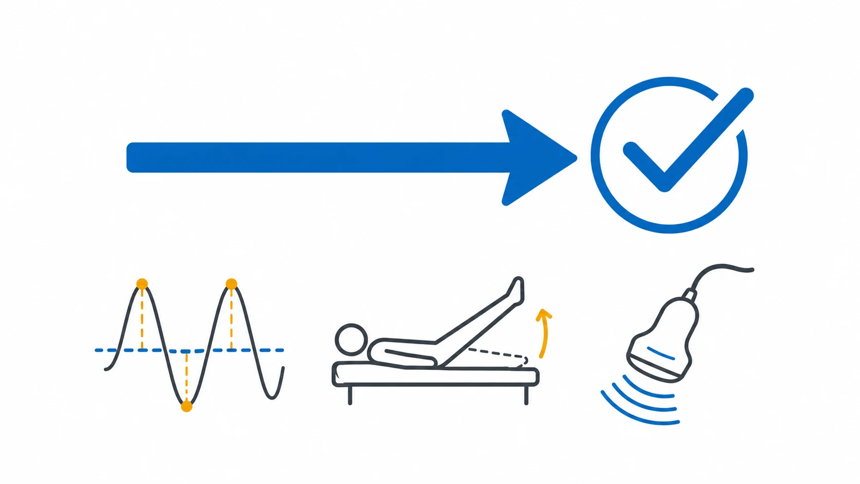
Before giving ANY fluid in the optimisation or stabilisation phases, assess fluid responsiveness. A fluid challenge to a non-responsive patient does not increase cardiac output — it only increases venous congestion, oedema, and harm. The principle: only give fluid if the heart is operating on the steep portion of the Frank-Starling curve (i.e., the ventricles are preload-responsive). [1]
Passive leg raise (PLR)
Best overall test
- Self-volume challenge: elevating legs to 45° auto-transfuses ~300 mL venous blood from legs to heart
- Positive if cardiac output/stroke volume increases >10% (measure with echocardiography LVOT VTI, pulse contour, or even pulse pressure if arterial line present)
- Advantages: reversible, repeatable, does not require fluid, works in spontaneous breathing and arrhythmia
- Limitations: requires real-time CO measurement (not just BP), cannot do with leg amputation/compression, patient must be in semi-recumbent start position
- The gold-standard dynamic test — use it FIRST before any fluid bolus in the optimisation phase
Pulse pressure variation (PPV) / SVV
Best for ventilated patients
- In mechanically ventilated patients with controlled ventilation, positive pressure inspiration reduces RV preload and increases RV afterload → cyclic variation in LV stroke volume → variation in pulse pressure
- PPV >13% (or SVV >10%) predicts fluid responsiveness with good sensitivity/specificity
- REQUIREMENTS: fully controlled ventilation (no spontaneous breaths), tidal volume >8 mL/kg, sinus rhythm (no AF), closed chest, no right heart failure
- LIMITED by: spontaneous breathing, arrhythmia, low tidal volume ventilation, open chest, severe RV failure, low lung compliance
- Requires arterial line — use PPV from the monitor. If PPV is HIGH, patient is fluid-responsive. If LOW, giving fluid will NOT help
IVC collapsibility
Simplest bedside test
- Subcostal POCUS: measure IVC diameter and collapsibility with respiration
- Collapsible IVC (CI >50%, small diameter) → likely volume-responsive (low right atrial pressure)
- Plethoric/fixed IVC (CI <50%, large diameter >2.5 cm) → NOT volume-responsive (high right atrial pressure) — DO NOT give fluid
- In ventilated patients: IVC DISTENSIBILITY (increase with inspiration) >18% suggests responsiveness
- Advantages: quick, non-invasive, repeatable. Limitations: semi-quantitative, operator-dependent, less reliable in spontaneous breathing with variable effort
Static markers (limited use)
Unreliable alone
- CVP (central venous pressure): traditionally used, but POOR predictor of fluid responsiveness — do NOT use CVP alone to guide fluid
- Heart rate, blood pressure, capillary refill: LATE and non-specific markers — tachycardia may be pain, fever, anxiety, not hypovolaemia
- Lactate: marker of hypoperfusion, not volume status — elevated lactate needs investigation, not automatic fluid
- Urine output: oliguria may be volume-responsive, but also occurs with AKI, abdominal hypertension, or as a stress response
- PRINCIPLE: dynamic tests (PLR, PPV, IVC) are always superior to static markers for fluid responsiveness
Fluid balance targets by phase
The target fluid balance CHANGES with the phase of illness. The most common error is applying a one-size-fits-all approach — either always positive (fluid "for low BP") or always negative. [1]
Resuscitation phase
POSITIVE balance target
- INDICATION: active shock, hypotension, overt hypoperfusion
- TARGET: positive balance — give fluid boluses to fluid-responsive patients
- TYPICAL VOLUME: up to 30 mL/kg crystalloid in first hours of septic shock (SSC guideline), titrated to response
- CAVEAT: even in this phase, assess responsiveness — do NOT blindly give 30 mL/kg as a single bolus. The FEAST trial showed that routine bolus therapy in patients without true hypovolaemic shock can be harmful
- Transition out of this phase when: shock resolves, lactate clearing, vasopressors being titrated, patient no longer fluid-responsive
Stabilisation phase
ZERO balance target
- INDICATION: shock resolved, on decreasing vasopressors or off them, stable haemodynamics
- TARGET: zero (even) balance — neither gain nor loss
- ACTION: stop maintenance IV fluids (enteral nutrition provides water), concentrate drug infusions, minimise flush volumes
- MONITOR: daily weights (should plateau), IVC, lung ultrasound, fluid balance chart
- This is a transitional phase — do not linger here. Move to de-resuscitation when stable
De-escalation phase
NEGATIVE balance target
- INDICATION: haemodynamically stable, cumulative fluid balance significantly positive, signs of overload (oedema, pulmonary congestion, weight above dry weight)
- TARGET: NEGATIVE balance — actively remove fluid
- TYPICAL TARGET: −1 to −3 L/day, guided by haemodynamic tolerance (watch BP, lactate, urine output — do not over-diurese into hypovolaemia)
- METHOD: furosemide (IV bolus or infusion), add thiazide if loop-resistant, albumin 20% for oncotic pull, CRRT if diuretic-resistant or concurrent AKI
- ENDPOINT: return to admission dry weight, resolution of oedema, normalisation of IVC, improvement in oxygenation
Management of fluid overload: de-resuscitation toolkit
De-resuscitation: stepwise approach to fluid removal
Step 1 — Stop fluid input
The simplest and most effective intervention. Review ALL IV fluids: stop maintenance fluids (enteral nutrition provides water), concentrate all drug infusions (noradrenaline in 50 mL not 250 mL, use double-strength preparations), minimise flush volumes, review blood product indications (do not transfuse for numbers alone). A "fluid stewardship" ward round with the pharmacist can eliminate 1–2 L/day of unnecessary fluid.
Step 2 — Furosemide (loop diuretic) — first-line
FUROSEMIDE IV is the mainstay of de-resuscitation. Start with bolus 20–40 mg IV, assess response in 30–60 min (urine output should increase). If response inadequate, double dose (40 → 80 → 120 mg). For sustained removal, use CONTINUOUS INFUSION (5–20 mg/h) — more predictable, less ototoxic, easier to titrate than repeated boluses. Target net negative balance 1–3 L/day. MONITOR: electrolytes (hypokalaemia, hypomagnesaemia, hypocalcaemia, metabolic alkalosis), serum creatinine (avoid over-diuresis into prerenal AKI), blood pressure, and urine output. A furosemide stress test (1–1.5 mg/kg IV bolus with urine output measured over 2h) can assess renal reserve — UO >200 mL/2h suggests viable tubular function.
Step 3 — Sequential nephron blockade (thiazide)
If loop diuretic alone fails (diuretic resistance — common in chronic kidney disease, heart failure, hypoalbuminaemia), add a THIAZIDE diuretic to block the distal convoluted tubule: METOLAZONE 5–10 mg PO/NG (preferred — long-acting, potent even in CKD) or CHLORTHALIDONE or IV CHLOROTHIAZIDE. The combination (loop + thiazide) produces synergistic natriuresis. CAUTION: this can cause MASSIVE diuresis and electrolyte derangement — start low (metolazone 2.5–5 mg), monitor closely (K, Mg every 6–12h). Hypokalaemia and hypomagnesaemia are near-universal — replace aggressively.
Step 4 — Albumin 20% (oncotic therapy)
In hypoalbuminaemic patients (albumin <25 g/L), fluid leaks into interstitium and furosemide is less effective (it binds albumin to reach its site of action). Giving ALBUMIN 20% (100–200 mL) followed by furosemide increases oncotic pressure, pulls fluid back into the intravascular space, and potentiates diuresis. The SAFE trial sub-study suggested albumin may benefit severe sepsis. Use selectively: do NOT give albumin routinely for resuscitation (SAFE: equivalent to saline). Albumin 20% (not 4%) is preferred for oncotic pull. Limited evidence for long-term outcome benefit — use as an adjunct, not primary therapy.
Step 5 — CRRT (for refractory overload)
If diuretics fail or the patient has concurrent AKI preventing diuresis, initiate CRRT (continuous renal replacement therapy — CVVHDF or CVVH) for slow, controlled fluid removal. Advantages of CRRT: precise fluid removal (set net ultrafiltration target, e.g. −100 mL/h = −2.4 L/day), haemodynamic stability (slow continuous removal better tolerated than intermittent HD), concurrent solute clearance (urea, creatinine, potassium). Set net UF based on haemodynamic tolerance — start at −50 to −100 mL/h, increase as tolerated. Monitor: MAP (avoid hypotension from excessive UF), electrolytes (CRRT removes K, Mg, PO4 — replace), filter life (anticoagulation). CRRT for fluid overload alone (without uraemia/electrolyte indication) is justified in severe refractory overload.
Step 6 — Monitor and titrate
Track response DAILY: weights (aim for downward trend toward dry weight), fluid balance (confirm net negative), IVC (should become more collapsible), lung ultrasound (B-lines should decrease), oxygenation (should improve). Adjust diuretic/CRRT prescription. Watch for over-diuresis: hypotension, rising lactate, worsening AKI, electrolyte derangements. The endpoint is return to admission weight, resolution of clinical overload signs, and improved organ function.
Key trials in fluid overload and fluid strategy
FACTT — Fluid and Catheter Treatment Trial (Wiedemann, NEJM 2006)
Multicentre RCT: 1000 patients with ALI/ARDS (ARDSNet)
Population: Mechanically ventilated adults with acute lung injury
Key finding
No significant difference in 60-day mortality (25.5% vs 28.4%, p=0.30). BUT conservative strategy: MORE ventilator-free days (14.6 vs 12.1, p<0.001), MORE ICU-free days (13.4 vs 11.2, p=0.003), improved oxygenation, NO increase in non-pulmonary organ failure, NO increase in shock. Conservative fluid management in ALI/ARDS is SAFE and BENEFICIAL.
Practice change
A conservative (restricted) fluid strategy in ALI/ARDS shortens ventilation and ICU stay without causing harm. Fluid overload in ARDS worsens pulmonary outcomes. Target a negative or even fluid balance once shock resolves.
CLASSIC — Conservative vs Liberal Approach to fluid therapy of Septic Shock (Meyhoff, NEJM 2022)
International multicentre open-label RCT: 1554 adults with septic shock
Population: ICU patients with septic shock who had received at least 1L IV fluid
Key finding
No significant difference in 90-day mortality (42.3% restrictive vs 42.1% standard, adjusted RR 1.00, 95% CI 0.89–1.13, p=0.96). The restrictive group received significantly less IV fluid (mean 1798 mL vs 3811 mL during ICU stay). No increase in serious adverse events. Pre-specified subgroup analysis: signal toward benefit with restrictive strategy in patients with less severe shock at enrolment and in patients without metastatic cancer.
Practice change
A restrictive fluid strategy in septic shock is SAFE — it does not increase mortality despite giving ~2L less fluid. The trial was underpowered for the modest effect size. The signal favours restriction, supporting modern fluid stewardship. Less fluid = less overload = less harm, with equivalent outcomes.
FEAST — Fluid Expansion As Supportive Therapy (Maitland, NEJM 2011)
Multicentre open-label RCT: 3170 children in Uganda, Kenya, Tanzania
Population: Children (60 days–12 years) with severe febrile illness and impaired perfusion (resource-limited setting)
Key finding
BOLUS INCREASED MORTALITY. 48-hour mortality: 10.5% (saline bolus), 10.6% (albumin bolus) vs 7.3% (no bolus). Relative risk of death with bolus: 1.45 (95% CI 1.13–1.86, p=0.003). Trial STOPPED EARLY for harm. The mechanism: bolus fluids in children without hypovolaemic shock caused cardiogenic pulmonary oedema, intracranial hypertension, and haemodynamic compromise.
Practice change
Fluid bolus therapy is NOT benign — in patients without true hypovolaemic shock, it can be HARMFUL. This landmark trial revolutionised paediatric and adult fluid resuscitation thinking: assess fluid responsiveness before giving boluses, do not give fluid reflexively. Fluid is a drug with a therapeutic window — too little is dangerous, but too much is equally dangerous.
Clinical pearls
Red flags
Prognosis
[1]Exam practice — SAQ
SAQ — Fluid overload and de-resuscitation
10 minutes · 10 marks
A 68-year-old is day 4 after septic shock. Off vasopressors. Cumulative balance +9 L. Increasing oxygen need, B-lines bilaterally, creat 160, still making 0.6 mL/kg/h urine.
References
- [1]Cordemans C, De Laet I, Van Regenmortel N, et al. Fluid management in critically ill patients: the role of extravascular lung water, abdominal hypertension, capillary leak, and fluid balance Ann Intensive Care, 2012.PMID 22873410
- [2]Maitland K, Kiguli S, Opoka RO, et al. Mortality after fluid bolus in African children with severe infection N Engl J Med, 2011.PMID 21615299
- [3]Silversides JA, Fitzgerald E, Manu VS, et al. Highly selective solid-phase extraction sorbents for chloramphenicol determination in food and urine by ion mobility spectrometry Anal Bioanal Chem, 2016.PMID 27734138
- [4]Wiedemann HP, Wheeler AP, Bernard GR, et al. (ARDSNet/FACTT). Comparison of two fluid-management strategies in acute lung injury N Engl J Med, 2006.PMID 16714767
- [5]Vincent JL, Sakr Y, Sprung CL, et al. (SOAP Study). Sepsis in European intensive care units: results of the SOAP study Crit Care Med, 2006.PMID 16424713
- [6]Meyhoff TS, Hjortrup PB, Wetterslev J, et al. (CLASSIC). Restriction of Intravenous Fluid in ICU Patients with Septic Shock N Engl J Med, 2022.PMID 35709019