ICU · Renal / acid-base
Lactic Acidosis — Type A, B & D, MALA, and Lactate Clearance
Also known as Lactic acidosis · Lactate · Type A lactic acidosis · Type B lactic acidosis · D-lactic acidosis · Metformin-associated lactic acidosis · MALA · Lactate clearance · Cori cycle · Propofol infusion syndrome · PRIS
The lactic acidosis is the high-anion-gap metabolic acidosis from the accumulation of the lactate. The Cohen and Woods classification: the Type A — the tissue hypoxia or the impaired oxygen delivery (the shock, the seizures, the mesenteric ischaemia, the carbon monoxide, the severe anaemia) — the commonest; the Type B — no overt hypoxia (the B1 disease — the liver failure, the malignancy; the B2 drugs — the metformin, the linezolid, the NRTIs, the propofol infusion syndrome, the cyanide; the B3 inborn errors); the Type D — the D-lactate from the gut bacteria in the short-bowel syndrome (NOT measured by the standard L-lactate assay). The lactate is cleared by the liver (the Cori cycle); the impaired clearance worsens the accumulation. The prognostic: the lactate over 4 severe; the clearance over 10 per cent per hour in the sepsis equals the better survival. The management: the treat the underlying cause (the cornerstone — restore the oxygen delivery for A, stop the drug for B2, the liver failure for B1), the supportive care, the metformin-associated lactic acidosis (the haemodialysis), the bicarbonate controversial (the pH under 7.1), the methylene blue emerging.
On this page & tools
Your progress
Saved locally on this device.
8 MCQs with explanations
Target exams
Overview & definition
The lactic acidosis is the high-anion-gap metabolic acidosis from the accumulation of the lactate. The lactate is the product of the anaerobic glycolysis — the pyruvate is converted to the lactate (by the LDH) when the oxygen is scarce or the mitochondria are overwhelmed. The lactate is normally cleared by the liver (the Cori cycle — the lactate to the glucose) and the kidney. The accumulation causes the high-AG acidosis, and the lactate level and the clearance are the prognostic markers in the critical illness.[1]
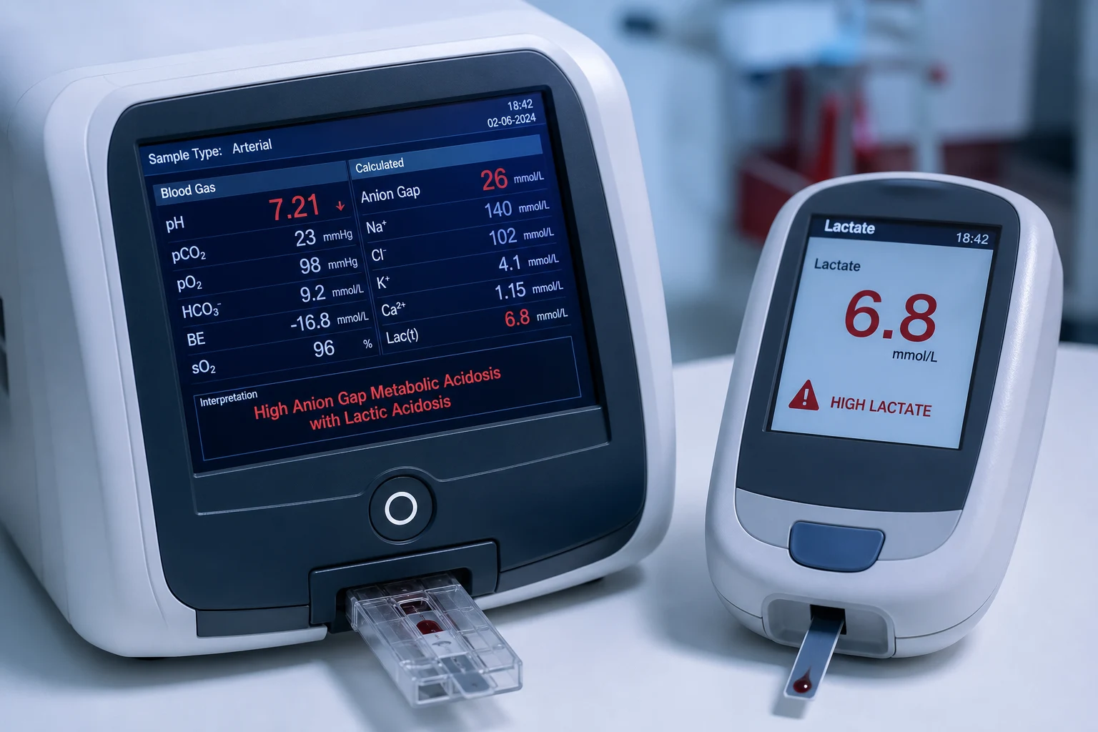
The classification (the Cohen and Woods)
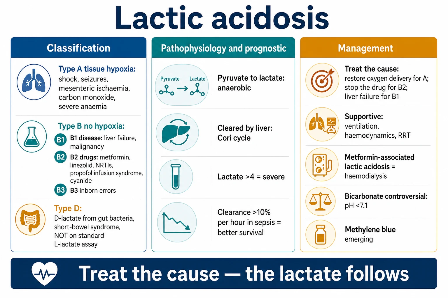
Type A — the tissue hypoxia (the commonest)
The Type A is the impaired oxygen delivery. Causes:[1]
- The shock — the septic, the hypovolaemic, the cardiogenic, the obstructive.[1]
- The severe hypoxaemia, the carbon monoxide, the severe anaemia.[1]
- The mesenteric ischaemia (the bowel hypoperfusion).[1]
- The seizures, the severe exercise, the shivering.[1]
Type B — no overt tissue hypoxia
- B1 — the underlying disease: the liver failure (the impaired clearance), the malignancy (the Warburg effect, the leukaemia, the lymphoma), the diabetes, the renal failure, the sepsis (a B-component, though it overlaps the A).[1]
- B2 — the drugs and the toxins: the metformin (the biguanide inhibits the mitochondrial complex I), the linezolid, the propofol infusion syndrome (the PRIS), the NRTIs (the mitochondrial toxicity), the salicylate, the ethylene glycol, the cyanide (the mitochondrial poisoning), the isoniazid, the beta-agonists.[1]
- B3 — the inborn errors of metabolism (the mitochondrial disorders, the glycogen storage diseases).[1]
Type D — the D-lactic acidosis
A special form: the D-lactate produced by the gut bacteria in the short-bowel syndrome (or the blind-loop). The D-lactate is absorbed and causes the high-AG acidosis with the neurological features (the confusion, the ataxia). The D-lactate is NOT measured by the standard L-lactate assay — a special D-lactate assay is required. The treatment: the antibiotics (to reduce the bacterial load), the low-carbohydrate diet, the thiamine.[1]
The pathophysiology and the prognostic
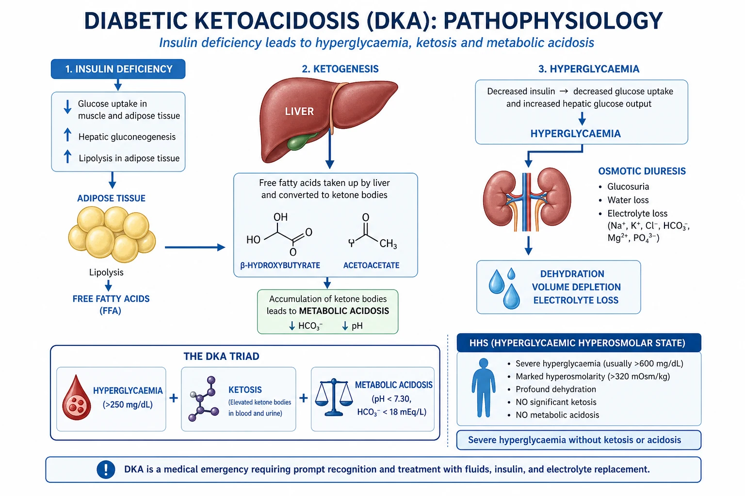
- The pyruvate → lactate (the LDH) in the anaerobic conditions; the lactate-to-pyruvate ratio rises.[1]
- The lactate is cleared by the liver (the Cori cycle) and the kidney. The liver failure impairs the clearance and worsens the accumulation.[1]
- The lactate over 4 mmol/L is the severe lactic acidosis; over 2 is the hyperlactataemia.[1]
- The lactate clearance of over 10 per cent per hour in the sepsis correlates with the better survival (the Jansen 2010; the lactate-guided resuscitation). The rising or the persistent lactate is the poor prognostic sign.[1]
The management
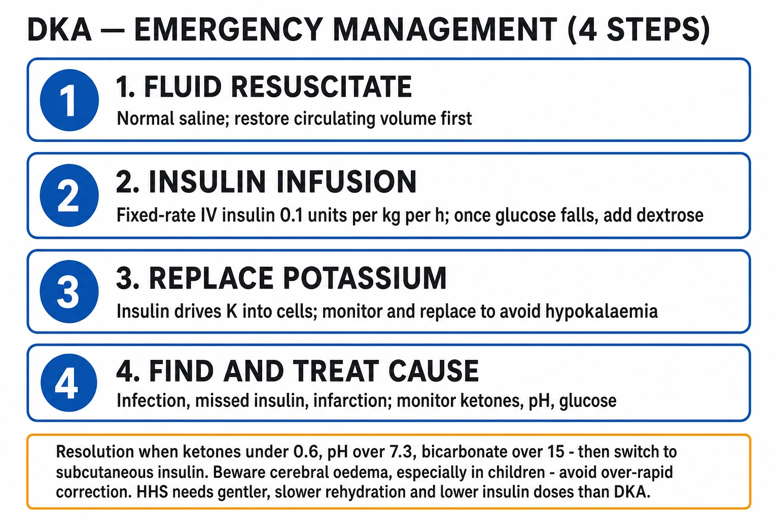
1. Treat the underlying cause (the cornerstone)
- The Type A — restore the oxygen delivery: the fluids, the blood, the inotropes, the vasopressors, the source control (the mesenteric ischaemia, the sepsis), the oxygen, the seizure control.[1]
- The Type B1 — the liver failure (the supportive, the transplant), the malignancy (the chemotherapy).[1]
- The Type B2 — stop the offending drug (the metformin, the linezolid, the propofol); the specific antidote where applicable (the hydroxocobalamin or the sodium thiosulfate for the cyanide, the fomepizole for the ethylene glycol).[1]
- The Type D — the antibiotics, the low-carbohydrate diet.[1]
2. The supportive care
- The ventilation and the haemodynamic support.[1]
- The renal replacement therapy — clears the lactate and the metformin; the high-flux haemodialysis for the MALA.[1]
- The bicarbonate — controversial; the pH under 7.1 (the severe acidosis with the haemodynamic compromise) may benefit transiently, but the bicarbonate generates the CO2 and worsens the intracellular acidosis. Use selectively, not routinely.[1]
- The thiamine for the suspected deficiency (the alcoholism, the malnutrition).[1]
3. The emerging and the specific
- The methylene blue — the mitochondrial electron carrier; emerging for the vasoplegic/septic lactic acidosis (the mitochondrial dysfunction). Controversial.[1]
- The MALA (the metformin-associated lactic acidosis) — the high-flux haemodialysis to clear the metformin and the lactate. The high mortality; the early recognition and the dialysis are the key.[1]
Exam practice
SAQ — Septic shock with persistent hyperlactataemia
10 minutes · 10 marks
A 58-year-old man is admitted to ICU with septic shock from a urinary source. He has received 30 mL/kg crystalloid, meropenem 1 g IV, and is now on noradrenaline 0.35 mcg/kg/min for MAP 65 mmHg. Lactate has risen from 3.2 to 6.8 mmol/L over 6 hours despite source control (nephrostomy) and an apparently adequate MAP. pH 7.25, HCO3 14, ScvO2 72%. He is 3.5 L in positive balance.
SAQ — Toxic alcohol poisoning (methanol / ethylene glycol)
10 minutes · 10 marks
A 44-year-old man is brought to the emergency department 8 hours after ingesting an unknown quantity of windshield washer fluid and antifreeze in a suicide attempt. He is confused and tachypnoeic (RR 32). ABG: pH 7.08, HCO3 8, pCO2 24, BE -22. Na 142, K 5.1, Cl 100, glucose 7.5, urea 6. Measured osmolality 360 mOsm/kg. Lactate reported as 9.2 mmol/L.
Red flags
The Cohen and Woods classification — the deep dive
The Cohen and Woods (1976) classification is the framework the examiners expect — it divides the lactic acidosis by the aetiology rather than the biochemistry.[1] The Type A is the tissue hypoxia (the impaired oxygen delivery or the impaired oxygen utilisation); the Type B is the no overt tissue hypoxia (sub-divided into the B1 — the underlying disease, the B2 — the drugs and the toxins, the B3 — the inborn errors of the metabolism); the Type D is the D-lactate from the gut bacteria. The reality in the ICU is that most of the critically ill have a mixed picture — the sepsis, for example, has the Type A component (the hypoperfusion) AND the Type B1 component (the mitochondrial dysfunction, the impaired clearance) at the same time.[1]
The Cohen and Woods — the comparison table
| Type | The mechanism | The representative causes | The key exam point |
|---|---|---|---|
| A | The impaired oxygen delivery or the utilisation | The shock (the septic, the cardiogenic, the hypovolaemic, the obstructive), the severe hypoxaemia, the carbon monoxide, the severe anaemia, the mesenteric ischaemia, the seizures, the severe exercise, the shivering | The commonest; restore the oxygen delivery; the lactate follows the cause |
| B1 | The underlying disease (the impaired clearance or the over-production) | The sepsis (a B-component), the liver failure, the malignancy (the leukaemia, the lymphoma — the Warburg effect), the diabetes, the renal failure | The mitochondrial dysfunction; the impaired hepatic clearance; overlaps the Type A in the sepsis |
| B2 | The drugs and the toxins | The metformin (the complex I), the linezolid (the mitochondrial toxicity), the propofol infusion syndrome (the PRIS), the NRTIs, the salicylate, the ethylene glycol, the cyanide (the cytochrome c oxidase), the isoniazid, the beta-agonists, the biguanides | Stop the offending drug; the specific antidote where applicable |
| B3 | The inborn errors of the metabolism | The pyruvate dehydrogenase deficiency, the pyruvate carboxylase deficiency, the mitochondrial disorders (the MELAS, the MERRF), the glycogen storage diseases (the McArdle, the von Gierke), the fructose-1,6-diphosphatase deficiency | The paediatric or the lifelong; the congenital lactic acidosis |
| D | The D-lactate from the gut bacteria | The short-bowel syndrome, the jejuno-ileal bypass, the blind-loop syndrome | NOT measured by the standard L-lactate assay; the special D-lactate assay |
The pathophysiology — the anaerobic glycolysis, the pyruvate-to-lactate, and the Cori cycle
The lactate is produced from the pyruvate by the lactate dehydrogenase (the LDH). In the normal aerobic metabolism, the pyruvate enters the mitochondria and is converted to the acetyl-CoA (by the pyruvate dehydrogenase, PDH) → the Krebs cycle → the oxidative phosphorylation → 36 ATP. When the oxygen is scarce (the anaerobic conditions) OR the mitochondrial function is impaired, the pyruvate cannot enter the mitochondria and is shunted to the lactate (by the LDH) → only 2 ATP per glucose (the anaerobic glycolysis is energetically inefficient).[1]
The biochemical pathway
flowchart LR
    A[Glucose] -->|glycolysis| B[Pyruvate]
    B -->|aerobic: PDH| C[Acetyl-CoA]
    C -->|Krebs cycle + OxPhos| D[36 ATP + CO2 + H2O]
    B -->|anaerobic: LDH| E[Lactate + 2 ATP]
    E -->|Cori cycle: liver/kidney| F[Gluconeogenesis]
    F --> A
    E -.->|impaired clearance: liver failure| G[Accumulation → acidosis]
The key enzymes and the rate-limiting steps
- The pyruvate dehydrogenase (PDH) — the gatekeeper. The PDH converts the pyruvate to the acetyl-CoA (the aerobic path). The PDH is inhibited by the NADH (the high NADH/NAD ratio pushes the pyruvate to the lactate). The thiamine is the cofactor for the PDH — the thiamine deficiency impairs the PDH and causes the lactic acidosis.[8]
- The lactate dehydrogenase (LDH) — converts the pyruvate ↔ the lactate (reversible). The equilibrium favours the lactate when the NADH is high.
- The pyruvate carboxylase — the alternative aerobic path; converts the pyruvate to the oxaloacetate (the gluconeogenesis). Requires the biotin.
The lactate-to-pyruvate ratio — the diagnostic clue
| The L/P ratio | The interpretation | The examples |
|---|---|---|
| High (over 10–13) | The anaerobic glycolysis — the NADH excess; the impaired oxidation | The shock, the seizures, the mesenteric ischaemia, the cyanide, the CO |
| Normal (around 10) | The aerobic over-production or the impaired clearance — the pyruvate and the lactate rise together | The malignancy (the Warburg), the PDH deficiency, the thiamine deficiency, the D-lactate |
The clearance — the liver and the kidney
The lactate is cleared at approximately 0.5–1.0 mmol/L/hour in the healthy adult. The liver handles about 50–70 per cent of the clearance (the Cori cycle — the gluconeogenesis); the kidney handles the rest (the gluconeogenesis and the oxidation). The liver can clear the lactate up to a maximum of about 4 mmol/L/hour before it is overwhelmed. The hepatic clearance is impaired by the liver failure, the hypoperfusion (the liver ischaemia), the hypoxia, and the severe acidosis (the pH under 7.1 inhibits the hepatic gluconeogenesis — a vicious cycle).[1]
The D-lactic acidosis — the short-bowel syndrome and the gut bacteria
The D-lactic acidosis is the special form. The mammalian tissues produce ONLY the L-lactate (the L-isomer). The D-lactate is produced by the bacteria in the colon. In the short-bowel syndrome (or the jejuno-ileal bypass, or the blind-loop syndrome), the unabsorbed carbohydrates reach the colon in excess; the bacteria (the lactobacilli, the bifidobacteria, the eubacteria) ferment the carbohydrates to the D-lactate; the D-lactate is absorbed and causes the high-AG acidosis.[1]
The clinical features — the neurological and the acidotic
The D-lactate crosses the blood-brain barrier (the D-lactate is a neuromodulator) and causes the neurological features: the encephalopathy, the confusion, the ataxia, the slurred speech, the nystagmus, the irritability — these can mimic the alcohol intoxication. The picture: the short-bowel patient with the unexplained high-AG acidosis + the neurological features + the normal L-lactate.[1]
The KEY — the D-lactate is NOT measured by the standard assay
The standard point-of-care and the laboratory lactate assays use the L-lactate oxidase (or the L-LDH), which is specific for the L-isomer and does NOT detect the D-lactate. The D-lactate requires a special D-lactate assay (the D-LDH enzymatic assay). This is the classic exam trap: the high-AG acidosis with the normal L-lactate in the short-bowel patient → order the D-lactate assay.[1]
The MALA — the metformin-associated lactic acidosis
The metformin (the biguanide) is the first-line oral hypoglycaemic for the type 2 diabetes. The metformin inhibits the mitochondrial complex I → the impaired oxidative phosphorylation → the increased anaerobic glycolysis → the lactic acidosis. The metformin is renally cleared; in the renal failure (the AKI, the CKD) the metformin accumulates and the lactic acidosis supervenes. The MALA is uncommon but the high mortality (the reported mortality of 30–50 per cent).[5]
The MALA — the risk factors
The MALA occurs almost exclusively in the setting of the accumulated metformin — the precipitating factor is usually the acute kidney injury (the AKI from any cause — the sepsis, the dehydration, the contrast, the surgery) in the patient taking the metformin. The other risk factors: the liver failure, the heart failure, the sepsis, the hypoperfusion, the chronic kidney disease, the age over 80, and the iodinated contrast (the held before the contrast). The metformin itself is rarely the sole cause in the patient with the normal renal function — the Cochrane review found no increased risk of the lactic acidosis with the metformin versus the other oral hypoglycaemics in the well-selected patients.[5]
The MALA — the management
- The stop the metformin — the first step.
- The supportive care — the ventilation, the haemodynamics, the correction of the acidosis.
- The high-flux haemodialysis — clears BOTH the metformin (the MW 129, the small, the water-soluble, the low protein binding — the highly dialysable) AND the lactate. The bicarbonate-based dialysate corrects the acidosis. The CVVHDF is the alternative if the haemodynamically unstable. The indications for the dialysis: the lactate over 20 mmol/L, the pH under 7.0–7.1, the metformin level over 5–10 mg/L, the failure of the standard therapy, or the severe AKI.[5]
- The bicarbonate — the controversial; the MALA is the severe acidosis and the bicarbonate (the high-flux bicarbonate dialysate) is used to correct the pH to over 7.1 to support the haemodynamics while the metformin clears. Avoid the over-correction (the sodium overload, the hypernatraemia, the alkalaemia).[5]
The PRIS — the propofol infusion syndrome
The propofol infusion syndrome (the PRIS) is the rare but the fatal complication of the prolonged, the high-dose propofol infusion. The PRIS is a Type B2 lactic acidosis (the mitochondrial toxicity). The pathophysiology: the propofol impairs the mitochondrial fatty-acid oxidation and the oxidative phosphorylation → the lactic acidosis, the rhabdomyolysis, the myocardial failure, the hepatomegaly, the lipaemia.[6]
The PRIS — the risk factors and the threshold
- The high dose — over 4 mg/kg/hour (the infusions over 5 mg/kg/hour for over 48 hours are the high risk).
- The prolonged duration — over 48 hours.
- The concurrent catecholamines (the vasopressors) and the corticosteroids — the additive mitochondrial toxicity.
- The severe head injury, the severe sepsis, the paediatric patient, the poor nutrition — the higher susceptibility. [1]
The PRIS — the clinical features
The PRIS presents with the metabolic acidosis (the high lactate), the rhabdomyolysis (the rising CK), the cardiac failure (the bradycardia, the low-output, the arrhythmias — the classical the Brugada-like ECG), the hepatomegaly (the fatty liver), the lipaemia (the milky serum), the hyperkalaemia, and the renal failure. The mortality is the high (over 50 per cent).[6]
The PRIS — the management
The stop the propofol — the first and the most important step. The supportive care — the ventilation, the haemodynamics (avoid the high-dose catecholamines — they worsen the mitochondrial toxicity), the haemodialysis for the AKI and the acidosis. The carbohydrate replacement (the dextrose — to suppress the lipolysis and the fatty-acid overload). The alternative sedation — the midazolam, the dexmedetomidine, the volatile anaesthetic. The avoid the propofol re-challenge.[6]
The thiamine deficiency and the lactic acidosis
The thiamine (the vitamin B1) is the cofactor for the pyruvate dehydrogenase (the PDH) and the alpha-ketoglutarate dehydrogenase. The PDH converts the pyruvate to the acetyl-CoA. The thiamine deficiency → the impaired PDH → the pyruvate cannot enter the Krebs cycle → the pyruvate is shunted to the lactate → the lactic acidosis. This is a Type B lactic acidosis (the impaired aerobic metabolism — the pyruvate accumulates and becomes the lactate).[8]
The thiamine deficiency — the ICU population
The thiamine deficiency is under-recognised in the ICU. The risk factors: the chronic alcoholism (the poor intake AND the impaired absorption — the Wernicke encephalopathy), the malnutrition, the refeeding syndrome (the thiamine is consumed in the refeeding), the hyperemesis gravidarum, the bariatric surgery, the chronic diuretic use (the loop diuretics increase the thiamine excretion), the dialysis (the thiamine is lost in the dialysate), the sepsis (the Donnino study — over 20 per cent of the septic patients at the ICU admission had the thiamine deficiency).[8]
The thiamine deficiency — the clinical features
The thiamine deficiency causes the Wernicke encephalopathy (the triad — the confusion, the ataxia, the ophthalmoplegia — though only about 10 per cent have the full triad), the high-output cardiac failure (the wet beriberi), the lactic acidosis, and the refeeding syndrome (the thiamine is consumed in the refeeding; the lactic acidosis worsens). The Donnino study found the thiamine supplementation in the thiamine-deficient septic patients improved the lactate and the mortality.[8]
The beta-hydroxybutyrate and the DKA — the ketoacidosis and the lactate
The diabetic ketoacidosis (the DKA) is a ketoacidosis — the high-AG acidosis from the ketone bodies (the beta-hydroxybutyrate, the acetoacetate, the acetone). The DKA also causes the lactic acidosis in about 30–50 per cent of the cases (the volume depletion, the impaired tissue perfusion, and the impaired lactate clearance from the insulin deficiency).[9]
The beta-hydroxybutyrate — the dominant ketone in the DKA
The beta-hydroxybutyrate (the BOHB) is the dominant ketone body in the DKA — the ratio of the beta-hydroxybutyrate to the acetoacetate is approximately 3:1 in the DKA (and rises to 7:1 or higher in the severe DKA or the lactic acidosis). The reason: the beta-hydroxybutyrate is produced from the acetoacetate (by the beta-hydroxybutyrate dehydrogenase) and the equilibrium favours the beta-hydroxybutyrate when the NADH is high (the high NADH/NAD ratio pushes the acetoacetate to the beta-hydroxybutyrate).[9]
The nitroprusside test and the beta-hydroxybutyrate trap
The classical ketone test (the nitroprusside / the Acetest) detects the acetoacetate (and the acetone) but NOT the beta-hydroxybutyrate. This is the classic exam trap: the severe DKA with the minimal or the absent ketones on the nitroprusside because the ketones are predominantly the beta-hydroxybutyrate (the nitroprusside-negative). The treatment (the insulin) shifts the beta-hydroxybutyrate BACK to the acetoacetate → the nitroprusside may transiently turn MORE positive (the "paradoxical" rise in the ketones during the treatment). The bedside beta-hydroxybutyrate meters are now the preferred test.[9]
The lactate clearance as the resuscitation marker — the Jansen 2010 and the ANDROMEDA-SHOCK
The serial lactate measurement is the cornerstone of the goal-directed resuscitation. The lactate falls when the tissue perfusion is restored; the lactate clearance (the percentage fall in the lactate over the time) is the surrogate marker of the adequate resuscitation. The two landmark trials: [1]
The Jansen 2010 — the lactate-guided therapy in the ICU
Jansen 2010 (the LACTATE study) — the early lactate-guided therapy in the ICU
- The design: the multicentre, the open-label, the randomised controlled trial (the Netherlands). 363 ICU patients with the lactate over 3 mmol/L.
- The intervention: the early lactate-guided therapy (the protocolised reduction of the lactate by the 20 per cent per 2 hours for the first 8 hours) versus the control (the standard care without the lactate-guided protocol).
- The result: the reduced hospital mortality when the lactate was reduced (the 18.9 per cent in the lactate-guided versus the 23.9 per cent in the control; the OR 0.84) but the increased 90-day mortality in the subgroup that did not achieve the lactate reduction. The signal of the benefit; not the definitive.
- The exam point: the early lactate-guided therapy is the reasonable but the over-aggressive resuscitation chasing the lactate (the over-resuscitation, the fluid overload) is the harmful. The lactate is the guide, not the target.[2]
The Jones 2010 — the lactate clearance vs the ScvO2
Jones 2010 (the EMShockNet) — the lactate clearance vs the central venous oxygen saturation
- The design: the randomised, the non-inferiority trial. 300 patients with the severe sepsis or the septic shock in the emergency department.
- The intervention: the early goal-directed therapy targeting the lactate clearance of over 10 per cent (the measurement at the baseline and the 2 hours) versus the central venous oxygen saturation (the ScvO2) over 70 per cent (the classical Rivers protocol).
- The result: the lactate clearance was the non-inferior to the ScvO2 (the in-hospital mortality of the 17 per cent in the lactate group versus the 23 per cent in the ScvO2 group). The lactate clearance is the simpler, the cheaper, and the non-inferior alternative to the ScvO2-guided therapy.
- The exam point: the lactate clearance of over 10 per cent per 2 hours is the established goal — and the non-inferior to the ScvO2-guided resuscitation. The blood gas + the lactate is all you need (no the central line, no the ScvO2).[3]
The Hernandez 2019 — the ANDROMEDA-SHOCK (the capillary refill vs the lactate)
Hernandez 2019 (the ANDROMEDA-SHOCK) — the peripheral perfusion vs the lactate
- The design: the multicentre, the randomised, the controlled trial. 424 patients with the septic shock.
- The intervention: the resuscitation targeting the peripheral perfusion (the capillary refill time of under 3 seconds) versus the lactate normalisation (the reduction of the lactate by the 20 per cent per 2 hours).
- The result: the capillary refill group had the lower 28-day mortality (the 34.9 per cent versus the 43.4 per cent; the OR 0.69) — the trial was the stopped early for the futility (the lactate group was the higher). The capillary refill was the superior (and the less fluid, the less RRT).
- The exam point: the capillary refill time is the cheapest, the simplest perfusion marker and the non-inferior or the superior to the lactate-guided resuscitation. The bedside clinical assessment (the capillary refill, the mottling, the skin temperature) is the high-yield. The lactate is the guide, not the sole target.[4]
The methylene blue — the emerging therapy for the vasoplegic lactic acidosis
The methylene blue is the mitochondrial electron carrier — it bypasses the inhibited complex IV and the cytochrome c oxidase (the cyanide, the CO, the sepsis-induced mitochondrial dysfunction). The methylene blue also inhibits the inducible nitric oxide synthase (the iNOS) and the soluble guanylate cyclase → the reduced nitric-oxide-mediated vasoplegia → the increased the SVR and the MAP.[7]
The methylene blue — the indications and the dose
The methylene blue is the salvage therapy for the refractory vasoplegic shock (the septic shock, the post-cardiac-surgery vasoplegia, the anaphylaxis) with the high lactate. The dose: the 1–2 mg/kg IV over 20–30 minutes (the bolus), the repeated as needed, or the 0.25–0.5 mg/kg/h infusion. The onset is the rapid (the minutes). The Zhao 2022 meta-analysis found the increased MAP and the reduced vasopressor requirements but the no clear mortality benefit.[7]
The methylene blue — the cautions
- The serotonin syndrome — the methylene blue is the monoamine oxidase inhibitor (the MAOI); the avoid the SSRIs, the SNRIs, the TCAs, the linezolid, the tramadol, the pethidine (the serotonin syndrome).
- The methaemoglobinaemia paradox — the methylene blue (at the low dose) reduces the methaemoglobinaemia (it is the treatment), but at the high dose (over 7 mg/kg) it can oxidise the haemoglobin to the methaemoglobin.
- The haemolysis in the G6PD deficiency — the screen the G6PD if possible.
- The pulse oximetry artefact — the methylene blue (the blue dye) spuriously lowers the SpO2 (the 85–90 per cent) for the 1–2 minutes after the bolus.
- The urine and the skin discolouration (the blue-green urine, the blue skin) — the benign but the alarming. [1]
The FlowSteps — the structured approach to the lactic acidosis
The structured approach to the lactic acidosis in the ICU
- The recognise — the blood gas (the high-AG acidosis) + the lactate. The lactate over 2 mmol/L = the hyperlactataemia; over 4 mmol/L = the severe lactic acidosis. The calculate the anion gap and the delta-delta.
- The classify (the Cohen and Woods) — the Type A (the shock, the seizures, the ischaemia, the hypoxaemia, the CO, the anaemia)? the Type B1 (the sepsis, the liver failure, the malignancy, the renal failure)? the Type B2 (the metformin, the linezolid, the propofol, the NRTIs, the salicylate, the ethylene glycol, the cyanide, the isoniazid)? the Type B3 (the inborn errors)? the Type D (the short-bowel, the normal L-lactate)?
- The history and the examination — the medication history (the metformin, the linezolid, the propofol), the alcohol history (the thiamine), the surgical history (the short-bowel), the malignancy, the recent procedures, the source (the fever, the focus).
- The investigations — the blood gas (the venous and the arterial), the lactate (the serial), the ketones (the beta-hydroxybutyrate — the DKA), the venous ammonia (the liver failure), the CK (the PRIS, the rhabdomyolysis), the salicylate level, the ethylene glycol (the calcium oxalate crystals, the osmolar gap), the carboxyhaemoglobin (the CO), the methaemoglobin, the cultures, the LFTs, the lipase.
- The treat the cause (the cornerstone) — the Type A: the fluids, the blood, the inotropes, the vasopressors, the source control, the oxygen, the seizure control. The Type B2: the stop the drug, the antidote (the hydroxocobalamin or the sodium thiosulfate for the cyanide, the fomepizole for the ethylene glycol, the NAC for the paracetamol). The Type B1: the liver failure (the supportive, the transplant), the malignancy (the chemotherapy). The Type D: the antibiotics, the low-carbohydrate diet, the thiamine. The the thiamine empirical for the risk factor.
- The supportive care — the ventilation, the haemodynamics, the renal replacement therapy (the high-flux haemodialysis for the MALA, the CVVHDF if the unstable), the bicarbonate (the controversial — the pH under 7.1 with the haemodynamic compromise).
- The serial lactate — the every 2–6 hours; the clearance over 10 per cent per hour = the better prognosis; the rising or the persistent lactate = the re-assess.
- The capillary refill and the mottling (the ANDROMEDA-SHOCK) — the bedside clinical perfusion; the non-inferior to the lactate.
The lactate in the specific clinical scenarios — the comparison
| The scenario | The type | The key cause | The key action |
|---|---|---|---|
| The septic shock | A + B1 | The hypoperfusion + the mitochondrial dysfunction | The fluids, the vasopressors, the source control, the empirical thiamine |
| The cardiogenic shock | A | The low cardiac output | The inotropes, the MCS (the IABP, the Impella, the VA-ECMO) |
| The mesenteric ischaemia | A | The bowel hypoperfusion (the SMA occlusion, the low-flow) | The surgical source control; the high mortality |
| The post-cardiac-arrest | A | The anoxic injury, the ROSC hyperlactataemia | The TTM, the supportive; the lactate clears with the recovery |
| The DKA | A + B1 (keto) | The hypoperfusion + the insulin-deficient clearance | The fluids, the insulin, the potassium; the beta-hydroxybutyrate |
| The MALA | B2 | The metformin + the AKI | The high-flux haemodialysis |
| The PRIS | B2 | The prolonged high-dose propofol | The stop the propofol, the supportive, the dialysis |
| The short-bowel | D | The D-lactate from the gut bacteria | The antibiotics, the low-carb diet, the thiamine |
| The alcoholic/malnourished | B1 | The thiamine deficiency (the PDH) | The empirical thiamine (the 100–300 mg IV) |
| The cyanide | B2 | The cytochrome c oxidase | The hydroxocobalamin or the sodium thiosulfate |
| The CO poisoning | A + B2 | The carboxyhaemoglobin (the impaired oxygen delivery) + the mitochondrial | The high-flow O2 / the HBO therapy |
The high-yield exam summary
[1]References
- [1]Kraut JA, Madias NE Lactic acidosis N Engl J Med, 2015.PMID 25760366
- [2]Jansen TC, van Bommel J, Schoonderbeek FJ, et al Early lactate-guided therapy in intensive care unit patients: a multicenter, open-label, randomized controlled trial Am J Respir Crit Care Med, 2010.PMID 20463176
- [3]Jones AE, Shapiro NI, Trzeciak S, et al Lactate clearance vs central venous oxygen saturation as goals of early sepsis therapy: a randomized clinical trial JAMA, 2010.PMID 20179283
- [4]Hernandez G, Ospina-Tascon GA, Damiani LP, et al Effect of a Resuscitation Strategy Targeting Peripheral Perfusion Status vs Serum Lactate Levels on 28-Day Mortality Among Patients With Septic Shock: The ANDROMEDA-SHOCK Randomized Clinical Trial JAMA, 2019.PMID 30772908
- [5]Salpeter SR, Greyber E, Pasternak GA, Salpeter EE Risk of fatal and nonfatal lactic acidosis with metformin use in type 2 diabetes mellitus Cochrane Database Syst Rev, 2010.PMID 20091535
- [6]Kam PC, Cardone D Propofol infusion syndrome Anaesthesia, 2007.PMID 17567345
- [7]Zhao CC, Zhai YJ, Hu ZJ, et al Efficacy and safety of methylene blue in patients with vasodilatory shock: A systematic review and meta-analysis Front Med (Lausanne), 2022.PMID 36237547
- [8]Donnino MW, Carney E, Cocchi MN, et al Thiamine deficiency in critically ill patients with sepsis J Crit Care, 2010.PMID 20646908
- [9]Dhatariya KK, Umpierrez GE Guidelines for management of diabetic ketoacidosis: time to revise? Lancet Diabetes Endocrinol, 2017.PMID 28372975