ICU · Renal / acid-base
Renal Tubular Acidoses (Types I, II, IV) — The Hyperchloraemic Acidoses
Also known as Renal tubular acidosis · RTA · Type 1 RTA · Distal RTA · Type 2 RTA · Proximal RTA · Type 4 RTA · Hypoaldosteronism · Fanconi syndrome · Non-anion-gap metabolic acidosis · Hyperchloraemic acidosis · Nephrocalcinosis · Fludrocortisone
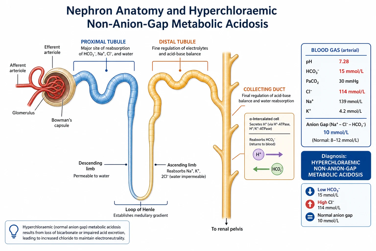
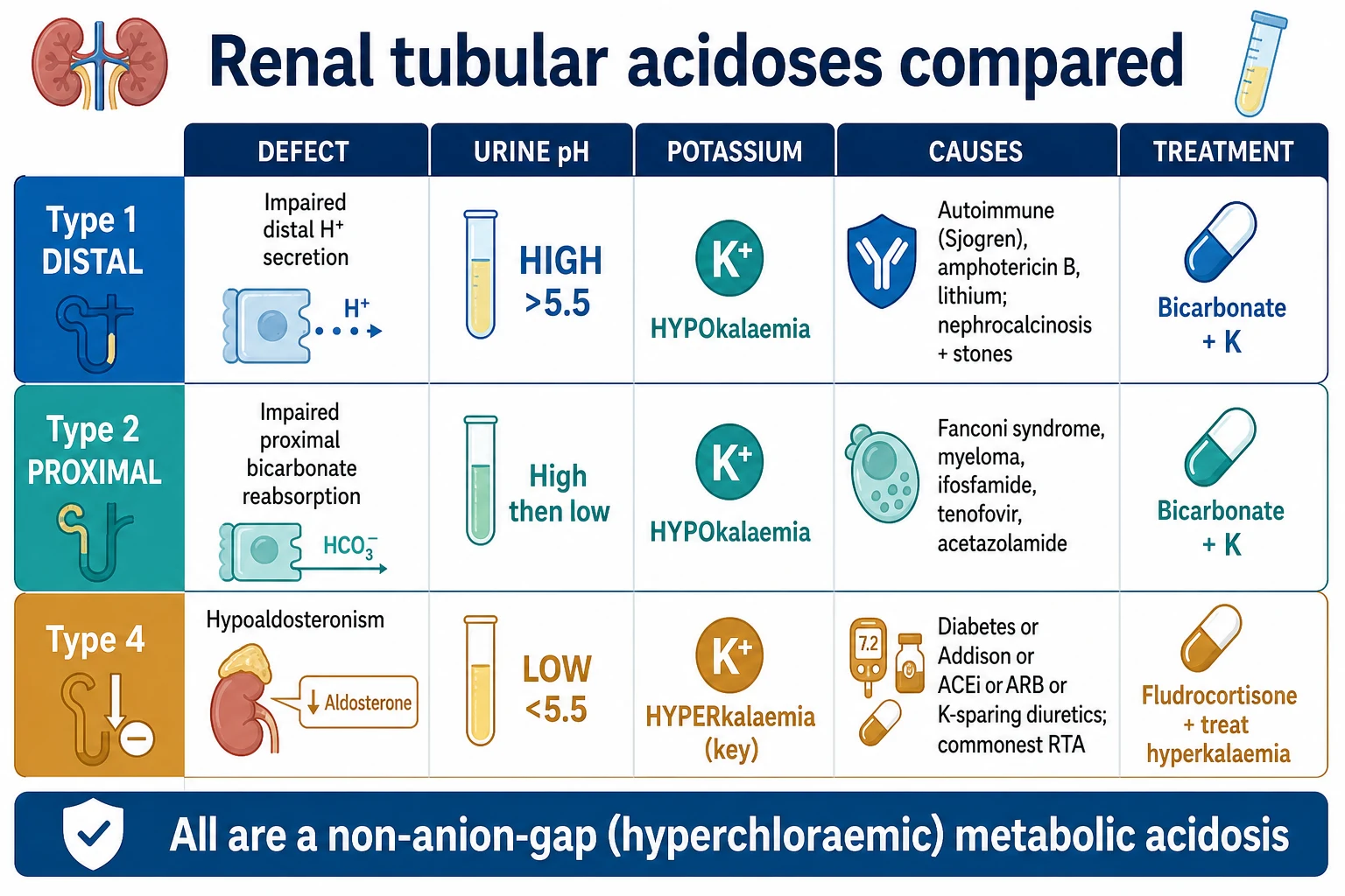
The renal tubular acidoses (RTA) are a group of disorders of the renal acid-base handling that produce a non-anion-gap (the hyperchloraemic) metabolic acidosis from the impaired renal acid excretion (the bicarbonate reabsorption or the acid secretion). The three types: the Type 1 (the distal) — the impaired distal H-plus secretion; the urine pH stays HIGH (over 5.5) despite the acidosis; the HYPOkalaemia; the causes the autoimmune (the Sjogren), the amphotericin B, the lithium; the nephrocalcinosis and the stones; the treatment the bicarbonate plus the potassium. The Type 2 (the proximal) — the impaired proximal bicarbonate reabsorption; the urine pH high then low; the HYPOkalaemia; the Fanconi syndrome, the myeloma, the ifosfamide, the tenofovir, the acetazolamide; the treatment the bicarbonate plus the potassium. The Type 4 — the hypoaldosteronism (or the aldosterone resistance); the urine pH LOW (under 5.5); the HYPERkalaemia (the key discriminator); the diabetes, the Addison, the ACEi, the ARB, the K-sparing diuretics; the commonest RTA; the treatment the fludrocortisone and the treat the hyperkalaemia.
On this page & tools
Your progress
Saved locally on this device.
8 MCQs with explanations
Target exams
Overview & definition
The renal tubular acidoses (RTA) are a group of disorders of the renal acid-base handling that produce a non-anion-gap (the hyperchloraemic) metabolic acidosis from the impaired renal acid excretion — either the impaired bicarbonate reabsorption (the proximal, the Type 2) or the impaired acid secretion (the distal, the Type 1) or the hypoaldosteronism (the Type 4). The normal anion gap (the hyperchloraemia) distinguishes the RTA from the lactic-acidosis, the ketoacidosis, and the toxin acidoses (the high-anion-gap acidoses).[1]
The three types are distinguished by the urine pH and the potassium:[1]
| Type | The defect | The urine pH | The potassium |
|---|---|---|---|
| 1 (the distal) | The impaired distal H-plus secretion | HIGH (over 5.5) | LOW (the hypokalaemia) |
| 2 (the proximal) | The impaired proximal bicarbonate reabsorption | HIGH then LOW | LOW (the hypokalaemia) |
| 4 | The hypoaldosteronism | LOW (under 5.5) | HIGH (the hyperkalaemia) |
Type 1 — the distal RTA
- The defect — the impaired H-plus secretion in the alpha-intercalated cells of the distal tubule and the collecting duct. The hydrogen cannot be excreted → the urine stays alkaline (the pH over 5.5) despite the systemic acidosis.[1]
- The urine pH — HIGH (over 5.5), inappropriately alkaline for the systemic acidosis. The urine anion gap is positive (the impaired NH4-plus excretion).[1]
- The potassium — LOW (the hypokalaemia from the increased distal K-plus secretion in exchange for the retained sodium).[1]
- The causes — the autoimmune (the Sjogren syndrome, the SLE, the RA), the amphotericin B, the lithium, the hereditary, the obstructive uropathy.[1]
- The complications — the nephrocalcinosis and the calcium-phosphate kidney stones (the alkaline urine plus the hypercalciuria precipitates the calcium phosphate).[1]
- The treatment — the bicarbonate (and the citrate) replacement, and the potassium supplementation. The bicarbonate corrects the acidosis and the hypokalaemia.[1]
Type 2 — the proximal RTA
- The defect — the impaired bicarbonate reabsorption in the proximal tubule. The bicarbonate is wasted in the urine until the plasma bicarbonate falls below the reduced threshold, then the urine pH drops (the distal reabsorption intact).[1]
- The urine pH — HIGH initially (the bicarbonaturia), then LOW once the plasma bicarbonate falls below the threshold.[1]
- The potassium — LOW (the hypokalaemia from the bicarbonate-driven distal K-plus wasting).[1]
- The causes — the Fanconi syndrome (the generalized proximal-tubule dysfunction: the glycosuria, the aminoaciduria, the phosphaturia), the myeloma, the ifosfamide, the tenofovir, the carbonic-anhydrase inhibitors (the acetazolamide), the cystinosis (the hereditary).[1]
- The treatment — the bicarbonate (the larger dose, the higher bicarbonaturia), the potassium supplementation. Treat the underlying cause.[1]
Type 4 — the hypoaldosteron RTA (the commonest)
- The defect — the hypoaldosteronism (or the aldosterone resistance) → the reduced H-plus and the K-plus secretion in the distal tubule.[1]
- The urine pH — LOW (under 5.5) — the intact H-plus secretion (the aldosterone-independent) keeps the urine acidified, but the total acid excretion is reduced.[1]
- The potassium — HIGH (the hyperkalaemia) — the key discriminator from the Type 1 and the Type 2 (which are the hypokalaemic). The hyperkalaemia itself impairs the ammoniagenesis, contributing to the acidosis.[1]
- The causes — the hyporeninaemic hypoaldosteronism (the diabetic nephropathy, the CKD — the commonest), the adrenal insufficiency (the Addison disease), and the drugs (the ACE inhibitors, the ARBs, the K-sparing diuretics — the spironolactone, the amiloride, the triamterene; the NSAIDs, the calcineurin inhibitors, the heparin).[1]
- The commonest RTA — the Type 4 is the most common of the three (the diabetic nephropathy and the ACE-inhibitor use are the prevalent).[1]
- The treatment — treat the hyperkalaemia (the patiromer, the sodium zirconium cyclosilicate, the loop diuretic), the fludrocortisone (the mineralocorticoid) if the aldosterone-deficient, the treat the underlying cause, and the stop the offending drugs.[1]
Type 3 (the rare, the obsolete)
A mixed proximal-distal defect, the rare, mostly the paediatric, associated with the osteopetrosis. Largely the historical; not the practical concern.[1]
[1]Exam practice
SAQ — Type 1 (distal) RTA in Sjogren syndrome
10 minutes · 10 marks
A 32-year-old woman with known Sjogren syndrome presents with proximal muscle weakness, polyuria and recurrent left flank pain. She is alert, RR 22, BP 110/70. ABG: pH 7.28, pCO2 28, HCO3 14, BE -12. Bloods: Na 138, K 2.7, Cl 116, glucose 5.0, urea 4.5, creatinine 74, albumin 40. Urine pH (catheterised) 6.2. A non-contrast CT abdomen shows bilateral medullary nephrocalcinosis.
SAQ — Type 4 RTA (hyperkalaemic) in a diabetic on perindopril and spironolactone
10 minutes · 10 marks
A 68-year-old man with type 2 diabetes, CKD stage 3b (eGFR 38), and HFrEF is admitted with malaise and palpations. Medications: metformin, perindopril, spironolactone 25 mg daily, frusemide. ECG shows peaked T waves. ABG: pH 7.30, pCO2 33, HCO3 18, BE -8. Bloods: Na 134, K 6.4, Cl 108, glucose 8.2, urea 12. Urine pH 5.3.
Red flags
Pathophysiology in depth — where the acid is handled
The three RTA types are best understood through the two nephron segments that handle acid, and the one hormone that governs distal cation handling. An examiner who hears "where in the nephron" wants these three answers.[1]
The proximal tubule — reclaims 80–85% of the filtered bicarbonate
The proximal tubule reabsorbs the bulk of the filtered load of bicarbonate. The sequence: the apical Na⁺/H⁺ exchanger (NHE-3) secretes H⁺ into the lumen; the H⁺ combines with filtered HCO₃⁻ to form H₂CO₃, which carbonic anhydrase IV (luminal, brush-border) cleaves to CO₂ + H₂O; the CO₂ diffuses back into the cell; intracellular carbonic anhydrase II re-forms H⁺ + HCO₃⁻; and the Na⁺/HCO₃⁻ co-transporter (NBCe1) on the basolateral membrane extrudes HCO₃⁻ into the blood. A defect anywhere in this chain (NHE-3, carbonic anhydrase, NBCe1) lowers the bicarbonate reabsorptive threshold. When the plasma HCO₃⁻ exceeds the new (lower) threshold, the unreabsorbed bicarbonate floods the distal nephron, producing bicarbonaturia and a high urine pH. As the plasma HCO₃⁻ falls below the threshold, distal reabsorption (intact in pure proximal RTA) resumes and the urine pH can acidify below 5.5 — hence the "high-then-low" urine pH of proximal RTA.[10][1]
The alpha-intercalated cell — secretes the H⁺ that titrates the daily acid load
The distal nephron (the late distal tubule and the outer-medullary collecting duct) secretes the H⁺ that titrates the daily non-volatile acid load (~1 mmol/kg/day of H⁺ from protein metabolism). The alpha-intercalated cell is the workhorse: intracellular carbonic anhydrase II generates H⁺ + HCO₃⁻; the apical H⁺-ATPase (and the H⁺/K⁺-ATPase) pumps H⁺ into the lumen against a steep gradient (up to 1000:1, i.e. urine pH 4.5 vs cell 7.5); the secreted H⁺ is buffered by the urinary buffers — primarily NH₃ (to NH₄⁺) and secondarily phosphate — and the HCO₃⁻ exits basolaterally via the Cl⁻/HCO₃⁻ exchanger (AE1, band 3). Distal RTA is, at its core, a failure of this alpha-intercalated cell — the gradient cannot be generated (the "secretory" defect), or the cell is lost to autoimmune destruction, or the membrane insert of AE1/H⁺-ATPase is mis-targeted (the genetic dRTAs). The net result: the urine cannot be acidified, NH₄⁺ excretion falls, and the systemic acid accumulates.[6][1]
The principal cell and aldosterone — the Type 4 territory
Aldosterone acts on the principal cell of the collecting duct: it opens the apical epithelial Na⁺ channel (ENaC) (amiloride-sensitive), generating the lumen-negative voltage that drives (a) K⁺ secretion through the ROMK channel and (b) H⁺ secretion by the neighbouring alpha-intercalated cell. Aldosterone also independently stimulates H⁺-ATPase and Na⁺/K⁺-ATPase. When aldosterone is absent (Addison's, hyporeninaemic hypoaldosteronism) or blocked (ACE inhibitors, ARBs, spironolactone, eplerenone, amiloride, triamterene), the lumen-negative voltage is lost, both K⁺ and H⁺ secretion fall, and the patient develops hyperkalaemia + a (usually mild) normal-gap acidosis — the Type 4 RTA. The urine pH is LOW (the alpha-intercalated H⁺-ATPase is intact; it is the aldosterone-driven amplification that is missing), which is the single feature that most reliably distinguishes Type 4 from Types 1 and 2.[8][9][7]
Type 1 (distal) RTA — the deep dive
The defect, restated
Type 1 is a failure of the alpha-intercalated cell to secrete H⁺ and hence to acidify the urine. The defining physiology: the urine pH stays above 5.5 despite a systemic metabolic acidosis — an inappropriately alkaline urine. (A normal kidney, faced with acidaemia, drives the urine pH to 5.0 and below; the inability to do so is the diagnostic signature.) The impaired NH₄⁺ excretion is reflected in a positive urine anion gap and a low urinary NH₄⁺. The hypokalaemia arises because the increased distal Na⁺ delivery and the high aldosterone (from volume depletion) drive K⁺ secretion through ROMK, and the bicarbonaturia itself drags K⁺ with it.[1][14]
Causes — autoimmune, drug, and genetic
The causes of distal (Type 1) RTA — autoimmune, drug, and genetic
| Category | Specific causes | Notes |
|---|---|---|
| Autoimmune (the commonest in adults) | Sjögren syndrome, SLE, rheumatoid arthritis, primary biliary cholangitis, thyroiditis | Sjögren is the single most-tested cause — autoimmune destruction of the intercalated cells. Look for the sicca syndrome |
| Drugs / toxins | Amphotericin B (a "permeability" defect — H⁺ back-leaks through the membrane), lithium, ifosfamide, foscarnet, toluene (late) | Amphotericin is the classic secretion-intact-but-gradient-lost mechanism — the drug inserts as a cation channel |
| Genetic | Autosomal dominant (AE1 / SLC4A1 mutation); autosomal recessive (H⁺-ATPase subunits ATP6V1B1, ATP6V0A4; carbonic anhydrase II with osteopetrosis + cerebral calcification) | Recessive dRTA presents in infancy with failure to thrive, vomiting, nephrocalcinosis, sensorineural deafness (ATP6V1B1) |
| Other | Obstructive uropathy, renal transplantation, sickle cell disease, medullary sponge kidney, hypercalcaemia/hyperparathyroidism | The secondary forms often coexist with nephrocalcinosis, making cause-vs-effect hard to disentangle |
Nephrocalcinosis and nephrolithiasis — the hallmark complication
Three converging mechanisms drive the calcium-phosphate stones and nephrocalcinosis of untreated Type 1 RTA: (1) the chronic acidosis leaches Ca²⁺ from bone (bone buffers the H⁺), producing hypercalciuria; (2) the alkaline urine (pH > 5.5) decreases the solubility of calcium phosphate (Ca₃(PO₄)₂ precipitates at high pH — the opposite of the uric-acid/cystine stones that favour acid urine); and (3) the hypocitraturia — citrate is the most important inhibitor of calcium crystallisation, and chronic acidosis drives intracellular citrate into the mitochondria, lowering urinary citrate. The result is bilateral medullary nephrocalcinosis and recurrent calcium-phosphate stones — the presentation that most often triggers the RTA work-up.[1][14]
Treatment
Replace alkali and potassium. Adult dosing: sodium bicarbonate (or the potassium citrate equivalent, especially when hypokalaemia predominates) titrated to 1–2 mmol/kg/day in divided doses, with the goal a plasma HCO₃⁻ of 22 mmol/L. The hypokalaemia must be corrected in parallel because alkali therapy drives K⁺ intracellularly and worsens the deficit. Children need higher doses (4–8 mmol/kg/day) because growing bone buffers a large acid load. Citrate preparations (e.g. potassium citrate) are preferred when stones are the issue — the citrate is metabolised to bicarbonate and raises urinary citrate, directly addressing the stone diathesis. Correcting the acidosis stops the bone-calcium release, lowers the hypercalciuria, and arrests the stone formation.[16][13]
Type 2 (proximal) RTA — the deep dive
The defect, restated
Type 2 is a failure of proximal bicarbonate reabsorption — the threshold falls (e.g. from a normal ~24 mmol/L to ~14–18 mmol/L). Above the new threshold, the unreabsorbed bicarbonate passes distally and is wasted; the urine pH is high (above 5.5) while the plasma HCO₃⁻ is above the threshold. As the plasma HCO₃⁻ equilibrates down to the threshold, the distal nephron (intact) reclaims what little bicarbonate reaches it and the urine pH can then fall below 5.5 — hence the "high-then-low" pattern that distinguishes Type 2 from the persistently high pH of Type 1. The hypokalaemia is driven by the bicarbonaturia (the non-reabsorbed HCO₃⁻ acts as a poorly reabsorbable anion, dragging K⁺ with it) and by secondary hyperaldosteronism.[10][1]
Causes — isolated or as part of Fanconi syndrome
The causes of proximal (Type 2) RTA — isolated vs generalised (Fanconi)
| Pattern | Specific causes | Notes |
|---|---|---|
| Generalised proximal tubulopathy (Fanconi syndrome) | Ifosfamide (chemotherapy — the chloroacetaldehyde metabolite), multiple myeloma (light-chain cast/tubular toxicity), tenofovir (mitochondrial toxicity, esp. the disoproxil fumarate), cystinosis (the commonest inherited cause in children), galactosaemia, Wilson disease, Lowe syndrome, paroxysmal nocturnal haemoglobinuria | Fanconi = RTA + glycosuria (normoglycaemic) + aminoaciduria + phosphaturia + uricosuria. The glycosuria with a normal serum glucose is a bedside clue |
| Carbonic anhydrase inhibition | Acetazolamide (the drug-induced paradigm), topiramate, sulfonamides | Acetazolamide reproduces isolated proximal RTA by blocking brush-border carbonic anhydrase — resolves on cessation |
| Isolated (genetic) | SLC4A4 (NBCe1) mutations — proximal RTA with ocular abnormalities (band keratopathy, glaucoma) | Rare; autosomal recessive |
Treatment
Proximal RTA is the hardest of the three to correct because the defective proximal tubule simply wastes whatever bicarbonate is given — the higher the plasma HCO₃⁻, the more is spilt into the urine, generating large bicarbonate doses, worsening hypokalaemia, and (in children) growth failure. Practical points: (1) give large alkali doses — 10–15 mmol/kg/day (much higher than Type 1), as a split sodium/potassium citrate or bicarbonate preparation; (2) aggressive potassium repletion — the bicarbonate load amplifies K⁺ wasting; (3) a thiazide diuretic (e.g. hydrochlorothiazide) induces mild volume depletion, lowers the GFR slightly, and reduces the filtered bicarbonate load, allowing a lower alkali dose to achieve the target HCO₃⁻; (4) treat the underlying cause (stop the offending drug, treat the myeloma). The acetazolamide- and tenofovir-induced forms are largely reversible on withdrawal.[10][11][13]
Type 4 (hyperkalaemic / hypoaldosteron) RTA — the deep dive
The defect, restated
Type 4 is hypoaldosteronism or aldosterone resistance. Without aldosterone's action on the principal cell, the lumen-negative voltage collapses, and both K⁺ secretion (via ROMK) and H⁺ secretion (via the intercalated H⁺-ATPase) fall. The result is hyperkalaemia and a usually mild (HCO₃⁻ 17–20 mmol/L) normal-gap acidosis. Crucially, the alpha-intercalated cell's intrinsic H⁺-ATPase is intact, so the urine can still be acidified to below 5.5 — the feature that most reliably separates Type 4 from Type 1. The hyperkalaemia itself further impairs acid excretion by suppressing ammoniagenesis in the proximal tubule (the high intracellular K⁺ inhibits the glutaminase/phosphate-dependent glutamate dehydrogenase pathway), so the NH₄⁺ available to buffer secreted H⁺ is reduced even though the pH-lowering machinery works. This is why the acidosis of Type 4 is "mild but real" and why treating the hyperkalaemia itself improves the acidosis.[8][7][12]
Causes — hyporeninaemic, adrenal, drug-induced
The causes of Type 4 (hyperkalaemic) RTA — the three buckets
| Mechanism | Examples | Notes |
|---|---|---|
| Hyporeninaemic hypoaldosteronism (the commonest overall) | Diabetic nephropathy (the paradigm — the juxtaglomerular apparatus is damaged and the sympathetic/prostaglandin renin trigger is blunted), chronic CKD, NSAIDs, HIV nephropathy | ~50% of all Type 4. The diabetic with a modestly high K⁺ and a mild acidosis is the textbook presentation |
| Adrenal insufficiency | Primary (Addison disease) — aldosterone and cortisol both low; secondary (pituitary) — renin and aldosterone low, cortisol low, but aldosterone preserved because it is renin-angiotensin driven (so usually less hyperkalaemic) | Add Addison whenever the K⁺ is high and the Na⁺ is low; check the 09:00 cortisol and the short Synacthen test |
| Drugs (very common in ICU) | ACE inhibitors / ARBs (block angiotensin II), renin inhibitors (aliskiren), K⁺-sparing diuretics (spironolactone, eplerenone — block the mineralocorticoid receptor; amiloride, triamterene — block ENaC), NSAIDs (block the prostaglandin renin trigger), calcineurin inhibitors (cyclosporin, tacrolimus), heparin (blocks aldosterone synthase), ketoconazole | Polypharmacy in the diabetic ICU patient (ACEi + spironolactone + NSAID) is the modern epidemic of Type 4 |
| Aldosterone resistance (pseudohypoaldosteronism) | Gordon syndrome (PHA2 — hypertension + hyperkalaemia + normal GFR), PHA1 (salt-wasting, paediatric) | Rare but examinable — Gordon syndrome is the "hypertension with hyperkalaemia" paradox |
Treatment
Treat the hyperkalaemia and replace the mineralocorticoid. (1) Stop the offending drugs wherever possible (the ACEi, the K⁺-sparing diuretic, the NSAID) — often sufficient. (2) Fludrocortisone (0.1–0.2 mg/day, the oral mineralocorticoid) for the aldosterone-deficient patient (Addison's, hyporeninaemic disease) — watch for fluid retention and hypertension. (3) Loop diuretics (frusemide) — promote distal Na⁺ delivery, generate a lumen-negative voltage, and enhance K⁺ and H⁺ secretion; the mainstay when fludrocortisone is contraindicated by fluid overload or hypertension. (4) K⁺-binders (patiromer, sodium zirconium cyclosilicate) for chronic K⁺ control. (5) Dietary K⁺ restriction. (6) Treat the underlying cause. Because the acidosis is driven by the hyperkalaemia, lowering the K⁺ corrects most of the acidosis without exogenous alkali.[8][12]
Distinguishing the three — the bedside algorithm
The urine pH and the serum potassium split the RTAs cleanly. The single most reliable discriminator is the potassium: hyperkalaemia → Type 4; hypokalaemia → Type 1 or 2. Then the urine pH: persistently high (> 5.5) → Type 1; high-then-low (able to fall below 5.5 once equilibrated) → Type 2; low (< 5.5) → Type 4.[1][4]
The three RTA types compared across every examinable axis
| Feature | Type 1 (distal) | Type 2 (proximal) | Type 4 (hypoaldosteron) |
|---|---|---|---|
| The defect | Impaired distal H⁺ secretion (alpha-intercalated cell) | Impaired proximal HCO₃⁻ reabsorption | Hypoaldosteronism / aldosterone resistance |
| Plasma HCO₃⁻ | 10–20 mmol/L | 12–20 mmol/L | 17–20 mmol/L (the mildest) |
| Urine pH (during acidosis) | > 5.5 (persistently alkaline) | Variable — > 5.5 early, < 5.5 once equilibrated | < 5.5 (acidified) |
| Serum K⁺ | LOW (hypokalaemia) | LOW (hypokalaemia) | HIGH (hyperkalaemia — the discriminator) |
| Urine anion gap / NH₄⁺ | Positive (low NH₄⁺) | Variable — negative once equilibrated | Positive (low NH₄⁺ — suppressed ammoniagenesis) |
| Anion gap | Normal | Normal | Normal |
| Stones / nephrocalcinosis | Yes — calcium phosphate | No | No |
| Osteomalacia / rickets | Possible (chronic acidosis) | Yes (with Fanconi — phosphaturia) | No |
| Cause archetype | Sjögren, amphotericin, lithium | Myeloma, ifosfamide, tenofovir, acetazolamide | Diabetes, Addison, ACEi, spironolactone |
| Treatment | Bicarbonate 1–2 mmol/kg/day + K⁺ | Bicarbonate 10–15 mmol/kg/day + K⁺ + thiazide | Fludrocortisone + loop diuretic + K⁺ restriction |
| Frequency | Less common | Least common | Commonest RTA |
The bedside algorithm — a non-anion-gap acidosis walks in
Confirm it is a non-anion-gap (hyperchloraemic) acidosis
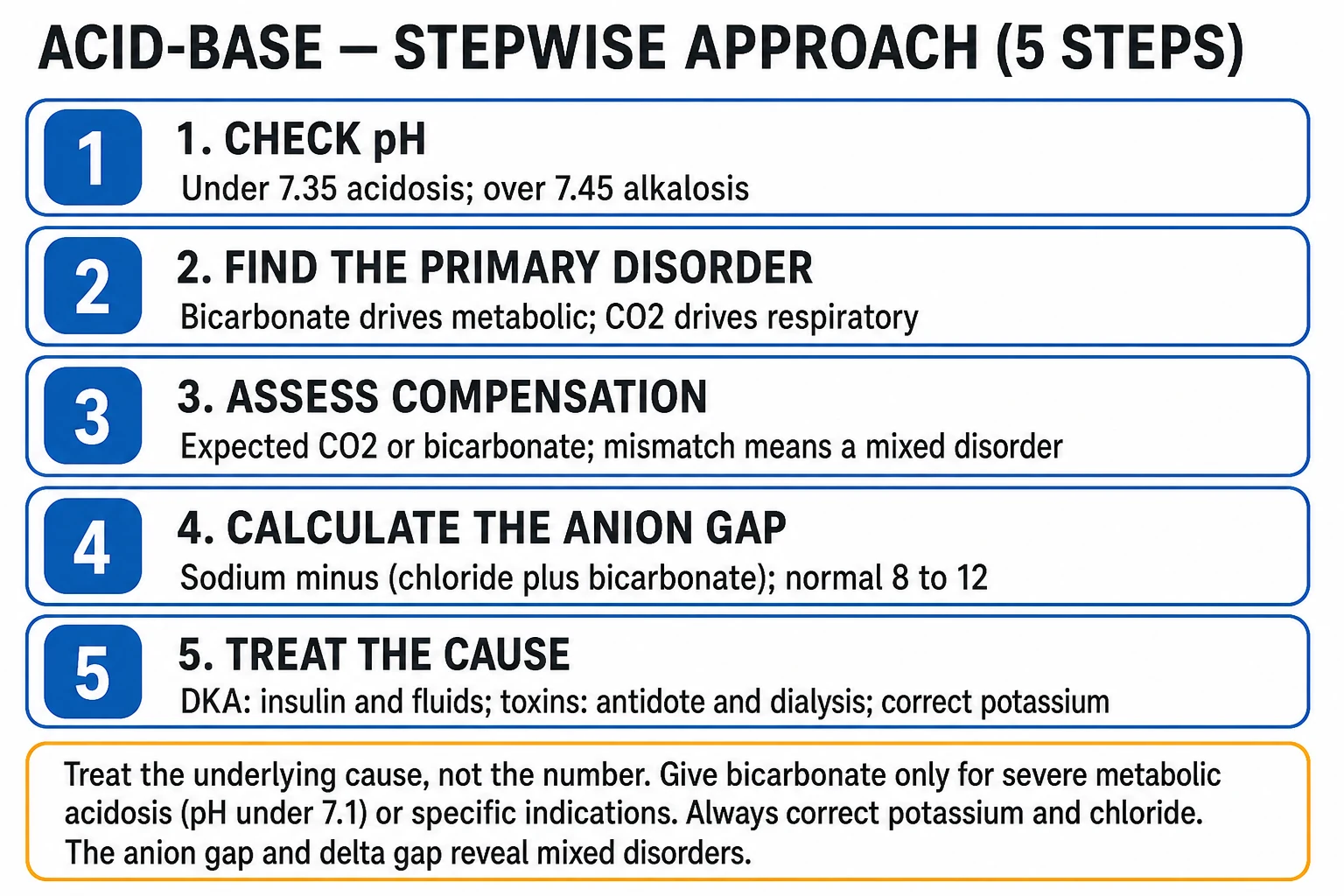
Anion gap = Na⁺ − (Cl⁻ + HCO₃⁻). If the AG is normal (8–12, albumin-corrected) and the HCO₃⁻ is low with a high Cl⁻, it is an NAGMA. Now ask "kidney or gut?"
Check the serum potassium
This single test splits the RTAs. K⁺ HIGH → Type 4 until proven otherwise. K⁺ LOW → Type 1 or Type 2. The hyperkalaemic NAGMA is Type 4; the hypokalaemic NAGMA is Type 1 or 2 (or diarrhoea, toluene, ureteroenteric).
Check the urine pH (on a fresh, catheterised sample)
If the K⁺ is LOW: a urine pH persistently above 5.5 → Type 1 (distal). A urine pH that can fall below 5.5 → Type 2 (proximal). If the K⁺ is HIGH: the urine pH should be below 5.5 → Type 4 (and the pH should be below 5.5 because the alpha-intercalated cell is intact).
Estimate the urinary NH₄⁺ via the urine anion gap (UAG)
UAG = (urine Na⁺ + urine K⁺) − urine Cl⁻. A NEGATIVE UAG (typically −20 to −50) means adequate NH₄⁺ excretion → an extrarenal cause (diarrhoea). A POSITIVE UAG (20 to 100) means impaired NH₄⁺ excretion → a renal cause (any RTA). The UAG is unreliable when the urine Na⁺ is very low, in volume depletion, or with unmeasured anions (ketoacids, hippurate from toluene) — then use the urine osmolar gap.
Confirm the type with the urine osmolar gap if needed
Urine osmolar gap = measured urine osmolality − 2(Na⁺ + K⁺ + urea + glucose). The osmolar gap ≈ 2 × NH₄⁺. A low NH₄⁺ (osmolar gap < 30) confirms an RTA; a high NH₄⁺ (osmolar gap > 100) confirms an extrarenal source. Use this whenever the UAG is misleading (toluene, ketoacidosis, low urine Na⁺).
Synthesise with the clinical context
Sjögren/amphotericin/lithium + nephrocalcinosis → Type 1. Myeloma/ifosfamide/tenofovir + Fanconi features → Type 2. Diabetes/Addison/ACEi/spironolactone + hyperkalaemia → Type 4. The context nearly always confirms the biochemistry.
The differential of the non-anion-gap (hyperchloraemic) metabolic acidosis
Once the AG is normal and the chloride is high, the differential is renal (RTA) vs extrarenal (gut/toxin). The discriminator is the urinary NH₄⁺ — high in the extrarenal causes (the kidney is excreting the acid normally), low in the renal causes (the kidney cannot). Because NH₄⁺ is not measured on a routine chemistry panel, it is inferred from the urine anion gap or, more reliably, the urine osmolar gap.[4][17]
The non-anion-gap metabolic acidosis — renal (low NH₄⁺) vs extrarenal (high NH₄⁺)
| Cause | Mechanism | Urine pH | Urine NH₄⁺ (UAG) | Key clue |
|---|---|---|---|---|
| Diarrhoea / GI loss | Loss of pancreatic/intestinal HCO₃⁻ | < 5.5 (kidney acidifies normally) | HIGH → UAG negative | History; low urine Na⁺ if volume-depleted |
| Type 1 RTA | Impaired distal H⁺ secretion | > 5.5 | LOW → UAG positive | Hypokalaemia, nephrocalcinosis, Sjögren |
| Type 2 RTA | Impaired proximal HCO₃⁻ reabsorption | Variable (high early, low late) | LOW → UAG positive (early) | Fanconi features, myeloma, tenofovir |
| Type 4 RTA | Hypoaldosteronism | < 5.5 | LOW (suppressed ammoniagenesis) → UAG positive | Hyperkalaemia, diabetes, ACEi |
| Ureteroenteric fistula / sigmoid loop | Chloride-bicarbonate exchange across the bowel mucosa (Cl⁻ absorbed, HCO₃⁻ lost) | < 5.5 | HIGH → UAG negative | Post-urological surgery, ileal conduit |
| Toluene (glue sniffing) | Metabolised to hippuric acid; hippurate is a poorly reabsorbable anion | Variable; early > 5.5, late < 5.5 | LOW → UAG positive (hippurate makes UAG unreliable — use the osmolar gap) | Solvent abuse history; osmolar gap low (low true NH₄⁺) |
| Hyperalimentation / amino-acid infusion | HCl load from arginine/lysine | < 5.5 | HIGH → UAG negative | TPN context |
| Resolving DKA + saline | Ketones cleared; saline-induced hyperchloraemia | < 5.5 | HIGH → UAG negative (kidney works) | Falling AG, persistent low HCO₃⁻, high Cl⁻ (delta ratio < 1) |
The mnemonic for the high-anion-gap acidosis is GOLD MARK (or MUDPILES); the mnemonic for the normal-gap acidosis is HARDUP / USED CARD — the most examinable items being diarrhoea (the commonest extrarenal), the three RTAs, the ileal conduit, toluene, and acetazolamide. The potassium and the urine NH₄⁺ (via the UAG/osmolar gap) split them at the bedside.[4]
The urine anion gap and the urine osmolar gap — the NH₄⁺ surrogates
Because the kidney excretes its daily acid load almost entirely as NH₄⁺ (not as free H⁺), measuring NH₄⁺ is the direct way to ask "is the kidney doing its job?" But NH₄⁺ is rarely measured. Two surrogates: [1]
- Urine anion gap (UAG) = (U_Na + U_K) − U_Cl. NH₄⁺ is excreted with Cl⁻ (and other anions). When NH₄⁺ excretion is high (an appropriate renal response, e.g. diarrhoea), the U_Cl rises and the UAG becomes negative (typically −20 to −50). When NH₄⁺ excretion is low (an RTA), the UAG is positive (20 to 100). The UAG assumes NH₄⁺ is excreted with Cl⁻; this fails when unmeasured anions are present (ketoacids in DKA, hippurate in toluene abuse, some drug anions) — then the UAG is falsely positive despite high NH₄⁺.
- Urine osmolar gap = measured urine osmolality − calculated [2(Na⁺ + K⁺) + urea + glucose]. The osmolar gap reflects all unmeasured urinary osmoles, of which NH₄⁺ is by far the dominant one in the acidotic patient. A high osmolar gap (> 100) = high NH₄⁺ (extrarenal cause); a low gap (< 30) = low NH₄⁺ (an RTA). The osmolar gap is more robust than the UAG when unmeasured anions are present, and is the preferred test when the diagnosis is unclear.[17][4]
Worked examples — the algorithm in action
Case 1 — the Sjögren patient with stones and weakness (Type 1)
The presentation
A 42-year-old woman with Sjögren syndrome presents with proximal muscle weakness and recurrent calcium-phosphate kidney stones. Blood: pH 7.28, HCO₃⁻ 14, PaCO₂ 28. Na⁺ 140, Cl⁻ 115, K⁺ 2.9, albumin 40.
Step 1 — the anion gap
AG = 140 − (115 + 14) = 11 (normal). A non-anion-gap (hyperchloraemic) metabolic acidosis. The chloride is high.
Step 2 — the potassium
K⁺ 2.9 (LOW) → Type 1 or Type 2 (or diarrhoea). The history of stones points away from diarrhoea.
Step 3 — the urine pH
Urine pH 6.8 (persistently above 5.5 despite the systemic acidosis). The kidney cannot acidify → Type 1 (distal) RTA.
Step 4 — the urine anion gap
UAG = (urine Na⁺ 40 + urine K⁺ 30) − urine Cl⁻ 40 = +30 (positive) → low NH₄⁺ excretion, confirming a renal (not gut) cause.
The synthesis and management
Type 1 (distal) RTA from Sjögren — autoimmune destruction of the alpha-intercalated cells. The nephrocalcinosis and calcium-phosphate stones are the hallmark complication (alkaline urine + hypercalciuria + hypocitraturia). Treat with oral bicarbonate (or potassium citrate) 1–2 mmol/kg/day plus potassium repletion. Citrate is preferred here because it raises urinary citrate and addresses the stone diathesis directly.
Case 2 — the diabetic with a mild acidosis and high potassium (Type 4)
The presentation
A 68-year-old man with type-2 diabetes (on an ACE inhibitor and trimethoprim for a UTI) is found on routine bloods to have a mild acidaemia. Blood: pH 7.33, HCO₃⁻ 19, PaCO₂ 36. Na⁺ 138, Cl⁻ 112, K⁺ 6.1, creatinine 140, glucose 7.5.
Step 1 — the anion gap
AG = 138 − (112 + 19) = 7 (normal). A non-anion-gap (hyperchloraemic) metabolic acidosis. The HCO₃⁻ is only mildly low (typical of Type 4).
Step 2 — the potassium
K⁺ 6.1 (HIGH) → Type 4 until proven otherwise. The diabetes, the ACE inhibitor, and the trimethoprim (an ENaC blocker like amiloride) are the triple hit.
Step 3 — the urine pH
Urine pH 5.2 (below 5.5 — the alpha-intercalated H⁺-ATPase is intact; only the aldosterone-driven amplification is missing). This is the key finding that separates Type 4 from Type 1.
Step 4 — the ammonium
UAG positive → low NH₄⁺ excretion. The hyperkalaemia itself has suppressed the proximal ammoniagenesis, contributing to the acidosis. (Treat the K⁺ and the acidosis will largely resolve.)
The synthesis and management
Type 4 (hypoaldosteron) RTA — hyporeninaemic hypoaldosteronism of diabetic nephropathy, amplified by the ACE inhibitor (blocks angiotensin II) and trimethoprim (blocks ENaC). Stop the trimethoprim; review the ACE inhibitor. Treat the hyperkalaemia (loop diuretic, K⁺ binder), add fludrocortisone if the K⁺ remains high, and dietary K⁺ restriction. The acidosis corrects as the K⁺ falls — no exogenous bicarbonate needed.
Case 3 — the myeloma patient on tenofovir (Type 2 / Fanconi)
The presentation
A 61-year-old man with multiple myeloma, on tenofovir for hepatitis-B prophylaxis, presents with bone pain and fatigue. Blood: pH 7.30, HCO₃⁻ 16, PaCO₂ 32. Na⁺ 140, Cl⁻ 113, K⁺ 3.1, phosphate 0.55, glucose 5.5. Urine: glucose 3+, amino acids present.
Step 1 — the anion gap
AG = 140 − (113 + 16) = 11 (normal). A non-anion-gap (hyperchloraemic) metabolic acidosis.
Step 2 — the potassium and the Fanconi features
K⁺ 3.1 (LOW). Glycosuria with a NORMAL serum glucose (5.5), phosphaturia (low serum phosphate), and aminoaciduria → generalised proximal tubulopathy = Fanconi syndrome → Type 2 (proximal) RTA. The myeloma (light-chain toxicity) and the tenofovir (mitochondrial toxicity) are the dual culprits.
Step 3 — the urine pH
Urine pH 6.0 initially (the plasma HCO₃⁻ is above the lowered threshold → bicarbonaturia). Once the plasma HCO₃⁻ falls to the new threshold (~14–16), the urine pH will fall below 5.5 (the distal nephron is intact). The "high-then-low" pattern.
The synthesis and management
Type 2 (proximal) RTA from Fanconi syndrome (myeloma + tenofovir). Treat the myeloma, stop the tenofovir (switch to entecavir). Replace alkali at high dose (10–15 mmol/kg/day) because the defective tubule wastes bicarbonate, add aggressive K⁺ and phosphate repletion, and consider a thiazide to reduce the filtered bicarbonate load. The hardest of the three to correct.
Case 4 — the RTA that is actually diarrhoea (the UAG saves you)
The presentation
A 35-year-old returns from travel with profuse watery diarrhoea for 5 days. Blood: pH 7.29, HCO₃⁻ 15, PaCO₂ 31. Na⁺ 138, Cl⁻ 116, K⁺ 3.0.
Step 1 — the anion gap
AG = 138 − (116 + 15) = 7 (normal). A non-anion-gap (hyperchloraemic) metabolic acidosis. The hypokalaemia could suggest Type 1 or 2 RTA.
Step 2 — the urine pH
Urine pH 5.0 (the kidney is acidifying normally). This already argues against Type 1 (which cannot acidify).
Step 3 — the urine anion gap — the key
Urine Na⁺ 25, K⁺ 35, Cl⁻ 90. UAG = (25 + 35) − 90 = −30 (NEGATIVE) → high NH₄⁺ excretion → the kidney is excreting acid appropriately → the cause is EXTRARENAL (the diarrhoea). A negative UAG rules out an RTA.
The lesson
The hypokalaemic, hyperchloraemic, normal-AG acidosis is diarrhoea (or another gut HCO₃⁻ loss), NOT an RTA, whenever the kidney is compensating with a high NH₄⁺ excretion (negative UAG, low urine pH). The history and the UAG distinguish the gut from the tubule.
Clinical pearls — high-yield points for the CICM / FFICM / EDIC exam
Trials and landmark evidence
Rodriguez-Soriano — Renal tubular acidosis: the clinical entity (PMID 12138150)
Source
Journal of the American Society of Nephrology, 2002 — the definitive modern clinical review of the RTAs
Scope
A unified framework for the three RTA types — the cellular defect, the urine pH, the potassium, the causes, and the treatment — written by the paediatric nephrologist who defined much of the field
Key teaching
Established the urine-pH-plus-potassium approach as the bedside discriminator: the distal (Type 1) and proximal (Type 2) are hypokalaemic, the Type 4 is hyperkalaemic; the distal cannot acidify the urine, the proximal can (once equilibrated), the Type 4 can
Enduring status
Still the single most-cited reference when teaching the RTAs; the framework every fellowship candidate is expected to reproduce
Adrogué & Madias — Management of life-threatening acid-base disorders (PMID 9414329, 9420343)
Source
New England Journal of Medicine, 1998 (two parts) — the authoritative clinical review of acid-base management
Part 1 (9414329)
The metabolic acidoses — the high-AG and the normal-AG (the RTAs and the gut losses), the diagnosis by the anion gap and the delta-delta, and the management (treat the cause; the role and the limits of bicarbonate)
Part 2 (9420343)
The metabolic alkaloses, the respiratory acidoses and alkaloses, and the mixed disorders — with the systematic compensation rules
Relevance to RTA
The reference for the anion-gap approach that places the RTA in its diagnostic position (the normal-AG / hyperchloraemic acidosis), and for the principle that bicarbonate is not given reflexively — only for the specific, acid-driven complications
Kraut & Madias — Differential diagnosis of nongap metabolic acidosis (PMID 22403272)
Source
Clinical Journal of the American Society of Nephrology, 2012 — the systematic approach to the normal-AG acidosis
Question
How do you distinguish the renal (RTA) from the extrarenal (gut) causes of a hyperchloraemic acidosis at the bedside?
The method
Use the serum potassium (high → Type 4; low → Type 1/2 or gut), the urine pH, and the urinary NH₄⁺ (estimated from the urine anion gap, or measured via the urine osmolar gap) to localise the cause
The lesson
The urine anion gap (and the more robust osmolar gap) is the bridge between the chemistry panel and the diagnosis — the test that turns non-anion-gap acidosis into a specific cause
Kraut & Nagami — Hyperkalemic forms of renal tubular acidosis (PMID 30139459)
Source
Advances in Chronic Kidney Disease, 2018 — the dedicated review of the Type 4 RTA and its mimics
Scope
The pathophysiology of hyporeninaemic hypoaldosteronism, adrenal insufficiency, the drug causes, and the aldosterone-resistance syndromes (Gordon, pseudohypoaldosteronism)
Key insight
The hyperkalaemia itself suppresses proximal ammoniagenesis — so the low NH₄⁺ of Type 4 is partly a consequence of the high K⁺, and treating the K⁺ restores the acid excretion. This is why the acidosis is mild and why it responds to K⁺-lowering therapy
Bottom line
The modern evidence base for the fludrocortisone / loop diuretic / K⁺-binder approach to Type 4 RTA, and for stopping the offending drugs first
Karet et al — AE1 mutations in autosomal dominant distal RTA (PMID 9600966)
Source
Proceedings of the National Academy of Sciences USA, 1998 — the molecular-genetic dissection of distal RTA
Finding
Mutations in the chloride-bicarbonate exchanger gene AE1 (SLC4A1, band 3) cause autosomal dominant but not autosomal recessive distal RTA — the recessive forms map to the H⁺-ATPase subunits (ATP6V1B1, ATP6V0A4)
Significance
Defined the molecular basis of the genetic dRTAs at the cellular level — the dominant form is a basolateral Cl⁻/HCO₃⁻ exchanger (AE1) defect; the recessive forms are apical H⁺-pump (H⁺-ATPase) defects, the latter often with sensorineural deafness
Exam relevance
The genetic answer when asked what causes hereditary distal RTA — AE1 (dominant) and the H⁺-ATPase subunits (recessive, with deafness and osteopetrosis if carbonic anhydrase II)
DeFronzo — Hyperkalemia and hyporeninemic hypoaldosteronism (PMID 6990088)
Source
Kidney International, 1980 — the classic description of the syndrome that underlies most Type 4 RTA
The syndrome
Hyperkalaemia, a mild hyperchloraemic acidosis, low plasma renin, and low plasma aldosterone, in patients with diabetic nephropathy (and other CKD) — despite a GFR that should be sufficient to excrete the K⁺ and the acid
The mechanism
Damage to the juxtaglomerular apparatus (the renin-secreting cells) and the blunted prostaglandin/sympathetic drive to renin release leave aldosterone deficient; the resulting loss of distal ENaC activity collapses the lumen-negative voltage and impairs both K⁺ and H⁺ secretion
Enduring relevance
The diabetic with the unexplained hyperkalaemia and mild acidosis is still the paradigm of Type 4 RTA, four decades on
Palmer & Clegg — Potassium homeostasis: Core Curriculum 2019 (PMID 31227226)
Source
American Journal of Kidney Diseases, 2019 — the modern core-curriculum review of potassium handling
Relevance to RTA
Explains why aldosterone deficiency produces BOTH hyperkalaemia AND a metabolic acidosis (the shared distal mechanism), and why the hyperkalaemia of Type 4 RTA is the primary driver of the acidosis through the suppression of ammoniagenesis
Clinical teaching
The framework for the K⁺-binder (patiromer, sodium zirconium cyclosilicate) and loop-diuretic management of the chronic hyperkalaemia of Type 4 RTA — the modern alternative to fludrocortisone in the fluid-overloaded or hypertensive patient
Sebastian & Morris — Sodium and chloride conservation during correction of dRTA (PMID 783200)
Source
Journal of Clinical Investigation, 1976 — the classic study of alkali therapy in distal RTA
Observation
During the sustained correction of the acidosis with bicarbonate in patients with distal RTA, the kidney fails to conserve sodium and chloride appropriately, necessitating careful volume and electrolyte management during treatment
Lesson
Bicarbonate therapy in distal RTA is not just give the alkali — the correction itself perturbs volume and chloride handling, which is why potassium must be repleted in parallel and why the patient is monitored, not just dosed
Alexander & Bitzan — Renal Tubular Acidosis and Management Strategies (PMID 33367987)
Source
Advances in Therapy, 2021 — a contemporary narrative review of all three RTA types and their practical management
Scope
The clinical features, the diagnostic algorithm (potassium → urine pH → NH₄⁺ surrogate), and the treatment of Types 1, 2, and 4, with the dosing of alkali, potassium, fludrocortisone, and the role of the loop diuretic and the K⁺-binder
Bottom line
The modern, dosing-level reference for the bedside management — the single source for how much bicarbonate for Type 1 vs Type 2, and when to add a thiazide
Palmer et al — Primary Distal RTA: toward an optimal correction (PMID 38967973)
Source
Clinical Journal of the American Society of Nephrology, 2024 — the contemporary update on alkali dosing in primary distal RTA
Question
What alkali dose and preparation best corrects the acidosis, the hypokalaemia, and the stone diathesis of primary distal RTA?
Finding
Reaffirms that alkali (1–2 mmol/kg/day in adults, 4–8 in children) titrated to a plasma HCO₃⁻ of 22 mmol/L corrects the acidosis, arrests the bone demineralisation, and reduces the hypercalciuria and hypocitraturia that drive the calcium-phosphate stones; potassium citrate is preferred when hypokalaemia or stones predominate
Bottom line
The current evidence that the goal of therapy is not just a normal HCO₃⁻ but the prevention of the long-term complications (stones, nephrocalcinosis, growth failure, osteomalacia)
Additional red flags
One-line summaries for the viva
- Type 1 (distal) RTA — impaired alpha-intercalated H⁺ secretion; urine pH > 5.5; HYPOkalaemia; nephrocalcinosis and calcium-phosphate stones; Sjögren, amphotericin, lithium; bicarbonate (citrate) 1–2 mmol/kg/day + K⁺.
- Type 2 (proximal) RTA — impaired proximal HCO₃⁻ reabsorption; urine pH high-then-low; HYPOkalaemia; Fanconi syndrome (myeloma, ifosfamide, tenofovir, acetazolamide); bicarbonate 10–15 mmol/kg/day + K⁺ + thiazide.
- Type 4 (hypoaldosteron) RTA — hypoaldosteronism or aldosterone resistance; urine pH < 5.5; HYPERkalaemia (the discriminator); diabetes, Addison, ACEi, spironolactone, ENaC blockers; the commonest RTA; fludrocortisone, loop diuretic, K⁺ restriction; treat the K⁺ and the acidosis resolves.
- The discriminator — the potassium (hyperkalaemia = Type 4) and the urine pH (persistently high = Type 1; high-then-low = Type 2; low = Type 4). The urine anion gap (negative = extrarenal; positive = renal) splits the gut from the tubule.[1][4][8]
References
- [1]Rodriguez-Soriano J Renal tubular acidosis: the clinical entity J Am Soc Nephrol, 2002.PMID 12138150
- [2]Adrogue HJ, Madias NE Management of life-threatening acid-base disorders. First of two parts N Engl J Med, 1998.PMID 9414329
- [3]Adrogue HJ, Madias NE Management of life-threatening acid-base disorders. Second of two parts N Engl J Med, 1998.PMID 9420343
- [4]Kraut JA, Madias NE Differential diagnosis of nongap metabolic acidosis: value of a systematic approach Clin J Am Soc Nephrol, 2012.PMID 22403272
- [5]Kraut JA, Madias NE Treatment of acute metabolic acidosis: a pathophysiologic approach Nat Rev Nephrol, 2012.PMID 22945490
- [6]Karet FE, Finberg KE, Nelson RD, et al Mutations in the chloride-bicarbonate exchanger gene AE1 cause autosomal dominant but not autosomal recessive distal renal tubular acidosis Proc Natl Acad Sci U S A, 1998.PMID 9600966
- [7]Batlle DC On the mechanism of impaired distal acidification in hyperkalemic renal tubular acidosis: evaluation with amiloride and bumetanide J Am Soc Nephrol, 1992.PMID 1450372
- [8]Kraut JA, Nagami GT Hyperkalemic Forms of Renal Tubular Acidosis: Clinical and Pathophysiological Aspects Adv Chronic Kidney Dis, 2018.PMID 30139459
- [9]DeFronzo RA Hyperkalemia and hyporeninemic hypoaldosteronism Kidney Int, 1980.PMID 6990088
- [10]Hannoud J, Trespalacios F, Tiwari A, et al Proximal renal tubular acidosis: a not so rare disorder of multiple etiologies Nephrol Dial Transplant, 2012.PMID 23235953
- [11]Foreman JW, Olbright T Drug-induced renal Fanconi syndrome QJM, 2014.PMID 24368854
- [12]Palmer BF, Clegg DJ Physiology and Pathophysiology of Potassium Homeostasis: Core Curriculum 2019 Am J Kidney Dis, 2019.PMID 31227226
- [13]Alexander RT, Bitzan M Renal Tubular Acidosis and Management Strategies: A Narrative Review Adv Ther, 2021.PMID 33367987
- [14]Palmer BF, Kelepouris E, Clegg DJ Primary Distal Renal Tubular Acidosis: Toward an Optimal Correction of Metabolic Acidosis Clin J Am Soc Nephrol, 2024.PMID 38967973
- [15]Kraut JA, Madias NE Adverse Effects of the Metabolic Acidosis of Chronic Kidney Disease Adv Chronic Kidney Dis, 2017.PMID 29031355
- [16]Sebastian A, Morris RC Jr Impaired renal conservation of sodium and chloride during sustained correction of systemic acidosis in patients with type 1, classic renal tubular acidosis J Clin Invest, 1976.PMID 783200
- [17]Batlle DC, Hizon M, Cohen E, et al Urinary Ammonium in Clinical Medicine: Direct Measurement and the Urine Anion Gap as a Surrogate Marker During Metabolic Acidosis Adv Kidney Dis Health, 2023.PMID 36868734