ICU · Respiratory
Acute respiratory failure: classification and approach
Also known as Type 1 respiratory failure (hypoxaemic) · Type 2 respiratory failure (hypercapnic) · A-a gradient · Respiratory failure approach
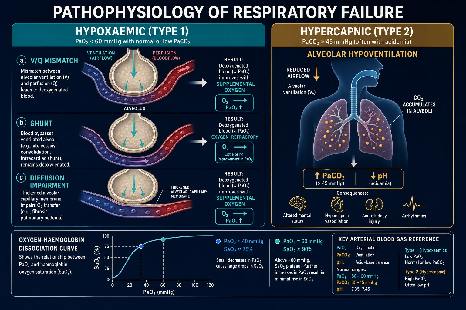
Acute respiratory failure is the inability of the respiratory system to maintain adequate gas exchange. Type 1 (hypoxaemic): PaO2 <60 mmHg (8 kPa) on room air, normal/low PaCO2. Causes: V/Q mismatch (1 — pneumonia, PE, pulmonary oedema, ARDS), shunt, diffusion impairment, hypoventilation. Type 2 (hypercapnic): PaCO2 45 mmHg (6 kPa). Causes: alveolar hypoventilation (COPD, neuromuscular disease, CNS depression), increased CO2 production (sepsis, burns), V/Q mismatch with fatigue. Approach: ABCDE, identify type (blood gas), treat underlying cause, oxygen therapy, ventilatory support (NIV or intubation). Key formula: A-a gradient (Alveolar-arterial oxygen gradient) distinguishes hypoxaemia causes.
On this page & tools
Your progress
Saved locally on this device.
Target exams
Red flags
Type 1 vs Type 2
Type 1 (Hypoxaemic)
PaO2 <60 mmHg, normal/low PaCO2
- A-a gradient ELEVATED (lung problem — gas exchange impaired)
- Mechanism: V/Q mismatch (#1), shunt, diffusion impairment
- Causes: pneumonia, ARDS, PE, pulmonary oedema, asthma, interstitial lung disease
- Treatment: oxygen therapy (high FiO2 for V/Q mismatch, PEEP for shunt)
- Ventilation: if severe — NIV or intubation with lung-protective ventilation
Type 2 (Hypercapnic)
PaCO2 >45 mmHg
- A-a gradient NORMAL (ventilation problem — alveolar hypoventilation)
- Mechanism: reduced alveolar ventilation OR increased CO2 production OR V/Q mismatch with fatigue
- Causes: COPD (#1), neuromuscular disease (MG, GBS, ALS), CNS depression (opioids, stroke), obesity hypoventilation, kyphoscoliosis
- Treatment: ventilatory support (increase minute ventilation) — NIV (BiPAP) first-line for COPD
- Oxygen: controlled (88-92% in COPD — avoid hyperoxia which worsens hypercapnia via Haldane effect)
A-a gradient
[2]Causes of hypoxaemia
5 mechanisms of hypoxaemia
V/Q mismatch (most common)
Ventilation-perfusion mismatch: some alveoli are perfused but not ventilated (pneumonia, atelectasis) or ventilated but not perfused (PE). Net effect: blood leaving lungs has less oxygen. RESPONDS to oxygen therapy (increased FiO2 corrects low V/Q units). Causes: pneumonia, COPD, asthma, pulmonary oedema.
Shunt
Blood bypasses ventilated alveoli completely → mixes with oxygenated blood → reduces overall PaO2. Does NOT respond to oxygen therapy (shunted blood never reaches alveoli). Causes: ARDS, lobar pneumonia, pulmonary AV malformation, intracardiac shunt (right-to-left). Treatment: PEEP (recruits collapsed alveoli), treating underlying cause.
Hypoventilation
Reduced alveolar ventilation → less fresh gas reaches alveoli → PaO2 falls AND PaCO2 rises. A-a gradient is NORMAL (lungs are fine — the pump is failing). Responds to oxygen AND to restoring ventilation. Causes: CNS depression (opioids, stroke), neuromuscular disease (MG, GBS), chest wall disorders.
Diffusion impairment
Thickened alveolar-capillary membrane → slower gas diffusion → less oxygen reaches blood. Rare acute cause. Causes: interstitial lung disease, pulmonary fibrosis. Responds to oxygen therapy.
Low inspired oxygen
High altitude, low FiO2 delivery, anaesthesia circuit disconnect. Corrects by increasing FiO2.
Clinical pearls
Red flags
Oxygen-haemoglobin dissociation curve
[2] [1]Work of breathing (WOB)
Work of breathing is the oxygen cost the respiratory muscles expend to move gas in and out. Normally ~2-5% of total O2 consumption, but in respiratory failure can rise to >30% — the muscle pump fatigues and fails.[2]
Components of WOB
Elastic work
~65% of total WOB
- Work done to overcome the elastic recoil of lung and chest wall
- Increased by: low lung compliance (ARDS, pulmonary oedema, fibrosis, atelectasis, pneumothorax)
- Reduced compliance means more pressure (and work) needed per unit volume
- Stiff lungs → rapid shallow breathing pattern to minimise elastic load per breath
Resistive work
~35% of total WOB
- Work done to overcome airway resistance (turbulent + lamininal flow)
- Increased by: bronchospasm (asthma, COPD), secretions, ETT/kink, upper airway obstruction
- High resistance → prolonged expiration, air-trapping, dynamic hyperinflation (auto-PEEP)
- Obstructed airways → slow deep breathing to minimise resistive load
Clinical assessment of increased WOB
Bedside markers of increased work of breathing
Respiratory rate (single most sensitive vital sign)
Normal 12-20. RR >30 consistently is a RED FLAG. RR rising over time is more ominous than a single value. A patient who is "breathing fast" is recruiting accessory muscles or fatiguing. Do not be reassured by a "normal" RR in a tiring patient — pre-terminal bradypnea can follow.
Accessory muscle use
Sternocleidomastoid, scalenes, intercostals, abdominal muscles visibly recruiting. Paradoxical abdominal motion (abdomen moves IN on inspiration) indicates diaphragm fatigue — diaphragm no longer generates negative intrathoracic pressure. This is a pre-arrest sign.
Inability to speak in full sentences
Patient who can only manage single words or syllables is markedly tachypnoeic. The "count to 10 in one breath" test is a quick bedside WOB assessment.
Tracheal tug and intercostal recession
Inward drawing of suprasternal notch (tracheal tug) and intercostal spaces during inspiration signals markedly negative intrathoracic pressures — high elastic or resistive load. Common in upper airway obstruction and severe asthma.
Diaphoresis, agitation, altered mental status
Sympathetic surge from hypoxaemia/hypercapnia. Agitation and confusion precede somnolence. A "calm" tachypnoeic patient who becomes drowsy is FATIGUING — CO2 is rising and narcotising the CNS.
Tidal volume pattern
Rapid shallow breathing index (RSBI = RR / Vt in litres): RSBI >105 predicts weaning failure. Watch for decreasing tidal volume with stable/increasing RR — the muscle pump is fatiguing.
ROX index (for patients on HFNC)
ROX = SpO2 / FiO2 ÷ RR. ROX <2.75, 2 hours after starting HFNC, predicts need for intubation. ROX <3.85 at 12 hours also predicts intubation. A simple bedside score to triage HFNC failures early.<Cite id="6" />
Gas exchange trajectory
Serial ABGs matter more than any single value. A rising PaCO2 (especially with falling pH) is the hallmark of ventilatory failure. Falling PaO2/FiO2 ratio indicates worsening oxygenation failure (lung failure).
WOB by pathology — pattern recognition
| Pathology | Breathing pattern | Reason |
|---|---|---|
| ARDS / pulmonary oedema | Rapid, shallow | Minimise elastic work in stiff lungs |
| Asthma / COPD exacerbation | Slow, prolonged expiration | Minimise resistive work in obstructed airways |
| Neuromuscular weakness (GBS, MG) | Rapid shallow → bradypnoea | Pump failure — progressive respiratory muscle weakness |
| Metabolic acidosis (DKA, sepsis) | Deep, rapid (Kussmaul) | Compensatory hyperventilation to blow off CO2 |
Severity of respiratory failure
Severity by gas exchange and clinical status (click each)
PaO2/FiO2 <100
Severe hypoxaemia. RR >35 or bradypnoeic. Single-word dyspnoea, agitation or somnolence. Intubate unless rapid reversible cause. Consider NIV as bridge while preparing for RSI.
When to intubate
The decision to intubate is clinical, not numeric. A patient can have "acceptable" numbers and still need intubation because of fatigue; equally, a patient can have poor numbers that respond rapidly to NIV/oxygen.[1][5]
Indications for endotracheal intubation in respiratory failure
Failure to maintain oxygenation
PaO2/FiO2 <150 despite optimised non-invasive support (HFNC at 60 L/min, FiO2 1.0, or NIV with EPAP/IPAP maxed) for 30-60 minutes. Or refractory SpO2 <88% on maximal non-invasive therapy.
Failure to ventilate (rising CO2)
PaCO2 rising with falling pH <7.30 despite NIV (especially COPD where NIV is first-line). Or pH <7.25 with PaCO2 >70 — impending cardiovascular collapse.
Failure to protect the airway
GCS <8 (cannot protect airway from aspiration), copious secretions the patient cannot clear, loss of gag/cough reflex, recurrent vomiting. Common in CNS depression (opioid overdose, stroke, hepatic encephalopathy).
Respiratory muscle fatigue
Paradoxical (see-saw) abdominal breathing, falling tidal volume despite increasing RR, declining RSBI, exhaustion. Fatigue = impending arrest — intubate BEFORE the arrest, not after.
Haemodynamic instability
Hypotension unresponsive to fluids/vasopressors, severe shock requiring multiple agents, cardiac arrest. Positive pressure ventilation reduces afterload and the work cost of breathing, redistributing cardiac output to vital organs.
Need for controlled ventilation / transport
Severe traumatic brain injury (to control PaCO2 and ICP), status epilepticus (to abolish motor activity), need for CT/MRI/inter-hospital transfer, anticipated prolonged course (severe ARDS).
Failure of / unsuitable for NIV
Patient unable to tolerate mask, facial deformity/trauma precluding mask fit, copious secretions, agitated and removing mask, NIV failure (rising CO2 after 1-4 hours of BiPAP in COPD). Note: NIV failure requiring conversion to intubation has higher mortality than initial intubation — do not persist with failing NIV.
Rapid sequence intubation (RSI)
RSI is the standard technique for intubating critically ill patients — simultaneous induction + paralysis to create optimal intubating conditions while minimising aspiration risk (full stomach assumed).[5]
The 7 Ps of RSI
1. Preparation (5-10 min before)
Assemble team (intensivist, nurse, RT, second intubator). Check equipment: ETT (size 8.0 male / 7.0 female, one size smaller as backup), laryngoscope (direct + video), bougie, suction, supraglottic airway, crash airway kit. Confirm IV access x2, on monitor (HR, SpO2, NIBP, ECG), defibrillator nearby.
2. Preoxygenation (3 min of tidal volume breathing or 8 vital-capacity breaths of FiO2 1.0)
Denitrogenates the FRC, building an O2 reservoir. In diseased lungs (shunt, V/Q mismatch) preoxygenation is INEFFECTIVE — SpO2 will fall fast. Use HFNC or NIV (PS 10, PEEP 5) during preoxygenation in hypoxaemic patients to extend apnoea time. Apnoeic oxygenation via HFNC at 60 L/min should CONTINUE during the attempt.
3. Pre-treatment (optional, hold if crashing)
Consider fentanyl/blunt sympathetic surge in head injury or aortic disease (avoid if hypotensive). Consider lidocaine in asthma/raised ICP (controversial). Largely abandoned in modern RSI except for specific indications.
4. Paralysis + induction (push simultaneously)
Induction: **propofol 1-2 mg/kg** (avoid if hypotensive), **ketamine 1-2 mg/kg** (bronchodilator, preserves BP — preferred in asthma, shock), **etomidate 0.2-0.3 mg/kg** (haemodynamically neutral — beware adrenal suppression), **thiopentone 3-5 mg/kg** (avoid in hypotension/asthma). Paralysis: **suxamethonium 1-1.5 mg/kg** (fast onset 45s, offset 6-8 min — contraindicated in hyperkalaemia, burns >24h, neuromuscular disease, malignant hyperthermia) OR **rocuronium 1.0-1.2 mg/kg** (onset 60-90s, offset 30-60 min — reversible with sugammadex 16 mg/kg).
5. Positioning + protection
Head-elevated ("ramped") position improves view in obese patients — external auditory meatus level with sternum. Cricoid pressure (Sellick) is NO LONGER routine — does not prevent aspiration and may worsen view. Apply BURP (Backward Upward Rightward Pressure) on larynx if anterior cords.
6. Placement (laryngoscopy + tube)
Visualise cords, pass ETT through, inflate cuff, confirm with end-tidal CO2 (gold standard — colour change + waveform capnography). Bilateral chest rise, auscultate axillae. Secure tube, note depth at teeth (typically 23 cm male, 21 cm female). Confirm with CXR — tip should be 2-4 cm above carina.
7. Post-intubation management
Sedate and analgese (propofol/midazolam + fentanyl) — do not leave a paralysed patient un-sedated. Connect to ventilator with lung-protective settings (Vt 6-8 mL/kg, RR 12-16, PEEP 5, FiO2 titrate). Check post-intubation ABG at 15-20 min. NG tube to decompress stomach.
Non-invasive respiratory support
NIV and HFNC reduce the need for intubation, ventilator-associated complications, and mortality in selected patients. Choosing the right device is pathology-specific.[1][3][1]
HFNC (High-flow nasal cannula)
Heated, humidified O2 at up to 60 L/min
- Delivers FiO2 0.21-1.0 at flows 30-60 L/min via nasal cannula
- Mechanism: washout of nasopharyngeal dead space (reduces work of breathing), low-level PEEP (~3-5 cmH2O at 60 L/min), reduced inspiratory resistance, humidification aids secretion clearance, matches inspiratory flow demand
- Best evidence: **acute hypoxaemic respiratory failure (P/F <300)** without hypercapnia — FLORALI showed reduced 90-day intubation vs conventional O2 (though not vs NIV) and reduced mortality in P/F <200 subgroup.
- Also: immunocompromised pneumonia (avoids intubation), post-extubation (high-risk), peri-procedural, mild-moderate cardiogenic pulmonary oedema
- Contraindications: type 2 respiratory failure with hypercapnia (less effective than BiPAP), facial trauma, agitation, inability to protect airway
- Monitor with **ROX index** (SpO2/FiO2 ÷ RR): <2.75 at 2h predicts intubation; >4.88 predicts success.
CPAP (Continuous positive airway pressure)
Constant pressure throughout respiratory cycle
- Single pressure (e.g. 5-10 cmH2O) throughout inspiration and expiration — splints alveoli open
- Mechanism: increases FRC, recruits atelectatic alveoli, reduces shunt, reduces LV preload AND afterload (cardiogenic pulmonary oedema)
- Best evidence: **cardiogenic pulmonary oedema** (improves oxygenation, reduces work of breathing, lowers LV afterload). 3CPO showed no MORTALITY benefit of NIV over standard O2 but faster symptom improvement.
- Also: OSA, type 1 respiratory failure with recruitability, weaning in COPD
- Does NOT augment ventilation — useless in pure hypercapnic failure (need IPAP → use BiPAP)
- Contraindications: pneumothorax (without chest drain), facial trauma, vomiting/copious secretions, agitation
BiPAP (Bilevel positive airway pressure)
IPAP + EPAP (pressure support)
- Two pressures: IPAP (inspiratory, e.g. 10-20 cmH2O) and EPAP (expiratory/PEEP, e.g. 4-8 cmH2O). Pressure support = IPAP - EPAP.
- Mechanism: IPAP augments tidal volume → increases minute ventilation → blows off CO2. EPAP splints alveoli and overcomes intrinsic PEEP.
- Best evidence: **COPD exacerbation with respiratory acidosis (pH 7.25-7.35)** — first-line therapy, reduces mortality, intubation rate, and length of stay vs invasive ventilation. Cochrane meta-analysis (Lightowler 2003) confirmed benefit.
- Also: hypercapnic respiratory acidosis from any cause (neuromuscular, obesity hypoventilation), asthma exacerbation failing medical therapy, cardiogenic pulmonary oedema with hypercapnia
- Typical starting settings (COPD): IPAP 10-12, EPAP 4-5 — titrate up by 2 every 10 min to target PaCO2 and pH (aim IPAP 15-20).
- Contraindications: as for CPAP. NIV failure (rising CO2 at 1-4 h) mandates intubation — do not persist.
Choosing the right non-invasive device
| Indication | First choice | Rationale |
|---|---|---|
| COPD exacerbation, pH 7.25-7.35 | BiPAP | Augments ventilation to clear CO2 — proven mortality benefit |
| Cardiogenic pulmonary oedema | CPAP (or BiPAP) | Recruits alveoli, reduces preload/afterload, faster symptom relief |
| Hypoxaemic respiratory failure (P/F <300, normocapnic) | HFNC | FLORALI mortality benefit in P/F <200; better tolerated than NIV |
| Pneumonia in immunocompromised | HFNC | Avoids intubation, lower mortality vs NIV in trials |
| Neuromuscular hypercapnia (MG, GBS) | BiPAP | Augments ventilation, often nocturnal long-term |
| Asthma exacerbation failing medical Rx | BiPAP (cautiously) | May avoid intubation; risk of delayed intubation if failing |
| Do-not-intubate / palliative dyspnoea | HFNC or NIV | Symptom relief without escalation to ventilation |
Ventilator settings by pathology
Different lung pathologies need different ventilator strategies — there is no "one size fits all". The wrong settings can cause ventilator-induced lung injury (VILI) or fail to address the underlying problem.[1][2]
ARDS
Stiff, non-recruitable baby lung
- Vt **6 mL/kg PBW** (ARDSNet) — reduced mortality from 40% to 31%
- Plateau pressure **<30 cmH2O**, driving pressure **<15 cmH2O**
- PEEP moderate-high (10-15) using higher PEEP/FiO2 ladder for moderate-severe
- RR 18-35 (allow permissive hypercapnia, pH >=7.20)
- FiO2 titrated to SpO2 88-95%, avoid FiO2 >0.6 if possible (oxygen toxicity)
- Add prone positioning (P/F <150), consider VV-ECMO if refractory (P/F <80)
- KEY PROBLEM: overdistension of remaining healthy alveoli → volutrauma
COPD exacerbation
Obstructed, air-trapping, dynamic hyperinflation
- Vt 6-8 mL/kg PBW (moderate — need adequate ventilation to clear CO2)
- RR LOW (10-12) to allow **prolonged expiration** and avoid air-trapping
- PEEP set BELOW intrinsic PEEP (typically 5, to counterbalance auto-PEEP without stacking)
- I:E ratio 1:3 to 1:4 (long expiratory time)
- Target PaCO2 at patient's baseline (often 50-60 mmHg) — NOT normal. Correct pH only.
- KEY PROBLEM: dynamic hyperinflation → auto-PEEP → hypotension, barotrauma
- Watch for: hypotension on initiation (auto-PEEP reducing venous return) — disconnect circuit to vent off trapped gas
Severe asthma
Severely obstructed, prolonged expiratory time, high risk
- Vt LOW (4-6 mL/kg) — small breaths to minimise hyperinflation
- RR VERY LOW (8-10) — long expiration essential
- PEEP 0-5 (low — added PEEP can worsen hyperinflation in pure asthma)
- I:E ratio 1:4 to 1:6 (very long expiration)
- Accept **permissive hypercapnia** (pH >=7.15) — gas exchange is secondary to avoiding dynamic hyperinflation
- KEY PROBLEM: pneumothorax, gas trapping, cardiac arrest from auto-PEEP
- Neuromuscular blockade often needed (cisatracurium) + deep sedation (ketamine for bronchodilation) ± volatile anaesthetic (sevoflurane/isoflurane) for refractory bronchospasm
Cardiogenic pulmonary oedema
Wet, recruitable lungs, low afterload benefit
- Vt 6-8 mL/kg PBW
- PEEP moderate-high (8-12) — recruits flooded alveoli, pushes fluid back into capillaries, reduces LV afterload (improves cardiac output)
- RR 12-16, FiO2 to SpO2 >94%
- Treat the cause: diuresis (furosemide), vasodilators (nitrate) if hypertensive, inotropes/pressor if cardiogenic shock
- PEEP should be TITRATED UP cautiously in cardiogenic shock — high PEEP reduces venous return and can worsen hypotension
- Often rapidly wean-able once diuresed — early extubation trial
Approach to the patient — integrated algorithm
ABCDE approach to suspected respiratory failure
A — Airway (patent, protectable?)
Assess patency: listen for stridor/gurgling, look for secretions/vomit/blood. If GCS <8, airway is at risk — prepare for intubation. Apply jaw thrust, oropharyngeal/nasopharyngeal airway if needed. C-spine precautions if trauma.
B — Breathing (rate, effort, gas exchange)
RR, SpO2, effort (accessory muscles, paradoxical breathing, tracheal tug), chest auscultation (wheeze, crackles, silent chest), symmetry. ABG (arterial — gold standard) or VBG if arterial not available. CXR. Identify type 1 vs type 2 and mechanism (A-a gradient).
C — Circulation (perfusion, cause of failure)
HR, BP, capillary refill, lactate. Hypoxaemia/hypercapnia cause tachycardia then bradycardia (pre-arrest). Look for the cause: sepsis (warm shock), cardiogenic shock (cold), PE (high RV strain), haemorrhage. Target MAP >65, lactate clearance.
D — Disability (GCS, pupils, glucose)
GCS <8 = intubate (cannot protect airway). Hypoxaemia → agitation → confusion → somnolence. Hypercapnia → headache, asterixis, somnolence. Check glucose (hypoglycaemia mimics many presentations). Pupils for raised ICP.
E — Exposure + Examination (full assessment, causes)
Full skin exam (rashes, needle marks, cellulitis), temperature (sepsis, hypothermia), abdominal exam (pancreatitis, peritonitis), focal neurological (GBS, MG). Take a history from family/witnesses. Send investigations: FBC, U&E, LFT, CRP, lactate, troponin, BNP, cultures, CXR, ECG, CT if indicated.
Resuscitative therapy (concurrent with assessment)
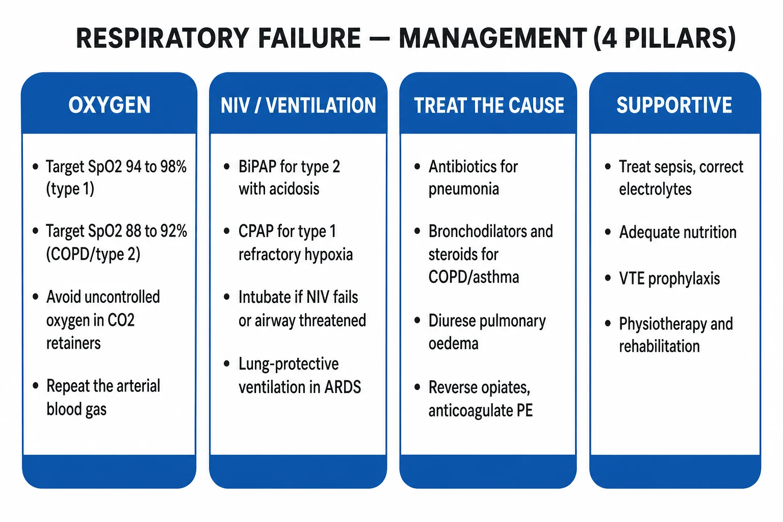
Oxygen: high-flow for type 1 (target SpO2 92-96%), controlled for COPD/type 2 risk (target 88-92%). Position upright. Nebulised bronchodilators if wheeze. Treat the cause empirically (antibiotics for sepsis, diuretic for oedema, naloxone for opioid). Escalate to HFNC / NIV / intubation as above.
Reassess and reassess again
Continuous SpO2, repeat ABG at 30-60 min after any change. Trajectory matters — improving or deteriorating? Set explicit thresholds for escalation (e.g. "if SpO2 <90% on HFNC 60 L/min FiO2 1.0 for 30 min, intubate"). Document the plan so the whole team shares it.
Oxygen therapy devices — escalation ladder
| Device | FiO2 range | Flow | Indication |
|---|---|---|---|
| Nasal cannula | 0.24-0.44 | 1-6 L/min | Mild hypoxaemia, chronic O2 |
| Simple face mask | 0.40-0.60 | 5-10 L/min | Moderate hypoxaemia (need >5 L/min to washout CO2 rebreathing) |
| Non-rebreather mask | 0.60-0.95 | 10-15 L/min | Severe hypoxaemia, bridge to definitive therapy |
| HFNC | 0.21-1.0 | 30-60 L/min | Hypoxaemic failure, recruitability, P/F <300 |
| CPAP / BiPAP | 0.21-1.0 | variable | Type 1 (CPAP), type 2 (BiPAP), pulmonary oedema |
| Invasive ventilation | 0.21-1.0 | set by ventilator | Failure of above, airway/ventilation failure, controlled ventilation needed |
Landmark evidence
FLORALI
NEJM 2015
310 pts with acute hypoxaemic respiratory failure (P/F <300) — HFNC vs standard O2 vs NIV
Key finding
Intubation rate: HFNC 38% vs standard 47% vs NIV 50%. 90-day mortality significantly LOWER with HFNC in P/F <200 subgroup. HFNC better tolerated.
Practice change
HFNC first-line for acute hypoxaemic respiratory failure, especially P/F <200
3CPO
NEJM 2008
1069 pts with acute cardiogenic pulmonary oedema — CPAP vs BiPAP vs standard O2
Key finding
No difference in 7-day or 30-day mortality. Faster symptom resolution and improvement in ABG with NIV. No harm.
Practice change
NIV reasonable in cardiogenic oedema for symptom/physiology benefit, but not a mortality-reducing intervention
Rochwerg meta-analysis
Intensive Care Med 2019
Systematic review of HFNC vs conventional O2 in acute hypoxaemic respiratory failure
Key finding
HFNC reduced need for intubation and post-hoc mortality vs standard O2; no significant difference vs NIV overall but HFNC better tolerated
Practice change
HFNC now standard first-line non-invasive support for hypoxaemic respiratory failure
Lightowler Cochrane (COPD)
BMJ 2003
Meta-analysis of NIV vs usual care for COPD exacerbation with respiratory acidosis
Key finding
NIV reduced mortality (RR 0.41), reduced need for intubation (RR 0.42), reduced treatment failure (RR 0.48), and shortened length of stay
Practice change
NIV (BiPAP) established as first-line for COPD exacerbation with pH 7.25-7.35
Outcomes by support strategy
Special situations
The COPD patient at risk of hypercapnia
The CO2-retaining COPD patient is the classic exam trap. Empirical high-flow O2 can precipitate hypercapnic coma via the Haldane effect and loss of hypoxic pulmonary vasoconstriction. [1]
- Target SpO2 88-92% from the moment of presentation.
- Use Venturi masks (fixed-performance, deliver known FiO2) over simple masks (variable FiO2 dependent on flow and breathing pattern).
- If hypercapnia develops despite controlled O2, start BiPAP (not more O2).
- Recheck ABG at 30-60 minutes. A rising PaCO2 with falling pH mandates BiPAP; if BiPAP fails, intubate.
- Do not be falsely reassured by a "normal" PaCO2 in a tachypnoeic COPD patient — they are compensating with high minute ventilation. As they fatigue, CO2 will rise rapidly. [1]
Neuromuscular respiratory failure
In Guillain-Barré syndrome, myasthenia gravis, ALS, periodic paralysis, the lungs are normal — the respiratory muscle pump fails. Type 2 pattern with normal A-a gradient. [1]
- Monitor FVC and MIP/MEP (maximal inspiratory/expiratory pressure). Electively intubate when FVC <15-20 mL/kg or <1 L, or MIP worse than -30 cmH2O, or bulbar weakness with aspiration risk.
- Do NOT wait for hypercapnia — by the time PaCO2 rises, the patient is in extremis. Hypercapnia is a LATE sign in neuromuscular failure.
- BiPAP can buy time in some (e.g. chronic neuromuscular disease, MG crisis pre-treatment) but most acute GBS need intubation.
- Treat the cause: IVIG/plasma exchange for GBS/MG, pyridostigmine, immunosuppression. [1]
Post-extubation respiratory failure
Patients at high risk of post-extubation failure (age >65, cardiac failure, COPD, obesity, prolonged ventilation, weak cough) benefit from prophylactic HFNC or BiPAP immediately on extubation. The HIGH/BIPOP-WEAN trials support NIV/HFNC to prevent reintubation. [1]
Exam practice
SAQ — Type 2 respiratory failure in COPD
10 minutes · 10 marks
A 68-year-old man with known COPD (FEV1 40% predicted) presents with 3 days of worsening dyspnoea, purulent sputum and confusion. RR 32, SpO2 84% on room air, BP 150/90, HR 110, GCS 14. Initial ABG on 2 L/min nasal cannula: pH 7.24, PaCO2 75, PaO2 52, HCO3 32. CXR: hyperinflation, no consolidation.
SAQ — Intubation decision in hypoxaemic failure
10 minutes · 10 marks
A 55-year-old woman with community-acquired pneumonia is on HFNC 60 L/min, FiO2 0.9. RR 34, SpO2 89%, accessory muscle use, speaking single words. ABG: pH 7.32, PaCO2 38, PaO2 58, HCO3 22. ROX index = 2.6 after 2 hours of HFNC. BP 95/60, HR 110, GCS 15.
Additional clinical pearls
Additional red flags
References
- [1]Papiris S, et al. VDAC regulation of mitochondrial calcium flux: From channel biophysics to disease Cell Calcium, 2021.PMID 33529977
- [2]West JB. Notum palmitoleoyl-protein carboxylesterase regulates Fas cell surface death receptor-mediated apoptosis via the Wnt signaling pathway in colon adenocarcinoma Bioengineered, 2021.PMID 34402722
- [3]Frat JP, Thille AW, Mercat A, et al. Biomarkers in Interstitial lung diseases Paediatr Respir Rev, 2015.PMID 26027849
- [4]Gray A, Goodacre S, Newby DE, et al. Spontaneous neural activity during human slow wave sleep Proc Natl Acad Sci U S A, 2008.PMID 18815373
- [5]Mosier JM, Hypes CD, Sakles JC. Better HHS Planning Needed for National Disaster Medical System JAMA, 2020.PMID 32720990
- [6]Roca O, Caralt B, Messika J, et al. Corrigendum to Outcomes associated with amiodarone and lidocaine in the treatment of in- hospital pediatric cardiac arrest with pulseless ventricular tachycardia or ventricular fibrillation [Resuscitation (2014) 85 381-386] Resuscitation, 2019.PMID 31356765
- [7]Lightowler JV, Wedzicha JA, Elliott MW, Ram FS. Spontaneous re-expansion of a collapsed thoracic endoprosthesis: case report J Vasc Surg, 2008.PMID 19118739