ICU · Advanced respiratory support
Advanced Respiratory Support — NIV, Weaning and Tracheostomy
Also known as Non-invasive ventilation · CPAP · BiPAP · Weaning · Spontaneous breathing trial · Tracheostomy · Extubation · Respiratory muscle fatigue
Advanced respiratory support covers the three skills that frame the respiratory failure beyond the ventilator — the non-invasive ventilation (the CPAP and the BiPAP, their indications and their evidence), the liberation from the mechanical ventilation (the spontaneous breathing trial, the weaning protocol, the difficult-to-wean patient and the role of the NIV-facilitated extubation), and the tracheostomy (the timing, the technique, the decannulation). This topic builds the examiner's framework on the NIV physiology (the positive pressure that unloads the respiratory muscles, the CPAP for the oxygenation, the BiPAP for the ventilation), the weaning evidence (the Esteban comparison of the four methods, the role of the NIV in the post-extubation failure, the Burns review of the NIV-assisted weaning), and the tracheostomy timing question.
On this page & tools
Your progress
Saved locally on this device.
Target exams
Overview & definition
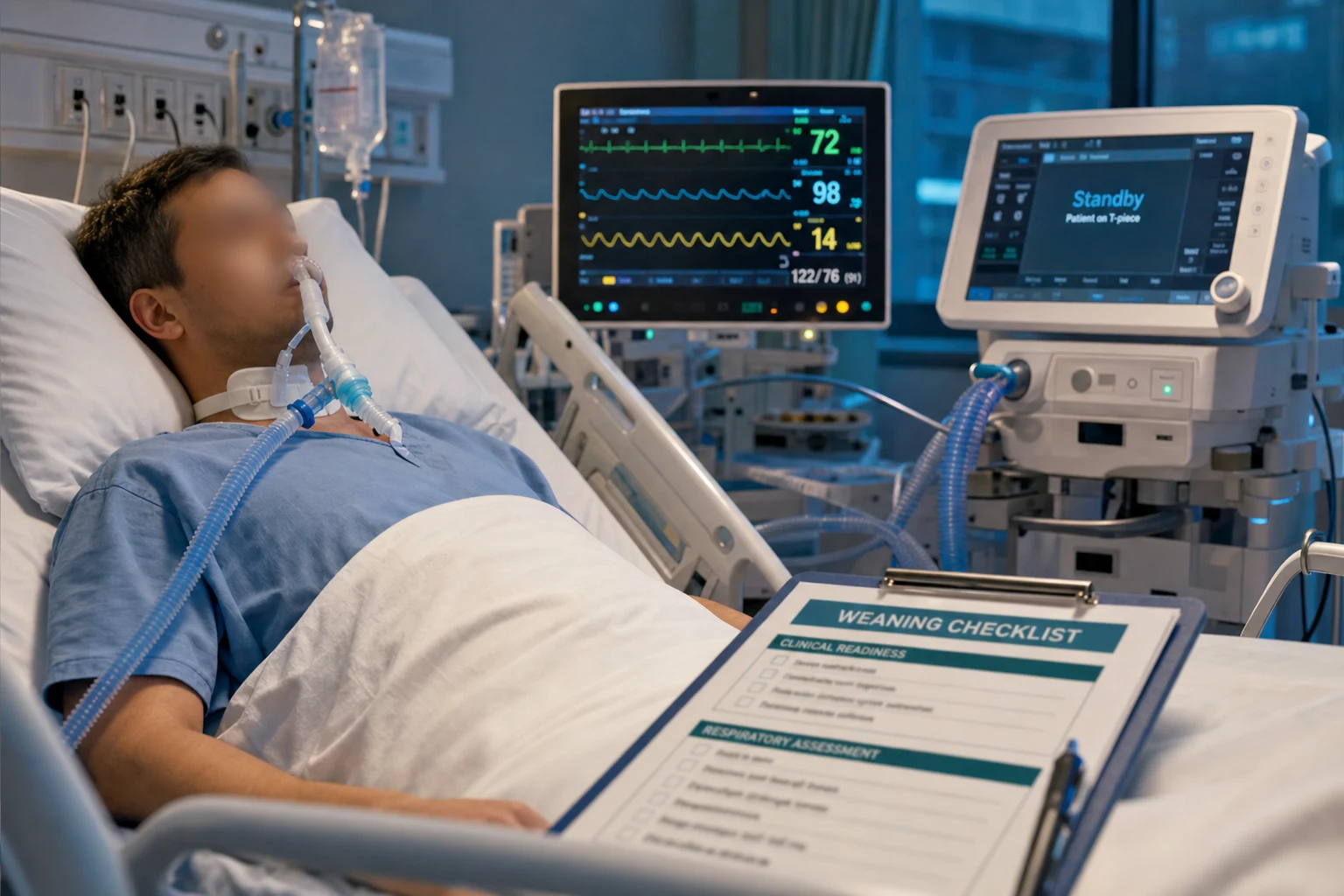
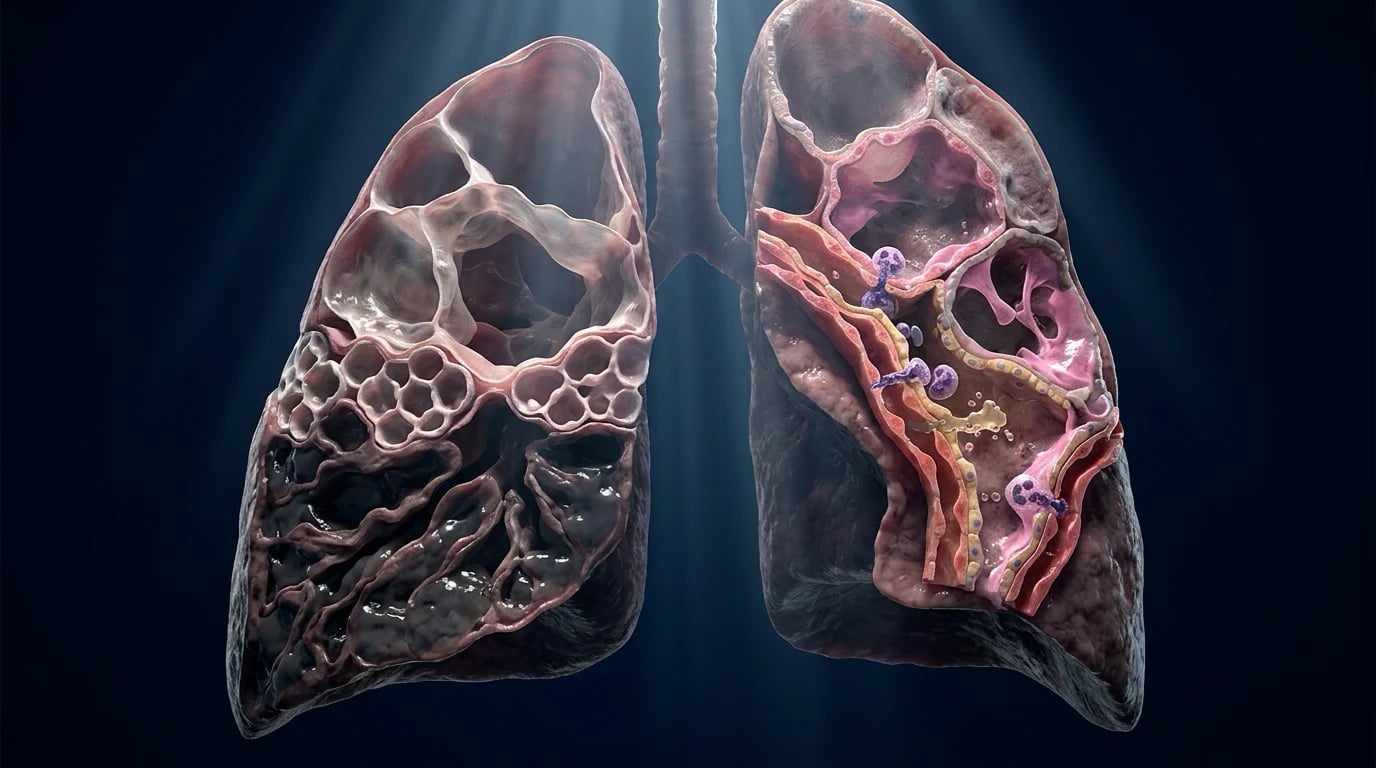
Advanced respiratory support is the set of skills that frame the respiratory failure beyond the intubation and the mechanical ventilation — the non-invasive ventilation (the CPAP and the BiPAP that avert or follow the intubation), the liberation from the mechanical ventilation (the spontaneous breathing trial, the weaning protocol, the difficult-to-wean patient), and the tracheostomy (the long-term airway for the patient who cannot be extubated). Together they are the higher respiratory skills the CICM examinor expects.[1][1]
The over-riding principle is that the prompt, the safe, the successful liberation is the goal of the mechanical ventilation — for every additional day on the ventilator raises the risk of the ventilator-associated pneumonia, the weakness and the mortality — and the NIV and the tracheostomy are the tools that facilitate it or that provide the alternative.[1]
Non-invasive ventilation: CPAP and BiPAP
Non-invasive ventilation is the positive-pressure ventilation delivered via a mask (or a helmet), without an endotracheal tube. It has two modes: the CPAP (the continuous positive airway pressure — a single positive pressure throughout the respiratory cycle, which splints the alveoli, recruits the collapsed lung, reduces the preload and the afterload of the heart, and improves the oxygenation) and the BiPAP (the bilevel positive airway pressure — an inspiratory positive airway pressure, the IPAP, and an expiratory positive airway pressure, the EPAP, which augments the tidal volume and unloads the respiratory muscles, improving the ventilation and the CO2 clearance).[1][1]
The physiological mechanism of the NIV benefit is the unloading of the fatiguing respiratory muscles — the positive pressure reduces the work of breathing (the pressure support does the work the patient cannot), and the CPAP recruits the collapsed alveoli and improves the compliance. The result is the reduced respiratory rate, the improved gas exchange, and the averted (or the shortened) intubation.[1]
The indications and evidence for NIV
The NIV is the first-line therapy for three conditions, each with a strong evidence base.[1][1]
- The COPD exacerbation with the respiratory acidosis (the pH below 7.35). The NIV (the BiPAP) is the first-line — it reduces the intubation rate, the mortality and the length of the stay, and it is the standard of care. The target is the pH correction (to above 7.35) and the PaCO2 reduction, with the settings titrated (the IPAP of 10 to 15, the EPAP of 4 to 5, the FiO2 for the 88 to 92 per cent saturation).
- The cardiogenic pulmonary oedema. The NIV (the CPAP or the BiPAP) is the first-line — it reduces the preload and the afterload (the CPAP), it unloads the failing heart, and it reduces the intubation rate. The CPAP alone is often sufficient.
- The immunocompromised patient with the respiratory failure. The NIV is the first-line — it reduces the intubation rate and the mortality (by avoiding the invasive ventilation and its infections in the vulnerable host).
- The post-extubation and the weaning-facilitating NIV — the evidence is more nuanced (below). [1]
Pathophysiology: the work of breathing and the respiratory muscle fatigue
The respiratory muscles behave like any skeletal muscle — they fatigue under a sustained load, and the fatigue leads to the rapid, shallow breathing (the low tidal volume, the high rate), the rising CO2 and the eventual failure. The work of breathing has three components: the elastic (the compliance of the lung and the chest wall — raised by the ARDS, the oedema, the fibrosis), the resistive (the airway resistance — raised by the COPD, the asthma, the secretions), and the threshold (the intrinsic PEEP of the COPD — the positive end-expiratory pressure that must be overcome before the inspiratory flow begins).[1]
The NIV unloads the muscles by the pressure support (which does the work of the inspiration) and the CPAP/EPAP (which counteracts the intrinsic PEEP and the elastic load). The result is the reduced work, the recovered muscles, and the time to treat the cause.[1]
Weaning from mechanical ventilation: the principles
The weaning is the liberation from the mechanical ventilation, and the principle is that the earlier the safe liberation, the better — the prolonged ventilation increases the complications. The daily assessment of the readiness (the resolution of the cause, the adequate oxygenation, the haemodynamic stability, the ability to protect the airway) is the first step, and the spontaneous breathing trial (the SBT) is the test.[1][1]
The Esteban trial (NEJM 1995) compared four methods of weaning — the T-piece, the SIMV, the pressure-support and the daily SBT — and found that the pressure-support and the daily SBT (the once- or twice-daily trial of the spontaneous breathing) were the fastest, the SIMV the slowest. The modern practice is the daily SBT (the low pressure support of 5 to 7 cmH2O, for 30 to 120 minutes), and if the patient tolerates it (the stable respiratory rate, the adequate gas exchange, no distress), the extubation follows.[1]
The spontaneous breathing trial and the weaning protocol
The SBT is the low-pressure-support trial (the 5 cmH2O, for 30 to 120 minutes) that assesses the patient's ability to breathe without the ventilator. The tolerance is judged by the respiratory rate (under 35), the heart rate and the blood pressure (the stability), the gas exchange (the saturation above 90 per cent), and the absence of the distress (the diaphoresis, the accessory-muscle use, the agitation). The failure (the rising rate, the distress, the desaturation) returns the patient to the ventilator, and the cause is sought and treated.[1]
The weaning protocol — the nurse- or the respiratory-therapist-led protocol that performs the daily SBT and the readiness assessment — is superior to the physician-directed weaning (it shortens the duration). The rapid shallow breathing index (the RSBI, the f/VT ratio) — the respiratory rate divided by the tidal volume in litres — is a validated predictor of the weaning success (the RSPI below 105 predicts the success), though the clinical SBT is the definitive test.[1]
The post-extubation NIV question
The role of the NIV after the extubation is nuanced, and the evidence is mixed. The prophylactic NIV (for the high-risk patient — the COPD, the older, the cardiac) started immediately after the extubation reduces the reintubation and the mortality. The therapeutic NIV (for the patient who has developed the respiratory failure after the extubation) is more controversial — the Esteban trial (NEJM 2004) found it did not reduce the reintubation, and a concern of the delayed reintubation (with a higher mortality) was raised.[2]
The synthesis is: the prophylactic post-extubation NIV for the high-risk patient is beneficial; the therapeutic NIV for the established post-extubation failure must be used with caution (and the prompt reintubation if the NIV fails), because the delay to the reintubation is the harm.[2][1]
The difficult-to-wean patient and the NIV-assisted weaning
Some patients (the COPD, the neuromuscular weakness, the older with the cardiac comorbidity) fail the repeated SBTs and require the prolonged ventilation — the difficult-to-wean patient. The NIV-facilitated extubation (the extubation to the NIV, regardless of the SBT pass/fail) is a strategy for these patients, and the Burns systematic review (Thorax 2022) found it reduced the duration of the invasive ventilation, the mortality and the ventilator-associated pneumonia in the selected (mostly COPD) population.[3]
The principle is that the NIV-assisted weaning unloads the fatiguing muscles while avoiding the complications of the prolonged intubation — but it requires the patient to protect the airway and to tolerate the mask, and the failure is the prompt reintubation.[3][1]
Tracheostomy: timing, technique and decannulation
The tracheostomy is the long-term airway for the patient who cannot be extubated (the prolonged ventilation, the inability to protect the airway, the secretion clearance). The timing is the debated question — the early (within 2 to 4 days) versus the late (after 10 to 14 days) — and the TRACMAN trial (NEJM 2013) found that the early tracheostomy did not reduce the mortality or the ventilator-associated pneumonia, though it reduced the sedation and the ICU stay in some. The practice is individualised: the tracheostomy is considered for the patient expected to need the ventilation beyond 7 to 10 days, and the percutaneous dilatational technique (at the bedside, by the intensivist) is the standard.[1][1]
The decannulation (the removal of the tracheostomy) follows the successful weaning from the tracheostomy (the progressive downsizing, the tolerance of the capping, the effective cough and the secretion clearance), and the swallowing assessment. The principle is the graded, the assessed liberation from the tracheostomy, as from the endotracheal tube.[1]
Management: the structured liberation approach
The liberation from the mechanical ventilation is a structured, daily, protocolised process.[1][1]
- Daily assessment of the readiness — the cause resolved, the oxygenation adequate (the FiO2 at or below 0.4, the PEEP at or below 8), the haemodynamics stable (no or low vasopressors), the ability to protect the airway.
- The SBT — the 5 cmH2O pressure support for 30 to 120 minutes; the tolerance (the stable rate, the adequate gas, no distress) is the indication for the extubation.
- The extubation — the assessment of the airway (the cuff leak, the laryngeal oedema risk), the cough and the secretions, the neurological status; the prophylactic NIV for the high-risk.
- The NIV-assisted weaning for the difficult-to-wean — the extubation to the NIV, with the prompt reintubation for the failure.[3]
- The tracheostomy for the prolonged ventilation (7 to 10 days) — the percutaneous, the bedside, the individualised timing.
- The prevention of the failure — the treatment of the cause, the sedation minimisation, the early mobilisation, the nutritional support.
Monitoring the weaning patient
Monitoring divides into the SBT tolerance, the respiratory muscle reserve, and the complications.[1][1]
- The SBT tolerance — the respiratory rate (under 35), the heart rate and the blood pressure (the stability), the saturation (above 90 per cent), the absence of the distress.
- The respiratory muscle reserve — the RSBI (the f/VT, under 105 predicts the success), the maximal inspiratory pressure (the negative inspiratory force), the trend of the tidal volume and the rate.
- The complications — the laryngeal oedema (the cuff-leak test), the secretion retention, the cardiac ischaemia (the weaning stress), and the delirium. [1]
Prognosis and extubation failure
The prognosis of the weaning is the prognosis of the underlying disease, but the time to the successful liberation is itself a prognostic marker — the prolonged ventilation (beyond 7 days) carries a higher mortality and morbidity (the ventilator-associated pneumonia, the ICU-acquired weakness). The extubation failure (the reintubation within 48 to 72 hours) occurs in about 10 to 20 per cent of the patients, and it is associated with a higher mortality — particularly when the reintubation is delayed (the NIV failing, the patient tiring). The principle is the prompt, the safe, the protocolised liberation, and the prompt reintubation for the failure.[2][1]
Advanced mechanical ventilation modes
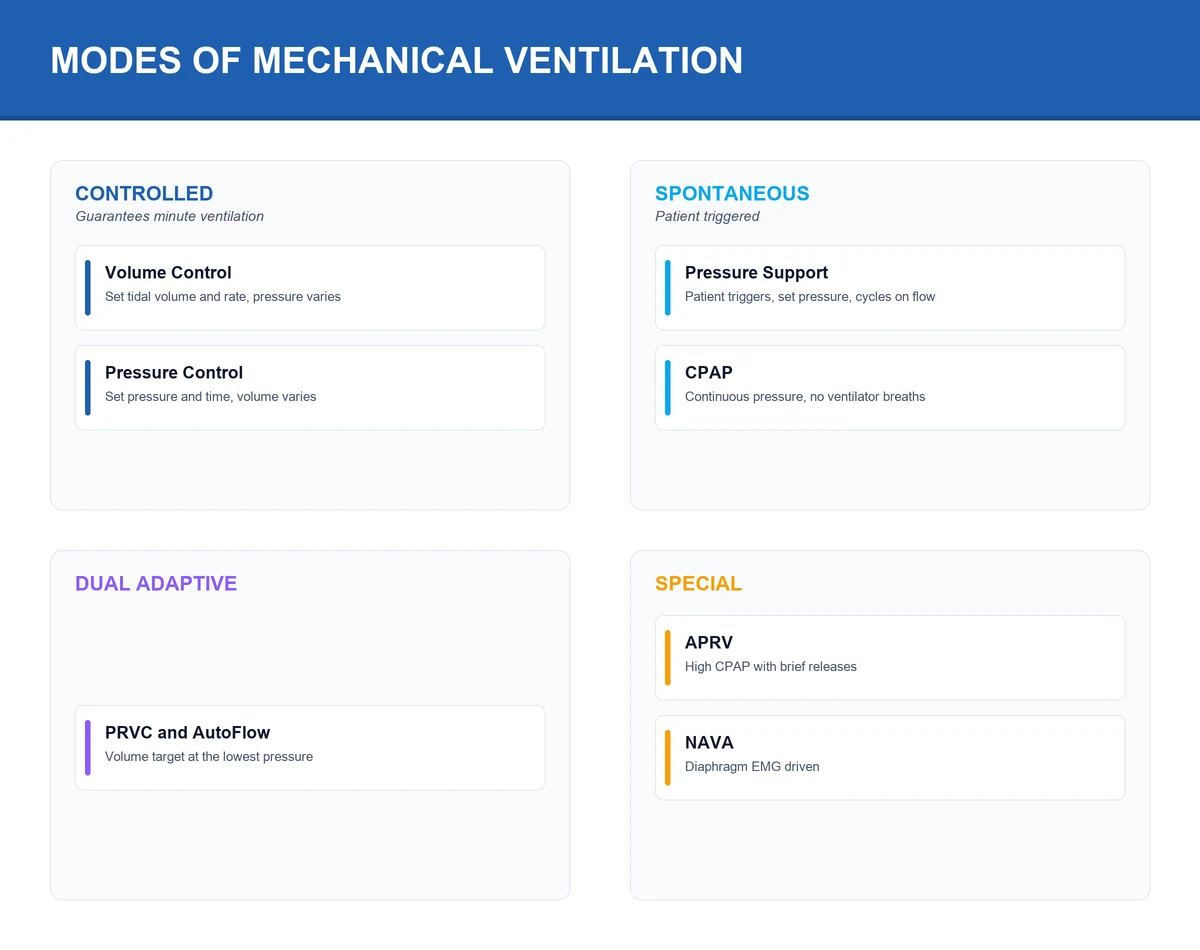
Beyond the NIV and the weaning lies the choice of the mode of the mechanical ventilation itself — the volume versus the pressure, the dual control, the closed-loop and the proportional modes, and the alarms and the troubleshooting that frame the ventilator at the bedside. The over-riding principle is that no advanced mode has proven a mortality benefit over the standard lung-protective ventilation (the volume control, the 6 mL/kg predicted body weight, the plateau below 30 cmH2O, the driving pressure below 15) — the advanced modes are the tools for the difficult-to-ventilate and the dyssynchronous patient, not a replacement for the fundamentals.[4][7]
The classification of a mode rests on three phase variables — the trigger (what starts the breath), the limit (what governs the delivered breath), and the cycle (what ends the breath). The volume control is the time/flow/volume; the pressure control is the time/pressure/time; the pressure support is the patient/pressure/flow. To name the three phase variables is to understand the mode.[7]
Volume control (VC)
The volume control guarantees the tidal volume (the set volume is delivered regardless of the lung compliance) and the pressure is the variable (it rises with the low compliance). The flow is constant (the square-wave pattern), the inspiration is time-cycled, and the peak airway pressure reflects the resistance and the compliance combined. The risk is the high pressure in the low-compliance lung (the ARDS) — managed by the inspiratory hold (the plateau pressure, below 30 cmH2O) and the upper pressure alarm. The VC is the standard, the safe and the easy-to-monitor mode, and it is the foundation of the lung-protective ventilation.[4][7]
Pressure control (PC)
The pressure control guarantees the inspiratory pressure (the set pressure is held throughout the inspiration) and the tidal volume is the variable (it changes with the compliance). The flow is decelerating (the high initial flow that falls toward the zero), the inspiration is time-cycled, and the peak pressure EQUALS the plateau pressure (the constant inspiratory pressure — no resistive overshoot, no need for an inspiratory hold). The advantage is the better gas distribution in the heterogeneous lung (the ARDS, the pneumonia, the lobar consolidation) and the lower peak pressure; the risk is the variable tidal volume (the falling compliance drops the Vt, the rising compliance overdistends). The PC requires the close monitoring of the delivered tidal volume.[7]
Pressure support ventilation (PSV)
The pressure support is the spontaneous mode — the patient triggers each breath (the trigger is the flow or the pressure drop at the airway), the ventilator delivers the set pressure support to augment the inspiration, and the breath cycles off when the inspiratory flow falls to the threshold (the cycling-off criterion, typically the 25 per cent of the peak flow). The PSV is the weaning mode (the SBT is the PSV at the 5 to 7 cmH2O) and the spontaneous-breathing mode of the ventilator. The advantage is the patient-controlled breathing (the rate, the Vt and the inspiratory time are patient-set); the risks are the inadequate support in the fatiguing patient (the fatigue → the failure) and the leak-related asynchrony (the NIV).[1]
Synchronised intermittent mandatory ventilation (SIMV)
The SIMV combines the mandatory breaths (the ventilator-delivered, the volume or the pressure, at the set rate) with the spontaneous breaths (the patient-triggered, supported by the pressure support). The mandatory breaths are synchronised to the patient effort (the ventilator waits for the trigger window before delivering the mandatory breath). The SIMV was historically popular but the evidence (the Brochard 1994, the Esteban 1995) showed it is the slowest and the least successful weaning method — the intermittent mandatory breaths impose the variable load on the diaphragm. The SIMV is now reserved for the initial support and the specific situations (the partial ventilatory support with the backup rate), not for the weaning.[1][8]
Dual control modes: PRVC, VC+ and AutoFlow
The dual control modes combine the volume guarantee with the pressure limitation — the volume-targeted, the pressure-limited ventilation. The operator sets the target tidal volume and the upper pressure limit; the ventilator delivers the pressure-control-type breath (the decelerating flow) and measures the resulting volume, then adjusts the inspiratory pressure breath-by-breath to deliver the target volume at the lowest possible pressure. The PRVC (the pressure-regulated volume control, Maquet/Getinge), the VC+ (the Hamilton) and the AutoFlow (the Dräger) are the proprietary names for the same concept. The advantage is the guaranteed volume with the pressure-minimised gas distribution — the modern ventilator default. The risk is the silent rise of the pressure to chase the falling compliance — the ventilator will raise the pressure toward the set limit breath-by-breath, and a high or default limit can allow the injurious pressures. Manage by the tight upper pressure limit (a few cmH2O above the working pressure) and the daily watching of the plateau and the driving pressure.[7]
APRV and BiLevel — the two-level pressure modes
The airway pressure release ventilation (the APRV) and the BiLevel (the DuoPAP, the Bilevel) are the two-level pressure modes. The APRV applies a continuous high pressure (the Phigh, the 25 to 35 cmH2O) held for a long time (the Thigh, the 4 to 6 seconds), with the brief releases to the low pressure (the Plow, the 0 to 5 cmH2O, for the short Tlow, the 0.4 to 0.8 seconds). The spontaneous breathing occurs throughout the cycle (the key feature — the diaphragm keeps working, the sedation is lighter, the venous return is preserved, the muscle atrophy is reduced). The high mean airway pressure recruits the alveoli (the oxygenation); the release generates the ventilation (the CO2 clearance). The Tlow is the single most important setting — titrate so the expiratory flow returns to the 50 to 75 per cent of the peak (the partial release). Too long → the derecruitment; too short → the air-trapping and the auto-PEEP.[7]
The BiLevel is the controlled variant with the set mandatory breaths (the pressure-controlled breaths at the high level, with the time-cycled transitions). The BiLevel is for the patient needing the recruitment and the synchrony benefits of the two-level mode but with the guaranteed minimum minute ventilation (the tiring patient, the marginal drive). The APRV is avoided in the obstructive disease (the COPD, the asthma) for the dynamic hyperinflation risk from the inverse ratio. No proven mortality benefit; the salvage or the adjunct for the recruitable, stiff lung of the ARDS.[7]
Closed-loop ventilation: ASV
The adaptive support ventilation (the ASV, Hamilton) is the closed-loop mode — the operator sets the target minute ventilation (as the percentage of the predicted, typically the 100 per cent) and the ventilator measures the compliance and the resistance each breath, then computes the rate-and-tidal-volume combination that achieves the target at the minimum work of breathing (the Otis optimum, based on the expiratory time constant). The ventilator continuously adapts the pattern to the changing mechanics and the patient drive — more support when the drive is low, the spontaneous breathing when the drive returns. The ASV is the automated mode — useful for the variable drive and the weaning (it reduces the clinician interventions and shortens the weaning in some trials), but it does not prove the mortality benefit over the protocolised conventional weaning.[7]
Proportional modes: PAV+ and NAVA
The proportional assist ventilation (the PAV+) and the neurally adjusted ventilatory assist (the NAVA) are the proportional modes — the support tracks the instantaneous patient effort. The PAV+ measures the elastance and the resistance each breath, the clinician sets the gain (the percentage of the work the ventilator does — typically the 50 to 80 per cent), and the support is the flow-and-volume-proportional — a small effort yields the small support, a large effort the large support. The patient governs the rate, the Vt, the flow and the inspiratory time. The NAVA uses the electrical activity of the diaphragm (the EAdi, via the specialised nasogastric electrode catheter) to trigger and to drive the support — the neural signal, the proportional pressure (the cmH2O per microvolt of EAdi). The advantage is the superior synchrony (the neural trigger eliminates the ineffective triggering and the auto-triggering, the cycling matches the neural inspiratory time) and the patient-governed breathing. The NAVA is the best mode for the NIV with the large leaks (the pneumatic triggers fail in the leak; the EAdi does not). The risks — the PAV+ requires the reliable elastance/resistance measurement (the coughing, the leaks disturb it); the NAVA depends on the intact neuromuscular drive (the high cervical cord injury, the phrenic nerve injury, the severe neuromuscular disease, the deep sedation or the paralysis) and the correctly placed catheter. Always set the backup pneumatic mode for the NAVA.[7]
Closed-loop oxygenation
The closed-loop oxygenation (the AutoMix, the O2-Closed Loop, the FreeO2) is the emerging automation of the FiO2 — the ventilator (or the HFNC) titrates the FiO2 to the target SpO2, reducing the hyperoxia and the hypoxia. The benefit is the reduced nursing workload and the lower mean FiO2 (the lung-protective and the oxygen-toxicity avoidance). The closed-loop oxygenation is not yet the standard, but it is a growing area — the closed-loop systems (the ASV with the auto-oxygenation, the SmartCare) move toward the fully automated ventilation. [1]
| Mode | Trigger | Limit | Cycle | Guarantees | Best use | Risk |
|---|
| Mode | Trigger | Limit | Cycle | Guarantees | Best use | Risk |
|---|---|---|---|---|---|---|
| Volume control (VC) | Time (or patient) | Flow | Time (volume) | Tidal volume | Standard lung-protective ventilation — most patients | High pressure in the low-compliance lung |
| Pressure control (PC) | Time (or patient) | Pressure | Time | Inspiratory pressure | Heterogeneous lung (ARDS, pneumonia); lower peak pressure | Variable Vt with changing compliance |
| Pressure support (PSV) | Patient (flow/pressure) | Pressure | Flow (cycle-off at 25%) | Pressure support | Spontaneous breathing; weaning; SBT | Inadequate support → fatigue; leak asynchrony |
| SIMV | Patient + time | Flow or pressure | Time or flow | Mandatory breaths + spontaneous | Initial partial support (rare) | Slowest, least successful weaning mode |
| PRVC / VC+ / AutoFlow | Time (or patient) | Pressure (auto-titrated) | Time (volume) | Tidal volume at lowest pressure | Modern default; changing compliance | Silent pressure rise to chase falling Vt |
| APRV | Time (release) + spontaneous | Pressure | Time (release) | Phigh + release Vt | Recruitable stiff ARDS lung; spontaneous breathing preserved | Dynamic hyperinflation in obstruction |
| BiLevel (DuoPAP) | Time + spontaneous | Pressure | Time | Mandatory breaths + spontaneous | Two-level recruitment with guaranteed minute ventilation | Less spontaneous-breathing benefit than APRV |
| ASV | Patient + closed loop | Closed loop | Closed loop | Target minute ventilation at min WOB | Variable drive; automated weaning | No mortality benefit; rely on the algorithm |
| PAV+ | Patient (flow/volume) | Proportional | Flow (patient) | % of the work of breathing | Dyssynchronous patient; load-sharing | Disturbed by leaks; needs reliable mechanics |
| NAVA | Patient (neural — EAdi) | Proportional | Neural (EAdi fall) | cmH2O per microvolt EAdi | Dyssynchrony; NIV with large leaks; diaphragm-protective | Fails without intact neuromuscular drive; catheter placement |
| Feature | Volume control (VC) | Pressure control (PC) |
|---|
| Feature | Volume control (VC) | Pressure control (PC) |
|---|---|---|
| What is guaranteed | Tidal volume | Inspiratory pressure |
| What is variable | Airway pressure | Tidal volume |
| Flow pattern | Constant (square wave) | Decelerating (descending ramp) |
| Peak vs plateau pressure | Peak > plateau (resistive overshoot); hold to read plateau | Peak = plateau (constant pressure) |
| Gas distribution in heterogeneous lung | Less uniform | More uniform (favours slow units) |
| Risk in falling compliance (worsening ARDS) | Pressure rises — alarm fires (visible) | Vt falls silently — must monitor Vt |
| Reading plateau pressure | Requires an inspiratory hold | Read directly from the airway pressure |
| Best for | Most patients; lung-protective ventilation standard | Stiff heterogeneous lung; desire for low peak pressure |
The dual-control adaptive loop — how PRVC keeps the volume guaranteed at the lowest pressure
Operator sets the target Vt, RR, inspiratory time, PEEP and the upper pressure limit
Enter target Vt (typically 6 mL/kg PBW), RR, I-time, PEEP, FiO2, and the upper pressure alarm limit (e.g. 40 cmH2O). The ventilator will not exceed this limit. The tightness of the limit is the safety mechanism.
The ventilator delivers a test pressure-control breath and measures the resulting Vt
A first decelerating-flow breath at a trial pressure lets the ventilator calculate the dynamic compliance (the Vt divided by the pressure above the PEEP). This is the baseline the algorithm works from.
It sets the working pressure just sufficient for the target Vt
The ventilator computes the inspiratory pressure that, delivered as a constant-pressure decelerating-flow breath, will yield the target Vt at the measured compliance. It uses the LOWEST pressure that achieves this — the volume guarantee at the pressure minimum.
It re-measures each breath and adapts breath-by-breath
On every subsequent breath the ventilator compares the delivered Vt to the target. If the Vt is too low (compliance fell), it raises the inspiratory pressure a few cmH2O; if too high (compliance rose), it lowers the pressure. The adaptation is gradual and continuous.
The danger — the silent chase of a falling compliance
If the lung collapses (rolling onto the bad side, plugging, oedema worsening), the compliance falls and the ventilator RAISES the pressure to maintain the Vt — up to the upper limit. A high or default limit allows the injurious pressures without an obvious alarm. Set the limit tight and watch the plateau and the driving pressure.
NAVA — from neural drive to proportional support
Place the EAdi catheter
A nasogastric tube carrying a ring of electrodes is passed into the stomach and withdrawn to the gastro-oesophageal junction, where the electrodes sit opposite the diaphragmatic crura. Correct position is confirmed by a characteristic phasic EAdi waveform (a peak with each inspiration) over a smaller ECG trace.
The EAdi is measured continuously
The electrical activity of the diaphragm (EAdi, in microvolts) is a direct real-time readout of the neural respiratory drive — brainstem to phrenic nerve to diaphragm — independent of the airflow, the airway pressure or the circuit leak. It rises with each inspiratory effort.
Inspiration is triggered by the rise in EAdi
Instead of a flow or pressure drop at the airway, the ventilator triggers on the rising EAdi — the neural signal itself. This eliminates the ineffective triggering (small efforts that fail to trigger) and the auto-triggering (false breaths from leaks or circuit motion).
Support is delivered proportionally to the EAdi
The clinician sets the NAVA level (cmH2O per microvolt of EAdi). Each microvolt of diaphragmatic activity is amplified into a proportional airway pressure. The harder the patient pulls, the more support the ventilator delivers — the patient governs every breath.
Cycling matches the neural inspiratory time
The breath cycles off when the EAdi falls to a fraction of the peak (typically 70 per cent) — the ventilator cycles exactly when the brain stops driving the diaphragm. This eliminates the delayed and the premature cycling that plague the pneumatic modes.
Ventilator alarms and troubleshooting
The ventilator alarms divide into the high-priority (the immediate threat — the apnoea, the disconnection, the high peak pressure, the low exhaled tidal volume, the low FiO2) and the lower-priority (the high respiratory rate, the low minute ventilation). The discipline is to NEVER silence and ignore an alarm — every alarm has a cause, and the bedside assessment (the patient, the circuit, the ventilator, the settings) is the method. The systematic approach is the DOPE mnemonic — the Displacement (the tube), the Obstruction (the tube or the airway), the Pneumothorax, the Equipment failure.[1][1]
The high peak airway pressure alarm — the systematic approach
1. Is the patient or the circuit the cause? — disconnect at the Y-piece
Disconnect the circuit at the Y-piece and ventilate by bag. If bagging is EASY and the pressure falls, the problem is the CIRCUIT or the TUBE (kink, obstruction, mucous plug, water in the circuit). If bagging is HARD and the chest barely moves, the problem is the PATIENT (bronchospasm, pneumothorax, mainstem intubation, ARDS worsening).
2. If the circuit/tube — find and fix the obstruction
Suction the tube (mucus plug), pass a suction catheter (if it will not pass, suspect tube obstruction or mainstem), check for kinks, drain the water from the circuit, check the cuff (herniation). Reposition or replace the tube if needed.
3. If the patient — distinguish resistance from compliance
After ensuring the tube is patent, perform an inspiratory hold. The PEAK minus the PLATEAU = the RESISTIVE component (bronchospasm, secretions, small tube). The PLATEAU above the PEEP = the ELASTIC component (low compliance — ARDS, oedema, pneumothorax, mainstem). A high peak with a normal plateau = resistance; a high plateau = compliance.
4. Auscultate and examine — bilateral air entry, wheeze, silence
Bilateral wheeze → bronchospasm (salbutamol). Unilateral silence → mainstem intubation (withdraw the tube) or pneumothorax (needle decompression if unstable). Decreased globally → worsening ARDS or oedema (increase PEEP, treat the cause).
5. Check the plateau and driving pressure — the lung-protection targets
The plateau must be below 30 cmH2O and the driving pressure below 15 cmH2O. If the plateau is high, reduce the Vt (to 4 mL/kg PBW), increase the sedation, treat the cause. A persistently high plateau despite the Vt reduction suggests the pneumothorax, the pleural effusion, the abdominal hypertension, or the chest wall restriction.
The low exhaled tidal volume / low minute ventilation alarm — the systematic approach
1. Look for the LEAK
The most common cause. Check the cuff (underinflation, rupture), the circuit connections (the HMEF, the humidifier, the catheter mount), the chest drains (bronchopleural fistula), and around the tube. In the NIV, the mask leak is expected (the intentional leak through the mask ports + the unintentional leak from the poor seal).
2. Distinguish the leak from the worsening compliance (PC/PSV)
In the pressure modes, a falling Vt can be the leak OR the worsening compliance. Check the inspired vs the exhaled Vt (the difference is the leak). If the inspired equals the exhaled and both are low, the compliance fell — recheck the plateau.
3. Check the patient — agitation, biting, coughing
The patient fighting the ventilator (the asynchrony, the biting the tube) generates the alarm. Sedate, analgesed, bronchodilate, suction, and address the trigger (the pain, the hypoxia, the delirium, the full bladder).
4. Confirm the tube position — extubation, mainstem, displacement
A displaced tube (the partial extubation into the pharynx, the oesophageal intubation) causes the low exhaled Vt. The end-tidal CO2, the auscultation and the chest movement confirm the tracheal position.
5. Reassess the settings — the leak compensation, the mode
In the NIV or the leaky circuit, enable the leak compensation (the PAV+, the NAVA, the PSV with the leak adjustment). If the leak cannot be managed, switch the mode or the interface.
| Alarm | Priority | Common causes | Immediate action |
|---|
| Alarm | Priority | Common causes | Immediate action |
|---|---|---|---|
| High peak airway pressure | High | Tube obstruction (plug, kink, bite), bronchospasm, worsening compliance, pneumothorax, mainstem, cough/bucking | Disconnect at Y; bag; DOPE; distinguish resistance (peak) from compliance (plateau) |
| Low exhaled Vt / minute ventilation | High | Leak (cuff, circuit), displaced tube, PC with falling compliance, apnoea | Check inspired vs exhaled Vt (leak); auscultate; confirm tube; sedate/analgesed |
| Apnoea | High | Disconnection, tube displacement, over-sedation, cardiac arrest, neurological deterioration | Bag the patient immediately; check the tube, the circuit, the pulse; ventilate manually |
| Low FiO2 / O2 supply failure | High | Wall/O2 cylinder empty, blender failure, tubing disconnect | Check the O2 supply and the blender; switch to the backup; bag with 100% O2 if unstable |
| High respiratory rate | Low-Medium | Pain, anxiety, hypoxia, fever, sepsis, metabolic acidosis, inadequate support, delirium | Treat the cause; increase the support; check the ABG; sedate/analgesed appropriately |
| Circuit disconnection | High | Accidental disconnect at the Y, the catheter mount, the humidifier, the water trap | Reconnect; verify the ventilation and the chest movement; check the tube |
Patient-ventilator asynchrony
The patient-ventilator asynchrony is the mismatch between the patient effort and the ventilator delivery — it occurs in roughly a quarter of the ventilated patients and is associated with the longer ventilation, the longer ICU stay and the worse outcomes. The major types are the ineffective triggering (the patient effort that fails to trigger the breath — the most common, due to the auto-PEEP, the over-sedation, the weak effort), the double triggering (the breath stacking — the second breath before the exhalation, due to the inspiratory time shorter than the neural time), the auto-triggering (the false breaths from the leak or the circuit motion), the flow starvation (the patient pulling harder than the ventilator delivers — the scalloped flow curve), and the delayed cycling (the ventilator cycles off after the patient has stopped inspiring). The discipline is to read the waveforms regularly and fix the cause — the trigger sensitivity, the cycling-off threshold, the rise time, the inspiratory time, the PEEP (for the auto-PEEP), or the mode switch (the PAV+, the NAVA).[7]
Reading the ventilator waveforms — five patterns and what they mean
Scalloped inspiratory flow curve (flow starvation)
In the pressure support or the PC, the inspiratory flow curve should be a smooth descending ramp. If it is CONCAVE (scalloped), the patient is pulling harder than the ventilator delivers — FLOW STARVATION (the patient is "fighting the ventilator"). Fix by increasing the rise time or the pressure support, lengthening the inspiratory time, or switching to a more responsive mode.
Expiratory flow not returning to baseline (auto-PEEP/air-trapping)
The expiratory flow curve should reach zero before the next breath. If it is still above zero at the end-expiration, there is the INCOMPLETE EXHALATION = the AIR TRAPPING (the intrinsic/auto-PEEP). Seen in the COPD, the asthma, the high RR, or the too-short expiratory time. Manage by reducing the RR, shortening the inspiratory time, applying the external PEEP about 80% of the auto-PEEP.
Double triggering (breath stacking)
Two breaths delivered with little or no exhalation between them — the patient triggers a second breath before the first has cycled off. Causes: the inspiratory time too short for the neural time (the delayed cycling), the high drive, the low support. Consequence: the stacked Vt, the high transpulmonary pressure, the VILI risk. Fix by lengthening the inspiratory time or raising the support or the cycling threshold.
Ineffective triggering (missed breaths)
The patient effort visible on the flow/pressure trace (a brief deflection) that does NOT trigger a ventilator breath. Causes: the auto-PEEP (the patient must first climb the intrinsic PEEP), the over-sedation, the weak respiratory muscle, the insensitive trigger. Manage by treating the auto-PEEP (bronchodilators, lower RR, external PEEP), reducing the sedation, setting a more sensitive trigger, or the NAVA.
Auto-triggering (false breaths)
The ventilator delivers breaths the patient did not initiate — the rapid cycling from the leak, the circuit motion, the water in the circuit, or the too-sensitive trigger. Manage by checking the circuit for the leaks/water, reducing the trigger sensitivity, or switching to the NAVA (the neural trigger is leak-proof).
Non-invasive ventilation: the interfaces and the contraindications
The NIV is delivered through an interface — the mask or the helmet that connects the circuit to the patient. The choice of the interface determines the tolerance and the leak. The oronasal mask (covering the nose and the mouth) is the standard first choice — the most effective for the acute respiratory failure, but the highest risk of the skin breakdown and the claustrophobia. The full-face mask (covering the nose, the mouth and the eyes) provides the best seal for the high pressure but is poorly tolerated. The nasal mask is comfortable but the mouth leak limits its use in the acute setting (better for the chronic NIV). The total face mask (covering the whole face) is an alternative for the claustrophobia and the skin-bridge breakdown. The helmet (the hood that covers the head and the neck) is the most comfortable and the lowest skin-breakdown, with the growing evidence (the HELMET trial) of the comparable or the superior outcomes in the ARDS — though it requires the higher pressures to overcome the larger internal volume.[1][1]
The contraindications to the NIV are the situations where the NIV cannot work or will harm: the cardiac or respiratory arrest, the inability to protect the airway (the reduced GCS, the copious secretions), the facial trauma or the surgery (the poor mask seal), the haemodynamic instability (the severe shock, the uncontrolled arrhythmia), the undrained pneumothorax (relative — the tension risk), the agitation or the non-cooperation that prevents the mask tolerance, and the recent upper GI surgery (the insufflation risk). The principle is that the NIV is for the patient who can protect the airway, can tolerate the mask, and whose failure of the NIV is not catastrophic — the prompt intubation for the worsening.[1]
| Interface | Seal | Tolerance | Leak | Best for | Limitation |
|---|
| Interface | Seal | Tolerance | Leak | Best for | Limitation |
|---|---|---|---|---|---|
| Oronasal mask | Good | Moderate | Moderate | The standard first choice for the acute respiratory failure (COPD, CPO) | Skin bridge breakdown; claustrophobia |
| Full-face mask | Best | Low | Low | The high-pressure requirement (the BiPAP IPAP > 20) | Poorly tolerated; eye irritation |
| Total face mask | Good | Moderate-High | Moderate | The claustrophobia; the skin breakdown over the nose bridge | Larger dead space; CO2 rebreathing risk |
| Nasal mask | Moderate | High | High (mouth leak) | The chronic NIV (the OSA, the neuromuscular) | Mouth leak limits the acute use |
| Helmet (hood) | Good (neck seal) | Highest | Low | The prolonged NIV; the ARDS (the HELMET trial); the difficult mask fit | Higher pressures needed; the CO2 clearance; the cost |
| Mouthpiece (lip seal) | Poor | Variable | High | The chronic NIV (the neuromuscular, the daytime ventilation) | Not for the acute; needs the intact bulbar function |
Starting and titrating BiPAP in the COPD exacerbation
Fit the oronasal mask and explain to the patient
Choose the oronasal mask (the standard for the acute). Hold the mask to the face for the first few breaths before strapping (lets the patient acclimatise, reduces the claustrophobia). Explain that the pressure will rise but the breathing will get easier. The initial acclimatisation is the key to the tolerance.
Set the initial EPAP at 4-5 cmH2O and the IPAP at 10-12 cmH2O
Start low and titrate up. The EPAP (the expiratory positive airway pressure) splints the airway and counteracts the intrinsic PEEP; the IPAP (the inspiratory positive airway pressure) augments the Vt and unloads the respiratory muscles. The difference (the IPAP minus the EPAP) is the pressure support that drives the ventilation.
Set the FiO2 for the SpO2 88-92% (the COPD target)
In the COPD, target the 88-92% (the CO2-retainer). In the pure type-1 respiratory failure, target the 92-96%. Avoid the hyperoxia (the Haldane effect, the absorption atelectasis, the oxygen toxicity).
Titrate the IPAP up by 2-3 cmH2O every 5-10 min to the target
Raise the IPAP (in 2-3 cmH2O steps) until the Vt is adequate (the 6-8 mL/kg), the respiratory rate falls (below 25), and the patient is comfortable. Most COPD patients need the IPAP of 15-20. Watch the leak, the skin, and the haemodynamics (the high intrathoracic pressure drops the venous return).
Check the ABG at 30-60 min — the pH and the PaCO2 trend
The NIV success is judged by the ABG at 1 hour: the pH rising (toward the 7.35), the PaCO2 falling, the respiratory rate falling, the patient more comfortable. The failure (the persistent acidosis, the rising RR, the distress) is the prompt intubation — do NOT persist with the failing NIV (the failure of the NIV has a higher mortality than the primary intubation).
Plan the discontinuation — the weaning as the cause resolves
As the bronchodilation, the steroids and the antibiotics work, titrate the IPAP down and increase the off periods. The COPD NIV is usually discontinued within 24-72 hours of the cause resolution. Do not abandon abruptly — the graded reduction prevents the rebound.
High-flow nasal cannula (HFNC)
The high-flow nasal cannula (the HFNC, the HFNO) delivers the heated, the humidified oxygen at the high flow (the 30 to 60 L/min) through the wide-bore nasal prongs. It sits between the standard oxygen (the low flow, the cold, the dry) and the NIV — it is NOT a ventilator (it does not guarantee the pressure or the Vt), but it delivers the physiological benefits beyond the FiO2: the low-level PEEP (the 3 to 5 cmH2O at the 30 to 50 L/min, proportional to the flow), the washout of the dead space (the anatomical dead space of the nose, the pharynx and the trachea is flushed, reducing the CO2 rebreathing and the Vt needed for the same alveolar ventilation), the reduced work of breathing (the lower inspiratory resistance, the matched flow to the patient demand), the heated humidification (the mucociliary clearance, the secretion mobilisation), and the reduced inspiratory resistance.[5][10]
The HFNC is the first-line for the acute hypoxaemic respiratory failure without the hypercapnia (the pneumonia, the ARDS, the pulmonary embolism, the post-operative) — the FLORALI trial (the Frat 2015, the NEJM) showed the HFNC reduced the intubation and the 90-day mortality compared with the standard oxygen and the NIV in the moderate-severe hypoxaemic respiratory failure (the PaO2/FiO2 below 300), particularly in the more severe subgroup (the PaO2/FiO2 below 200). The HFNC is NOT for the hypercapnic respiratory failure (the COPD with the CO2 retention — the BiPAP is the first-line) and it is not a substitute for the intubation in the failure of the airway protection or the severe distress.[5]
The ROX index (the SpO2/FiO2 divided by the respiratory rate, all as the fractions or the integers per the source) is the validated predictor of the HFNC success — a ROX of at least the 3.85 at the 2, 6 or 12 hours predicts the success; a ROX below the 3.59 (the 2 hours), the 3.45 (the 6 hours) or the 3.16 (the 12 hours) predicts the failure and the need for the intubation. The discipline is to set a time-bound HFNC trial (the 1 to 2 hours) and to intubate early if the ROX falls or the patient distress worsens — the delayed intubation after the prolonged HFNC failure is associated with the higher mortality.[6]
Starting and assessing the HFNC — the practical protocol
Set the initial flow at 30-40 L/min and titrate up to the patient tolerance
Start at 30-40 L/min and increase by 5-10 L/min every few minutes to the target (commonly 50-60 L/min). The flow is the PEEP and the dead-space-washout variable. Higher flow = more support, but the nasal dryness and the claustrophobia limit the tolerance in some.
Set the FiO2 to the target SpO2 (92-96% for the type-1 failure)
Start at the FiO2 of 0.5-1.0 and wean as the oxygenation improves. Target the 92-96% for the type-1, the 88-92% for the COPD-retainer. The closed-loop oxygenation (the FreeO2, the O2 closed-loop) is an emerging automation that reduces the mean FiO2.
Set the temperature at 34-37°C (the humidification)
The heated humidification is part of the mechanism (the mucociliary clearance). Lower temperature = more condensation in the tubing; higher = the patient discomfort. 34-37°C is the standard.
Reassess at 1-2 hours — the ROX index and the clinical picture
Calculate the ROX (SpO2/FiO2 ÷ RR) at 1-2 hours. A ROX above the 3.85 supports the continued HFNC. A falling ROX, the rising RR, the persistent distress, the worsening work of breathing, or the rising lactate is the intubation. Do not persist with the failing HFNC.
Decide the trajectory — the improvement, the stability, or the failure
The improvement → the wean of the FiO2 and the flow. The stability → the continued HFNC with the monitoring. The failure → the prompt intubation (the delayed intubation after the HFNC failure is the harm).
The ventilator liberation: the structured strategy
The liberation from the mechanical ventilation is the daily, the protocolised process — the earlier the safe liberation, the better. The structured strategy is the SAT + SBT (the spontaneous awakening trial and the spontaneous breathing trial) — the daily interruption of the sedation (the SAT), and if passed, the SBT. The SAT-SBT pairing (the ABC trial, the Girard 2008) reduced the ventilation days and the mortality compared with the usual care plus the SBT. The principle is that the sedation is the enemy of the liberation — the lighter the sedation, the earlier the patient can breathe.[1][1]
The weaning categories are the simple (the patient passes the first SBT and is extubated — about 70 per cent), the difficult (the patient passes the SBT after up to three attempts or within 7 days — about 20 per cent), and the prolonged (the patient fails the three attempts or the 7-day mark — about 10 per cent). The prolonged weaning carries the higher mortality — the cause is sought and treated (the cardiac, the respiratory-muscle weakness, the electrolyte, the sedation, the secretions, the delirium, the malnutrition, the over-ventilation from the high mandatory rate).[1][1]
| Category | Definition | Proportion | Prognosis | The focus |
|---|
| Category | Definition | Proportion | Prognosis | The focus |
|---|---|---|---|---|
| Simple weaning | Passes the first SBT and the extubation | ~70% | Good | The protocolised daily SBT |
| Difficult weaning | Passes the SBT after up to 3 attempts or within 7 days | ~20% | Moderate | The cause-finding (cardiac, sedation, weakness, secretions) |
| Prolonged weaning | Fails the 3 attempts or the 7-day mark | ~10% | Poor (higher mortality) | The multidisciplinary approach; the tracheostomy; the NIV-assisted extubation |
The causes of the difficult-to-wean patient — the systematic search
1. The respiratory load is too high (the load-capacity imbalance)
The increased load: the bronchospasm, the secretions, the pulmonary oedema, the pneumonia, the ARDS, the dynamic hyperinflation (COPD). The reduced capacity: the respiratory-muscle weakness (the ICU-acquired, the neuromuscular disease, the electrolyte — the hypophosphataemia, the hypomagnesaemia, the hypokalaemia), the malnutrition, the over-sedation.
2. The cardiac cause (the weaning-induced cardiac dysfunction)
The weaning raises the venous return (the preload) and the afterload (the systemic vascular resistance rises as the intrathoracic pressure falls) — the failing LV cannot cope, and the pulmonary oedema ensues. Suspect in the cardiac history, the rising filling pressures, the new crackles, the S3, the echocardiogram. Treat with the diuretics, the nitrates, the afterload reduction.
3. The neuropsychiatric cause
The delirium, the anxiety, the depression, the sleep deprivation. The delirium is the common, the under-recognised enemy of the weaning. Screen with the CAM-ICU; treat with the orientation, the sleep, the antipsychotic (the haloperidol, the quetiapine) only if the distress is severe.
4. The metabolic and the endocrine cause
The hypothyroidism (the respiratory-muscle weakness), the adrenal insufficiency (the hypotension, the fatigue), the hypophosphataemia and the hypomagnesaemia (the ATP depletion, the muscle weakness), the hypokalaemia. Check and correct.
5. The over-ventilation and the mode
A high mandatory rate or the too-high pressure support over-ventilates the patient and suppresses the drive — the patient "forgets" to breathe. Reduce the support to the SBT level (5 cmH2O) and let the patient take over. The SIMV is the worst weaning mode (the variable load on the diaphragm) — switch to the PSV.
6. The airway and the secretion cause
The copious secretions the patient cannot clear (the weak cough, the thick mucus), the laryngeal oedema (the cuff-leak), the tracheal stenosis (the prolonged intubation). The mucolytics, the physiotherapy, the suction, the cuff-leak test, and the consideration of the tracheostomy for the secretion clearance.
The tracheostomy in the ventilator liberation
The tracheostomy facilitates the liberation by reducing the dead space, lowering the work of breathing (the less resistance than the long endotracheal tube), improving the secretion clearance, and allowing the lighter sedation and the oral intake. The timing is the debated question — the early (within the 2 to 4 days) versus the late (after the 10 to 14 days) — and the TRACMAN trial (the Young 2013, the JAMA) found that the early tracheostomy did not reduce the mortality or the VAP compared with the late, though it reduced the sedation use and the ICU stay in some. The practice is individualised — the tracheostomy for the patient expected to need the ventilation beyond the 7 to 10 days, and the percutaneous dilatational technique (at the bedside, by the intensivist) is the standard. The decannulation follows the successful weaning (the progressive downsizing, the capping, the cough and the secretion clearance, the swallowing assessment).[1][9]
SAQ — Recruitment manoeuvre for refractory hypoxaemia in severe ARDS
10 minutes · 10 marks
A 48-year-old man with community-acquired pneumonia has severe ARDS. On day 3 he is ventilated in volume control at 6 mL/kg predicted body weight, PEEP 12 cmH2O, FiO2 1.0, with PaO2 56 mmHg (PaO2/FiO2 56), SpO2 88%, plateau pressure 32 cmH2O. He is deeply sedated and paralysed. The registrar asks whether a recruitment manoeuvre is appropriate.
SAQ — PEEP optimisation in moderate ARDS
10 minutes · 10 marks
A 55-year-old woman is admitted with aspiration pneumonitis evolving into moderate ARDS (PaO2/FiO2 145). She is ventilated in volume control at 6 mL/kg predicted body weight, PEEP 8 cmH2O, FiO2 0.7, with a plateau pressure of 24 cmH2O and a driving pressure of 16 cmH2O. Outline your approach to optimising her PEEP.
Red flags
Clinical pearls
Evidence and trials
ARMA trial (ARDSNet 2000, NEJM) — the low-tidal-volume ventilation
RCT: 861 patients with the ALI/ARDS. Vt 6 mL/kg PBW, plateau ≤30 cmH2O vs Vt 12 mL/kg, plateau ≤50.
- Mortality: 31% (low Vt) vs 40% (traditional) — the absolute risk reduction 9%, the NNT 11
- Stopped early for benefit
- Established the standard lung-protective ventilation that NO advanced mode has surpassed
- Take-home: the 6 mL/kg PBW, the plateau below 30, the driving pressure below 15 — the foundation that every advanced mode builds on.[4]
FLORALI trial (Frat 2015, NEJM) — the HFNC in the hypoxaemic respiratory failure
RCT: 310 patients with the acute hypoxaemic respiratory failure (the PaO2/FiO2 below 300, without the hypercapnia). HFNC vs standard oxygen vs NIV.
- Intubation rate: HFNC 38% vs standard 47% vs NIV 50% (NS overall)
- 90-day mortality: HFNC 12% vs standard 23% vs NIV 28% (NS overall, but the subgroup PaO2/FiO2 below 200: HFNC significantly better)
- The post-hoc subgroup (the PaO2/FiO2 below 200) drove the benefit — the HFNC superior in the moderate-severe hypoxaemia
- Take-home: the HFNC is the first-line for the moderate-severe hypoxaemic (type-1) respiratory failure; not for the hypercapnia.[5]
ROX index validation (Roca 2016, AJRCCM) — predicting the HFNC outcome
Multicentre prospective observational: 191 patients with the pneumonia on the HFNC. Derived and validated the ROX index = (SpO2/FiO2) ÷ RR.
- ROX ≥ 4.88 at 2 hours predicted the HFNC success (the sensitivity/specificity)
- ROX ≥ 3.85 at 2, 6, 12 hours predicts the success
- ROX < 3.59 (2 h), < 3.45 (6 h), < 3.16 (12 h) predicts the failure → the intubation
- Take-home: the ROX is the validated bedside predictor of the HFNC outcome; a falling ROX is the early intubation.[6]
Esteban 1995 (NEJM) and Brochard 1994 (AJRCCM) — the weaning-method comparison
- Esteban 1995: 546 patients, the 4 methods (the T-piece, the SIMV, the pressure-support, the daily SBT). The pressure-support and the daily SBT were the FASTEST; the SIMV was the SLOWEST.
- Brochard 1994: 456 patients, the 3 methods (the T-piece, the SIMV, the pressure-support). The pressure-support was the FASTEST; the SIMV was the SLOWEST.
- Combined take-home: the SIMV is the inferior weaning mode; the pressure-support and the daily T-piece/CPAP SBT are the standard. The modern practice is the daily SBT (the 5-7 cmH2O for 30-120 min).[1][8]
TRACMAN trial (Young 2013, JAMA) — the early vs the late tracheostomy
RCT: 909 patients expected to need the ventilation beyond 7 days. Early tracheostomy (within 4 days) vs late (after 10 days).
- Mortality: early 30% vs late 31.5% — no significant difference
- VAP, ICU stay, total hospital stay: no significant difference
- The early tracheostomy reduced the sedation use and the ICU stay in some (the subgroup of the patients who actually received the tracheostomy)
- Take-home: the early tracheostomy does not reduce the mortality; the timing is individualised (the 7-10 days); the percutaneous bedside technique is the standard.[9]
Esteban 2004 (NEJM) — the therapeutic NIV after the extubation failure
RCT: 221 patients who developed the respiratory failure within 48 hours of the extubation. NIV vs standard medical therapy (with the intubation as needed).
- Reintubation rate: NIV 48% vs standard 48% — no difference
- Mortality: NIV 25% vs standard 14% (NS, but the concerning trend)
- The median time to the reintubation was LONGER in the NIV group (the delay concern)
- Take-home: the therapeutic NIV for the ESTABLISHED post-extubation failure does not reduce the reintubation and may delay it; the prophylactic NIV for the HIGH-RISK is the beneficial strategy.[2]
Burns 2022 systematic review (Thorax) — the NIV-assisted weaning
Systematic review and meta-analysis: 20 RCTs, the critically ill adults on the invasive ventilation. Extubation to the NIV (the NIV-assisted weaning) vs the continued invasive weaning.
- Mortality: reduced with the NIV-assisted weaning (the mostly-COPD population)
- Ventilation duration, the VAP, the ICU stay: reduced
- The benefit was concentrated in the COPD and the difficult-to-wean; the mixed populations showed less benefit
- Take-home: the extubation to the NIV for the selected (mostly COPD) difficult-to-wean patient reduces the duration, the mortality and the VAP — with the prompt reintubation for the failure.[3]
ABC trial (Girard 2008, Lancet) — the SAT + SBT pairing
RCT: 336 mechanically ventilated patients. Daily SAT (sedation interruption) followed by SBT vs usual sedation + daily SBT.
- Ventilation days: reduced in the SAT-SBT group
- ICU and hospital stay, mortality at 1 year: reduced in the SAT-SBT group
- The SAT-SBT pairing was safe (no excess self-extubation) and effective
- Take-home: the daily sedation interruption paired with the SBT is the evidence-based liberation strategy; the sedation is the enemy of the liberation.[1]
References
- [1]Esteban A, Frutos F, Tobin MJ, et al. A comparison of four methods of weaning patients from mechanical ventilation. Spanish Lung Failure Collaborative Group N Engl J Med, 1995.PMID 7823995
- [2]Esteban A, Frutos-Vivar F, Ferguson ND, et al. Noninvasive positive-pressure ventilation for respiratory failure after extubation N Engl J Med, 2004.PMID 15190137
- [3]Burns KEA, Rizvi L, Au NHC, et al. Non-invasive ventilation versus invasive weaning in critically ill adults: a systematic review and meta-analysis Thorax, 2022.PMID 34716282
- [4]The Acute Respiratory Distress Syndrome Network (ARDSNet) Handling of hazardous materials Ann Emerg Med, 2000.PMID 10613956
- [5]Frat JP, Thille AW, Mercat A, et al. DNA double-strand breaks alter the spatial arrangement of homologous loci in plant cells Sci Rep, 2015.PMID 26046331
- [6]Roca O, Caralt B, Messika J, et al. Epidemiology, Patterns of Care, and Mortality for Patients With Acute Respiratory Distress Syndrome in Intensive Care Units in 50 Countries JAMA, 2016.PMID 26903337
- [7]Chatburn RL, El-Khatib M, Mireles-Cabodevila E A taxonomy for mechanical ventilation: 10 fundamental maxims Respir Care, 2014.PMID 25118309
- [8]Brochard L, Rauss A, Benito S, et al. [Foreign bodies in the appendix and videoceliosurgery] J Chir (Paris), 1994.PMID 7989419
- [9]Young D, Harrison DA, Cuthbertson BH, Rowan K, TracMan Collaborators Images in clinical medicine. Corneal-flap dehiscence after screwdriver trauma N Engl J Med, 2013.PMID 23282001
- [10]Spoletini G, Alotaibi M, Masala D, Wyncoll D, Grazioli S, Kho P, Kaltsakas G, Nava S, Hill NS, Carteaux GP An update on Ayurvedic herb Convolvulus pluricaulis Choisy Asian Pac J Trop Biomed, 2014.PMID 25182446