ICU · Respiratory
Acute respiratory failure: advanced ventilation modes (APRV and oscillator)
Also known as Airway pressure release ventilation · APRV · Biphasic positive airway pressure · BiPAP ventilation · High-frequency oscillatory ventilation · HFOV · Oscillator
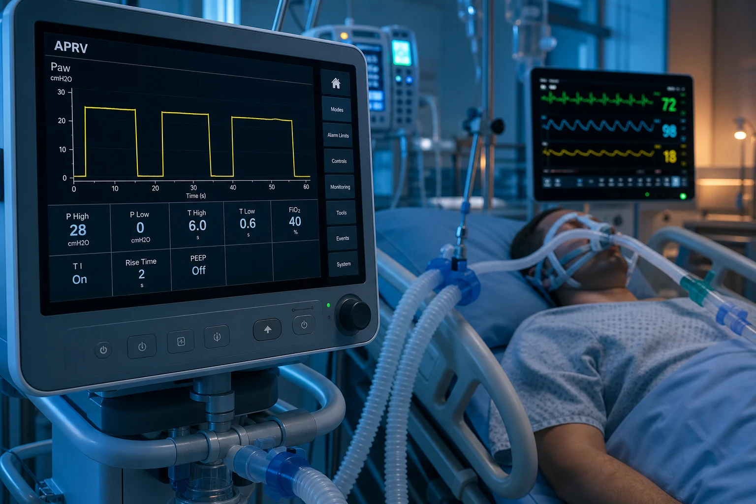
Advanced ventilation modes for severe/refractory ARDS. APRV (Airway Pressure Release Ventilation): continuous high pressure (P-high) with brief release to low pressure (P-low) — allows spontaneous breathing throughout. Theoretical benefits: maintains alveolar recruitment (continuous high PEEP), allows spontaneous breathing (less sedation, preserved diaphragm), improved oxygenation. HFOV (High-Frequency Oscillatory Ventilation): very high respiratory rate (3-15 Hz, 180-900 breaths/min) with very small tidal volumes (~1-3 mL/kg) around a constant mean airway pressure. Theory: lung protection (tiny Vt, constant recruitment). EVIDENCE: OSCAR and OSCILLATE trials (2013) — HFOV did NOT improve mortality (OSCILLATE: trend to HARM). HFOV now RESERVED for refractory cases. APRV: some evidence of improved oxygenation, but no clear mortality benefit.
On this page & tools
Your progress
Saved locally on this device.
Target exams
Red flags
APRV vs HFOV vs conventional ventilation
| Feature | Conventional (VC/PC) | APRV | HFOV (Oscillator) |
|---|---|---|---|
| Mechanism | Volume or pressure controlled, cycled | Continuous high pressure + brief releases | Oscillating diaphragm at 3-15 Hz |
| Tidal volume | 4-6 mL/kg (protective) | Variable (release-dependent) | 1-3 mL/kg (tiny) |
| PEEP | Moderate (from PEEP/FiO2 table) | HIGH (P-high 25-35 cmH2O) | Mean airway pressure (high) |
| Spontaneous breathing | Possible (if mode allows) | YES (throughout — key feature) | NO (paralysis usually needed) |
| Sedation | Moderate-deep | Less (spontaneous) | Deep + paralysis |
| Oxygenation | Good | Often IMPROVED | Often improved |
| Mortality benefit | YES (low Vt — ARMA) | NO clear benefit | NO (OSCILLATE: trend to harm) |
| Current role | FIRST-LINE | Selected (salvage) | REFRACTORY only |
When to consider advanced ventilation modes
- Optimise conventional ventilation FIRST — low tidal volume (4-6 mL/kg), plateau <30, PEEP/FiO2 table, POCUS-guided PEEP
- Try PRONE POSITIONING — PROSEVA: for severe ARDS (PaO2/FiO2 <150), >16h/day
- Consider neuromuscular blockade — early severe ARDS (cisatracurium 48h — ACURASYS, but ROSE was negative)
- If still refractory (PaO2/FiO2 <80):
- APRV: if patient can breathe spontaneously, may improve oxygenation and reduce sedation. Setting: P-high 25-30, P-low 0-5, T-high 4-6s, T-low 0.5-0.8s
- ECMO: for refractory hypoxaemia (VV-ECMO) — stronger evidence than HFOV
- HFOV: LAST RESORT — only if APRV and ECMO unavailable/failed. Oscillator at mean Paw 25-30, frequency 4-6 Hz, delta pressure 60-90
Exam practice
SAQ — Choosing and setting APRV in refractory ARDS (vs HFOV)
10 minutes · 10 marks
A 40-year-old man (height 175 cm, PBW ~71 kg) with severe ARDS from H1N1 influenza is on day 2 of mechanical ventilation. Optimised conventional ventilation: Vt 420 mL (6 mL/kg PBW), RR 30, PEEP 16, FiO2 0.95. Plateau pressure 30 cmH2O, driving pressure 14 cmH2O. Despite 18 hours of prone positioning, ABG: pH 7.29, PaCO2 52, PaO2 59, HCO3 24 (P/F = 62). Noradrenaline 0.15 mcg/kg/min, RASS -1, triggering the ventilator. You are considering an advanced ventilation mode.
SAQ — Liberation from APRV after recovery
10 minutes · 10 marks
A 58-year-old woman has been on APRV for 6 days for severe ARDS from aspiration pneumonitis. Current settings: P-high 26 cmH2O, P-low 0, T-high 5 s, T-low 0.7 s, FiO2 0.30. ABG: pH 7.42, PaCO2 40, PaO2 92, HCO3 26. She is triggering comfortably at a spontaneous rate of 20/min, RASS -1, on no vasopressors, with a productive cough and improving compliance. P/F ratio is now 307. You plan to liberate her from APRV.
Clinical pearls
Red flags
Prognosis
OSCILLATE trial (Ferguson 2013, NEJM) — HFOV in ARDS
RCT: 548 patients with moderate-severe ARDS (PaO2/FiO2 <200). HFOV vs conventional low-tidal-volume ventilation.
- In-hospital mortality: HFOV 47% vs conventional 35% (RR 1.33, p=0.005) — HFOV WORSE
- Trial STOPPED EARLY: for safety (HFOV clearly harmful)
- Vasopressor use: MORE with HFOV
- CONCLUSION: HFOV is HARMFUL in early ARDS. Should NOT be used routinely. Reserve for refractory cases only. [1]
OSCAR trial (Young 2013, NEJM): HFOV vs conventional in UK ARDS. No significant difference in 30-day mortality (41% vs 39%). No benefit. APRV systematic reviews: improved oxygenation in some studies, no clear mortality benefit. Insufficient evidence for routine use.
APRV in depth — mechanics, settings and tuning
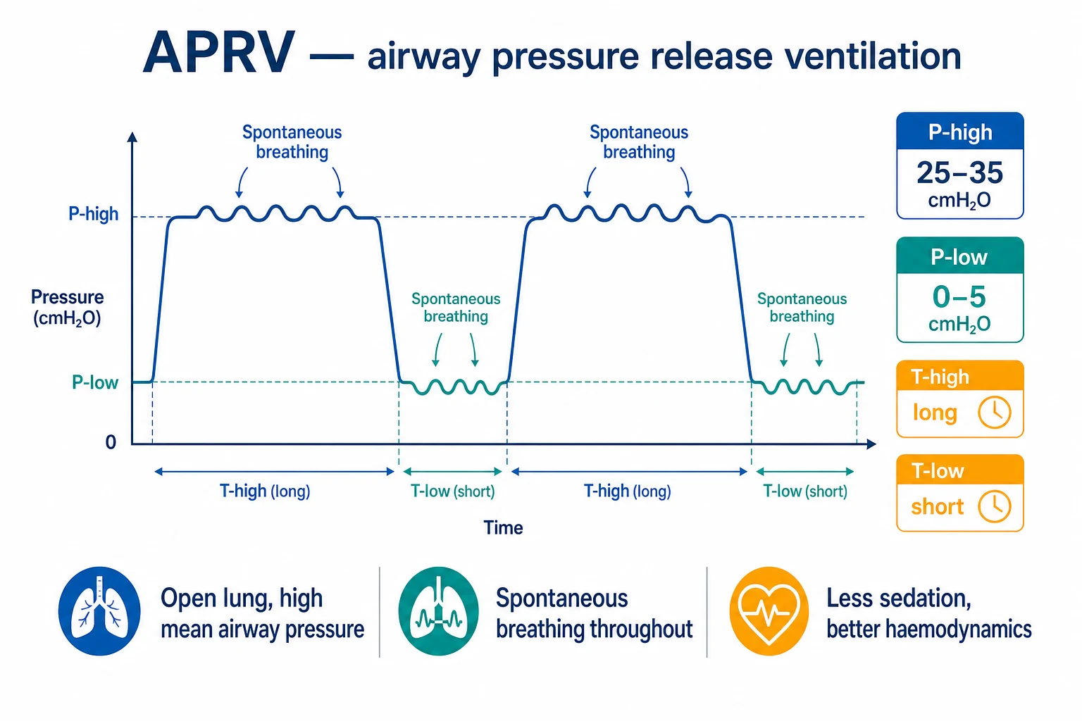
Airway pressure release ventilation (APRV) is best understood as two levels of continuous positive airway pressure (CPAP) with a time-cycled release. The ventilator holds a high pressure (P-high) for most of the respiratory cycle and briefly releases to a low pressure (P-low). The patient may breathe spontaneously at any point — during P-high and during the release — and these spontaneous breaths are superimposed on the pressure waveform. The defining feature is therefore not the release itself but the near-continuous mean airway pressure with preserved spontaneous effort (an "open-lung" strategy).[3] }
The four APRV variables
APRV settings — the four variables and how to set them
| Variable | Typical range | What it controls | How to titrate |
|---|---|---|---|
| P-high (high pressure) | 25-35 cmH2O | Mean airway pressure → recruitment & oxygenation | Start at plateau pressure from conventional mode (or 25-30). Limit by driving pressure / plateau <30 and haemodynamics |
| P-low (release pressure) | 0-5 cmH2O (often 0, i.e. atmospheric) | How much the lung is allowed to derecruit on release | Usually set to 0 (full release). Raising P-low reduces Vt and CO2 clearance but may stabilise haemodynamics |
| T-high (release interval) | 4-6 s (long) | Time at recruitment pressure; the longer T-high, the higher the mean Paw | Increase to improve oxygenation; reduce if hypercapnia/ventilation inadequate |
| T-low (release time) | 0.2-0.8 s (brief) | Time allowed for exhalation — must be SHORT to avoid derecruitment | Titrate to terminate at 50-75% of peak expiratory flow (T-PEF), NOT a fixed time |
The inverse I:E ratio is a consequence, not a setting: because T-high is long and T-low is brief, the ratio is typically 8:1 to 10:1 (sometimes 5:1 to 15:1). This keeps the alveolus open for most of the cycle, minimising tidal opening-and-closing (atelectrauma). Oxygenation is governed mainly by the mean airway pressure (dominated by P-high and the long T-high); CO2 clearance is governed by the release volume (the bigger the swing between P-high and P-low during T-low, the more gas exchanged) plus the patient's own spontaneous breaths.[5] }
The T-low concept — termination of peak expiratory flow (T-PEF)
This is the single most important and most misunderstood APRV concept for the exam. During the brief release, expiratory flow spikes then decays as the lung empties toward P-low. If T-low is too long, the lung fully empties → derecruitment (loss of the recruitment you spent T-high building). If T-low is too short, exhalation is incomplete → air trapping and intrinsic PEEP (auto-PEEP). The standard approach is to end the release when expiratory flow has fallen to 50-75% of its peak — this preserves an "open-lung" with a small residual alveolar pressure while still clearing CO2. In practice T-low is therefore 0.2-0.8 s in most adults, adjusted by watching the expiratory flow waveform.[3] }
Why spontaneous breathing matters
In conventional controlled ventilation the diaphragm is largely inactive; dependent (dorsal) lung — the most oedematous/collapsed region in ARDS — receives little ventilation and is poorly recruited. With active diaphragmatic contraction, pleural pressure falls preferentially in dependent zones, recruiting dorsal alveoli and improving V/Q matching. Spontaneous effort also generates negative intrathoracic pressure, which augments venous return and reduces the right-ventricular afterload increase seen with positive-pressure ventilation. This is the physiological rationale for preferring APRV over deep sedation + paralysis when the patient can breathe.[3] }
"True APRV" vs BiLevel / Biphasic / BiVent
Vendor names confuse the literature. APRV classically means a long T-high with one or two brief releases per minute (extreme inverse ratio). BiLevel / BiVent / DuoPAP use the same two-CPAP architecture but with more comparable times (e.g. P-high 4 s, P-low 2 s) and are often programmed like a pressure-targeted mode with pressure support added on top. For the exam the unifying concept is: all are pressure-targeted, time-cycled, two-level modes that allow spontaneous breathing throughout; the difference is the time ratio. The clinical claim of APRV (extreme inverse ratio, very brief releases) is the most recruitment-favourable.[3] }
Starting and titrating APRV — a practical protocol
- Set P-high at the plateau pressure of the preceding conventional mode (or 25-30 cmH2O if de novo). Respect a driving pressure <15 cmH2O and avoid P-high that drops the SpO2-corrected mean arterial pressure.
- Set P-low to 0 cmH2O (atmospheric) — full release maximises CO2 clearance. Add 3-5 cmH2O only if haemodynamically unstable or if release volume is excessive.
- Set T-high to 4-6 s (start 4 s). Increase if oxygenation poor; this raises mean Paw.
- Set T-low to 0.5-0.8 s, then titrate to terminate at 50-75% of peak expiratory flow on the flow waveform. Watch for auto-PEEP.
- Set FiO2 the same as on conventional; reduce as oxygenation improves.
- Monitor: arterial/venous blood gases at 30-60 min (expect a transient rise in PaCO2), mean arterial pressure, urine output, spontaneous effort (sedation to a Richmond score of −1 to −2 — the patient should be breathing).
- Re-tune: failing oxygenation → ↑ P-high (by 2 cmH2O) or ↑ T-high; failing ventilation → ↑ release gradient (lower P-low or lengthen T-low cautiously) or accept permissive hypercapnia (keep pH >7.20).
- Daily: lung-protective limits still apply (release Vt ~4-6 mL/kg PBW, plateau/driving pressure bounded); plan transition to conventional/PSV as lung injury resolves.
Weaning from APRV back to conventional ventilation
- Lower P-high by 2 cmH2O every 2-4 h as oxygenation and compliance improve (target P-high ~20-22).
- Shorten T-high toward a more conventional I:E ratio (e.g. 4 s → 2 s → 1 s) — the mode becomes BiLevel-like.
- When P-high is ~15-20 cmH2O with a near-symmetric ratio, switch to pressure support (PSV/CPAP) with PS 8-12 cmH2O and PEEP 5-8.
- Perform a spontaneous breathing trial; extubate if tolerated (rapid shallow breathing index <105, stable ABG, intact cough/mental status).
- Avoid prolonged APRV in a recovering patient — the very feature that recruits in early ARDS (high mean pressure) can delay liberation once compliance improves.
HFOV in depth — mechanism, tuning and why the trials failed
How the oscillator works
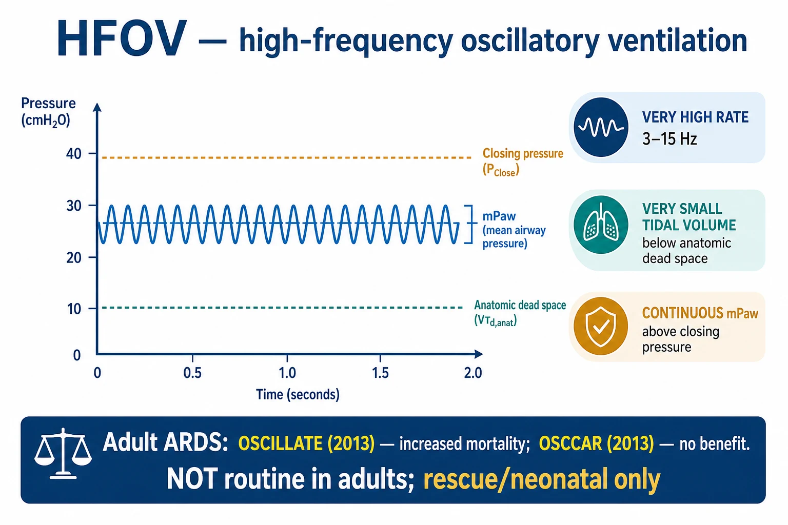
High-frequency oscillatory ventilation (HFOV) uses an oscillating piston/diaphragm (driven by an electromagnet in adult machines such as the SensorMedics 3100B) to generate a sinusoidal pressure wave around a high mean airway pressure. Crucially, both inspiration and expiration are active — the diaphragm actively pulls back during expiration, which differentiates HFOV from high-frequency jet or percussive ventilation where expiration is passive. A continuous bias flow washes CO2 from the circuit.[5] }
HFOV variables
HFOV settings — what each knob does
| Variable | Typical adult range | Effect of increasing it |
|---|---|---|
| Mean airway pressure (mPaw) | 25-30 cmH2O (above the conventional PEEP) | ↑ Oxygenation (recruitment) — but ↑ intrathoracic pressure → ↓ venous return, ↑ PVR |
| Frequency | 3-6 Hz (range 3-15 Hz; higher for small lungs) | ↑ Frequency → ↓ tidal volume → ↑ CO2 (counter-intuitive!) |
| ΔP (power/amplitude) | 60-90 cmH2O | ↑ ΔP → ↑ tidal volume → ↑ CO2 clearance (the "chest wiggle" should be visible to the iliac crests) |
| Inspiratory time % | ~33% | Minor effect on Vt |
| Bias flow | 20-40 L/min | Provides fresh gas; ↑ bias flow aids CO2 washout |
| FiO2 | As required | Oxygenation |
Gas exchange below dead space — the core paradox
HFOV delivers tidal volumes of ~1-3 mL/kg — less than anatomical dead space. Bulk convective flow therefore cannot explain gas exchange. Instead, gas transport in HFOV relies on: (1) direct alveolar ventilation of proximal, short-pathway units; (2) convective dispersion / Taylor dispersion (the oscillating axial flow smears gas along the bronchial tree); (3) pendelluft — asynchronous filling between lung units with different time constants, so gas moves between alveoli rather than in and out; (4) molecular diffusion in the alveolar region; and (5) cardiogenic mixing. The net effect is alveolar ventilation achieved without bulk tidal flow, keeping the lung open and minimising volutrauma and atelectrauma in theory.[5] }
Tuning logic — oxygenation vs ventilation are decoupled
On HFOV the two failure modes are managed with different knobs, unlike conventional ventilation:
- Hypoxaemia → increase mPaw (recruitment) and FiO2.
- Hypercapnia → decrease frequency (counter-intuitive — slower = bigger Vt) and/or increase ΔP (bigger swing = bigger Vt), or increase bias flow. [1]
Why OSCILLATE and OSCAR changed practice
Before 2013 HFOV was widely used in severe ARDS on the basis of physiological plausibility and oxygenation improvement. Two large RCTs published simultaneously in the NEJM in 2013 reversed this:
- OSCILLATE (Ferguson): stopped early for harm — in-hospital mortality 47% vs 35% (RR 1.33), more vasopressors, more sedation.[1] }
- OSCAR (Young): no mortality difference at 30 days.[2] }
The 2016 Cochrane review (Sud) concluded HFOV may increase in-hospital mortality and provides no survival benefit. HFOV is therefore now reserved for rescue in refractory hypoxaemia when all other options (lung-protective ventilation, prone, ECMO) have been exhausted or are unavailable.[12] }
Plausible mechanisms of harm
(1) Heavy sedation and prolonged paralysis required for HFOV → weakness, ICU-acquired weakness, prolonged ventilation, VAP. (2) High mean airway pressure → reduced venous return and increased pulmonary vascular resistance → haemodynamic instability and acute cor pulmonale / right heart failure. (3) The theoretical lung protection was never realised because mean airway pressure was often set above the upper inflection point of the pressure-volume curve, causing overdistension rather than protection. (4) Delay in instituting proven conventional lung-protective ventilation. (5) Inadequate humidification and airway complications in older oscillator circuits.[1] }
Conventional and dual-control modes — PCV, PSV, SIMV, PRVC
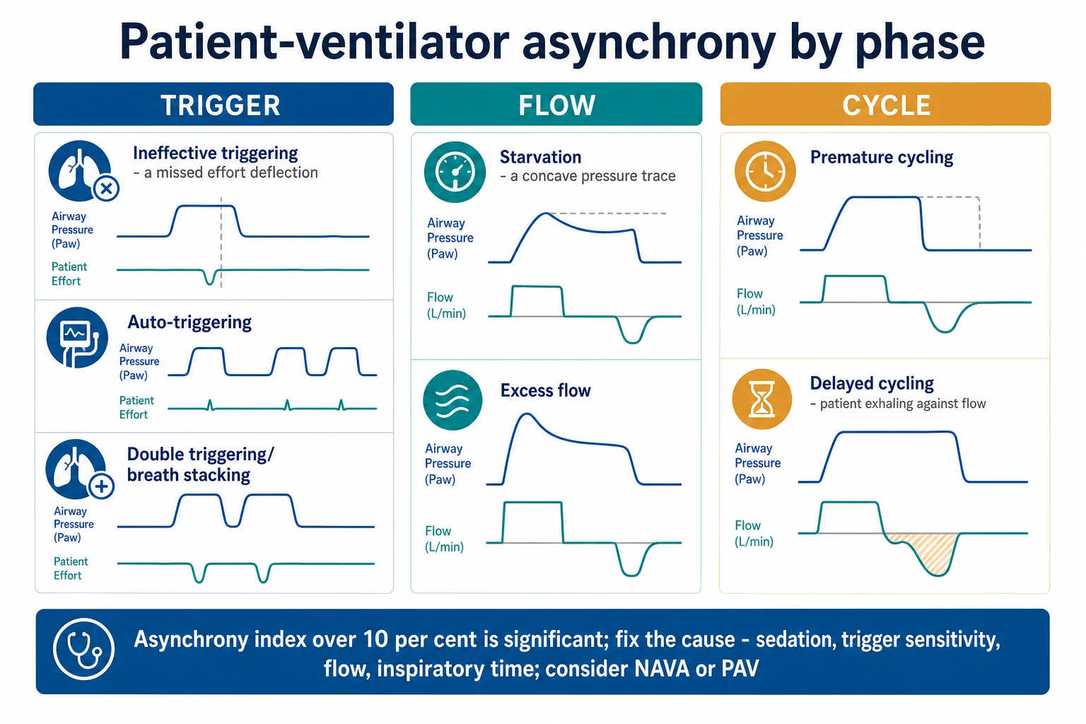
Advanced mode selection is built on three control variables (pressure, volume, flow) and three phase variables (trigger, limit, cycle). A mode is fully described by stating what triggers the breath, what limits it, and what cycles it off, plus whether spontaneous breaths are permitted. This "rational framework" is the CICM/FFICM-friendly way to compare modes.[6] }
PCV vs PSV vs SIMV vs dual-control (PRVC/VC+) — the four mode families
| Feature | PCV (pressure control) | PSV (pressure support) | SIMV (synchronised intermittent mandatory) | PRVC / VC+ (dual control, volume-targeted) |
|---|---|---|---|---|
| Trigger | Time (or patient) | Patient (flow/pressure) | Time for mandatory + patient for spontaneous | Time (or patient) |
| Limit | Set pressure (constant) | Set pressure support | Set pressure/volume for mandatory | Pressure, but auto-adjusted breath-to-breath |
| Cycle | Time (set I-time) | Flow (cycles at ~25% of peak flow) | Time (mandatory) / flow (spontaneous) | Time (decelerating flow to target volume) |
| Flow pattern | Decelerating | Decelerating | Variable | Decelerating |
| Vt delivered | Variable (depends on compliance/resistance) | Variable (patient + effort-dependent) | Variable for mandatory; variable for spontaneous | Guaranteed (auto-adjusts pressure to hit set Vt) |
| Key strength | Limits pressure; good leak tolerance; uniform gas distribution | Patient-ventilator synchrony; for weaning | Backup rate guaranteed | Volume guarantee with decelerating-flow benefit |
| Key risk | Hypoventilation if compliance falls (Vt drops silently) | Ineffective triggering; auto-cycling; apnoea if drive lost | Prolongs ventilation (superseded); stacking | Can still overdistend if set Vt too high |
| Typical use | ARDS (pressure-limited); severe dyssynchrony | Liberation/weaning; spontaneous breathing patients | Largely historical — avoid for weaning | Default volume-targeted mode on many ICU ventilators |
Why SIMV has fallen out of favour
Synchronised intermittent mandatory ventilation delivers a set number of mandatory, synchronised breaths and allows unsupported (or pressure-supported) spontaneous breaths in between. Two landmark RCTs — Brochard (1994) and Esteban (1995) — showed that SIMV-based weaning prolongs the duration of mechanical ventilation compared with pressure-support or T-piece weaning, likely because the mandatory rate buffers the work of breathing unevenly and the patient "fights" the mandatory breaths. SIMV is therefore now considered inferior for liberation and is largely retained only for initial ventilation or as a backup pattern.[6] }
Dual-control / closed-loop volume targeting
PRVC (Pressure Regulated Volume Control; vendor names: AutoFlow, VC+, APV) combines the decelerating flow and pressure-limiting benefits of pressure control with a volume guarantee. The ventilator delivers a "test" breath, measures compliance, and then breath-to-breath adjusts the inspiratory pressure (within a safety cap) to hit the set tidal volume. The clinician sets Vt, PEEP, FiO2, I-time/rate; the machine manages pressure. This is now the default volume-targeted mode on most modern ICU ventilators because it tolerates small leaks, compensates for changing compliance (e.g. worsening oedema), and still bounds peak pressure. It does not by itself guarantee lung protection — the clinician must still ensure Vt 4-6 mL/kg PBW and plateau/driving pressure limits.[6] }
Adaptive and closed-loop modes — NAVA and PAV+
Conventional and dual-control modes decide how much support to give based on set pressure or volume. The next generation decides based on the patient's own instantaneous effort — so-called "patient-driven" or adaptive modes. The unifying principle: support is proportional to demand, which improves synchrony, avoids both under- and over-assist, and aims to protect the diaphragm from disuse atrophy as well as the lung from overdistension.[4] }
NAVA vs PAV+ — patient-driven adaptive modes
| Feature | NAVA (neurally adjusted ventilatory assist) | PAV+ (proportional assist ventilation, load-adjustable) |
|---|---|---|
| Signal that drives support | Electrical activity of the diaphragm (EAdi) via a specialised nasogastric catheter with electrodes at the oesophago-gastric junction | Patient's inspiratory flow and volume; resistance & elastance estimated intermittently by the ventilator |
| How support scales | Pressure = NAVA level (cmH2O/µV) × EAdi — linear with neural drive | Pressure = (gain × elastance × volume) + (gain × resistance × flow) — proportional to instantaneous effort |
| Trigger | Neural (EAdi) — bypasses pneumatic trigger | Flow/pressure (pneumatic) |
| Cycle-off | Neural (when EAdi falls to 70% of peak) | Flow/volume (patient effort) |
| Strengths | Eliminates ineffective triggering; works despite auto-PEEP (COPD); prevents over-assist; patient fully controls rate, Vt, Ti | Amplifies patient effort in proportion to demand; auto-tunes gain to compliance; lung- and diaphragm-protective |
| Limitations | Catheter placement; EAdi affected by oesophageal position, electrocautery, sedation; cost | Needs reliable effort; runaway risk if gain > ~90% of elastance; not for profoundly weak drive |
| Backup | PSV backup if apnoea / EAdi lost | Apnoea backup (PSV/PCV) |
| Best evidence | Improved synchrony, reduced over-assist; mortality benefit not proven | Improved synchrony; trials ongoing for earlier liberation |
NAVA in detail
NAVA delivers pressure in direct proportion to the electrical activity of the diaphragm (EAdi), captured by an array of electrodes on a specialised feeding tube positioned at the level of the crural diaphragm. The breath is therefore triggered, limited, and cycled by the patient's neural output rather than by airway pressure or flow. Because the trigger is neural, it is immune to auto-PEEP — a major advantage in COPD/asthma where pneumatic triggering is delayed by intrinsic PEEP and most "ineffective efforts" occur. The clinician sets a NAVA level (cmH2O per µV of EAdi), which determines how aggressively neural drive is amplified. An intrinsic safety feature: if the patient stops breathing (deep sedation, brainstem injury), a backup pressure-support mode takes over. Randomised data show improved patient-ventilator synchrony and less over-assist; no mortality benefit has yet been demonstrated.[13] }
PAV+ in detail
PAV+ delivers pressure proportional to the patient's instantaneous inspiratory effort — it amplifies the work the patient is already doing ("you press, I push with you"). To do this the ventilator must know the patient's resistance and elastance, which PAV+ estimates by intermittently applying brief end-inspiratory and end-expiratory pauses (the "load-adjustable gain factors" method). The gain is then automatically tuned to a fraction (≤ ~90%) of the measured load. Because the patient controls rate, tidal volume, flow and inspiratory time entirely, PAV+ provides variable support that tracks changing demand (fever, agitation, sepsis) and may both protect the lung (less overdistension) and the diaphragm (less disuse atrophy). The principal hazard is runaway: if the gain approaches or exceeds 100% of the patient's own elastance/resistance, the system becomes self-exciting and generates very large tidal volumes — modern PAV+ limits the gain to prevent this and requires a preserved respiratory drive.[14] }
Adaptive Support Ventilation (ASV) — closed-loop targeting
ASV is a closed-loop mode that targets a clinician-set minute volume and uses the minimum work-of-breathing principle: it continuously adjusts rate, tidal volume, pressure support and I:E ratio toward the calculated least-effort pattern (a derivative of Otis' equation). If the patient is apnoeic it delivers pressure-controlled mandatory breaths; as the patient's effort increases, mandatory support falls away. ASV has been shown to reduce the number of manual ventilator adjustments and may shorten time to liberation, though — like NAVA and PAV+ — without a proven mortality benefit. It is best understood as an automated, lung- and diaphragm-aware version of SIMV-plus-PSV.[6] }
High-yield clinical pearls — advanced and adaptive modes
Additional trials — the evidence base
OSCAR trial (Young 2013, NEJM) — HFOV in UK ARDS
RCT: 795 patients with ARDS (PaO2/FiO2 <200) across 37 UK ICUs. HFOV vs conventional ventilation.
- 30-day mortality: HFOV 41.3% vs conventional 41.6% — no significant difference
- No difference in duration of ventilation, ICU or hospital stay
- CONCLUSION: HFOV provides no benefit in moderate-severe ARDS. Combined with OSCILLATE's harm signal, HFOV was demoted from routine to rescue.
ARMA trial (ARDSNet 2000, NEJM) — low vs traditional tidal volume
RCT: 861 patients with ALI/ARDS. Vt 6 mL/kg PBW, plateau ≤30 vs Vt 12 mL/kg, plateau ≤50.
- Mortality: 31% (low Vt) vs 40% (traditional) — absolute risk reduction 9%, NNT 11
- Stopped early for benefit
- CONCLUSION: Low tidal volume lung-protective ventilation is the foundation of ARDS care — the single best-evidenced ventilator intervention, against which all advanced modes are mere adjuncts.[7] }
PROSEVA trial (Guérin 2013, NEJM) — prone positioning in severe ARDS
RCT: 466 patients with severe ARDS (PaO2/FiO2 <150, FiO2 ≥60%, PEEP ≥5). Prone ≥16 h/day vs supine.
- 28-day mortality: 16% (prone) vs 33% (supine) — NNT 6
- 90-day mortality: 24% vs 41%
- CONCLUSION: Prolonged prone positioning reduces mortality dramatically in severe ARDS. Cheapest, most available and most effective intervention to try before any advanced mode.[8] }
EOLIA trial (Combes 2018, NEJM) — VV-ECMO in very severe ARDS
RCT: 249 patients with very severe ARDS (PaO2/FiO2 <50) — stopped early for futility at interim analysis.
- 60-day mortality: 35% (ECMO) vs 46% (conventional) — not statistically significant (RR 0.76, p=0.09)
- High crossover (28% control patients received rescue ECMO)
- Bayesian re-analysis suggested 77-96% probability of benefit
- CONCLUSION: VV-ECMO is the salvage modality of choice for refractory hypoxaemia — preferred over HFOV.[9] }
ACURASYS (Papazian 2010) vs ROSE (PETAL 2019) — early cisatracurium in ARDS
- ACURASYS: 340 patients, severe ARDS (PaO2/FiO2 <150). Cisatracurium 48 h reduced adjusted 90-day mortality (HR 0.44) and reduced barotrauma — benefit.
- ROSE: 1006 patients, moderate-severe ARDS (PaO2/FiO2 <150, PEEP ≥8). Stopped early for futility — no mortality difference (42.5% vs 42.8%), more ICU-acquired weakness with placebo-receiver handling.
- CONCLUSION: Routine 48-h cisatracurium is not standard. Short-course paralysis remains reasonable for dangerous transpulmonary pressures or severe dyssynchrony. HFOV requires paralysis — another mark against it.[10] }[11] }
Cochrane review (Sud 2016) — HFOV vs conventional ventilation in ARDS
- 9 RCTs, 1629 patients
- HFOV may increase in-hospital mortality (RR 1.15) and provides no 30-day mortality benefit
- No consistent improvement in barotrauma, oxygenation failure or ventilation duration
- CONCLUSION: HFOV should not be used routinely; at best a rescue option when all proven therapies have failed.[12] }
Red flags — expanded
Pitfalls — exam one-liners
[1]References
- [1]Ferguson ND, Cook DJ, Guyatt GH, et al.; OSCILLATE Trial Investigators; Canadian Critical Care Trials Group. High-frequency oscillation in early acute respiratory distress syndrome N Engl J Med, 2013.PMID 23339639
- [2]Young D, Lamb SE, Shah S, et al.; OSCAR Study Group. High-frequency oscillation for acute respiratory distress syndrome N Engl J Med, 2013.PMID 23339638
- [3]Facchin F, Fan E. Airway Pressure Release Ventilation and High-Frequency Oscillatory Ventilation: Potential Strategies to Treat Severe Hypoxemia and Prevent Ventilator-Induced Lung Injury Respir Care, 2015.PMID 26405188
- [4]Vaporidi K, Akoumianaki E, Telias I, et al. NAVA and PAV+ for lung and diaphragm protection Curr Opin Crit Care, 2020.PMID 31738231
- [5]Fan E, Needham DM, Stewart TE. New modalities of mechanical ventilation: high-frequency oscillatory ventilation and airway pressure release ventilation Clin Chest Med, 2006.PMID 17085250
- [6]Mireles-Cabodevila E, Hatipoglu U, Chatterjee A, et al. A rational framework for selecting modes of ventilation Respir Care, 2013.PMID 22710796
- [7]The Acute Respiratory Distress Syndrome Network (ARDSNet). Ventilation with lower tidal volumes as compared with traditional tidal volumes for acute lung injury and the acute respiratory distress syndrome N Engl J Med, 2000.PMID 10793162
- [8]Guérin C, Reignier J, Richard JC, et al.; PROSEVA Trial Investigators. Prone positioning in severe acute respiratory distress syndrome N Engl J Med, 2013.PMID 23688302
- [9]Combes A, Hajage D, Capellier G, et al.; EOLIA Trial Investigators. Extracorporeal Membrane Oxygenation for Severe Acute Respiratory Distress Syndrome N Engl J Med, 2018.PMID 29791822
- [10]Papazian L, Forel JM, Gacouin A, et al.; ACURASYS Study Investigators. Neuromuscular blockers in early acute respiratory distress syndrome N Engl J Med, 2010.PMID 20843245
- [11]National Heart, Lung, and Blood Institute PETAL Clinical Trials Network; Moss M, Huang DT, Brower RG, et al. Early Neuromuscular Blockade in the Acute Respiratory Distress Syndrome N Engl J Med, 2019.PMID 31112383
- [12]Sud S, Friedrich JO, Taccone P, et al. High-frequency oscillatory ventilation versus conventional ventilation for acute respiratory distress syndrome Cochrane Database Syst Rev, 2016.PMID 27043185
- [13]Sinderby C, Navalesi P, Beck J, et al. Neural control of mechanical ventilation in respiratory failure Nat Med, 1999.PMID 10581089
- [14]Xirouchaki N, Kondili E, Vaporidi K, et al. Proportional assist ventilation with load-adjustable gain factors in critically ill patients: comparison with pressure support Intensive Care Med, 2008.PMID 18607562