ICU · Respiratory
Mechanical ventilation: advanced modes and waveforms
Also known as Advanced ventilation modes · Pressure control vs volume control · Ventilator waveforms · Airway pressure release ventilation (APRV) · Dual control modes
Advanced mechanical ventilation modes offer additional options for difficult-to-ventilate patients. VOLUME CONTROL (VC): guaranteed tidal volume, variable pressure — safe, standard. PRESSURE CONTROL (PC): guaranteed inspiratory pressure, variable volume — better gas distribution in heterogeneous lung (ARDS). DUAL CONTROL (PRVC, AutoFlow): volume-targeted, pressure-limited — combines benefits. AIRWAY PRESSURE RELEASE VENTILATION (APRV): continuous positive pressure (Phigh) with brief release (Plow) — allows spontaneous breathing throughout, maintains alveolar recruitment. HIGH-FLOW NASAL OXYGEN (HFNO): not a ventilator but bridges NIV and intubation. PROPORTIONAL ASSIST VENTILATION (PAV): proportional support based on patient effort — near-physiological. NEURALLY ADJUSTED VENTILATORY ASSIST (NAVA): diaphragm EMG-triggered — most physiological synchrony. Waveform analysis: pressure, flow, volume curves — identify asynchrony, leaks, obstruction.
On this page & tools
Your progress
Saved locally on this device.
Target exams
Red flags
SAQ — APRV as a rescue mode in moderate-severe ARDS
10 minutes · 10 marks
A 55-year-old man with pneumococcal pneumonia is intubated for severe ARDS (PaO₂/FiO₂ 95 on FiO₂ 0.8, PEEP 14). On volume-control lung-protective ventilation (Vt 6 mL/kg PBW, plateau 29 cmH₂O, driving pressure 15) he remains hypoxaemic with SpO₂ 88%, and prone ventilation is only transiently effective. The consultant suggests a trial of airway pressure release ventilation (APRV). He is on no vasopressors and has an intact respiratory drive.
SAQ — HFOV in adult ARDS: why it was abandoned
10 minutes · 10 marks
You are asked to review a unit protocol that lists high-frequency oscillatory ventilation (HFOV) as a rescue option for severe ARDS. A junior colleague asks why the oscillators in the hospital appear to be unused, and whether HFOV should be offered to a patient with refractory hypoxaemia (PaO₂/FiO₂ 70) who has failed proning and optimised conventional ventilation.
Clinical pearls

Red flags
Phase variables — the building blocks of every mode
Every mechanical ventilation mode is assembled from the same phase variables: what triggers inspiration, what is the control/target variable, what limits the breath, and what cycles inspiration off. Knowing the phase variables lets you decode ANY mode, including proprietary brand-specific ones, instead of memorising marketing names.[1]
Trigger (starts inspiration)
Time vs patient effort
- Time trigger: the ventilator starts the breath on a timer (controlled/mandatory breaths)
- Pressure trigger: patient effort drops circuit pressure below a threshold (e.g. -1 to -2 cmH2O)
- Flow trigger: patient effort draws flow from the circuit above a threshold (e.g. 2 L/min) — more sensitive, less work
- Neural trigger (NAVA): the rise in diaphragm EMG starts the breath — the most synchronous of all
Control / target (the set variable)
Volume or pressure
- Volume control: Vt is set; pressure VARIES with compliance (constant flow, rising pressure)
- Pressure control: inspiratory pressure is set; Vt VARIES with compliance (decelerating flow, flat-top pressure)
- Dual control: Vt is the target, pressure is the variable that adjusts to reach it (PRVC, VC+/APV, AutoFlow)
- Adaptive / proportional (ASV, NAVA, PAV+): a higher-order target (minute ventilation, EAdi, patient effort) governs each breath
Limit (the ceiling during inspiration)
What cannot be exceeded
- Pressure limit: in pressure control the set inspiratory pressure is both target and limit (held constant)
- Volume limit: in volume control the set Vt is delivered but pressure is bounded by the alarm limit
- Dual-control pressure limit: the upper alarm ceiling that stops the auto-raise before injurious pressure
Cycle (ends inspiration)
Time, volume, or flow
- Time cycling: inspiration ends after the set inspiratory time (VC, PC, PRVC)
- Volume cycling: inspiration ends when the set Vt is delivered (classic constant-flow VC)
- Flow cycling: inspiration ends when inspiratory flow decays to a threshold (~25% of peak, PSV) — patient sets inspiratory time
- Neural cycling: inspiration ends when the EAdi falls (NAVA) — intrinsically synchronous with neural timing
Mode-by-mode: how each advanced mode works
Pressure control ventilation (PCV)
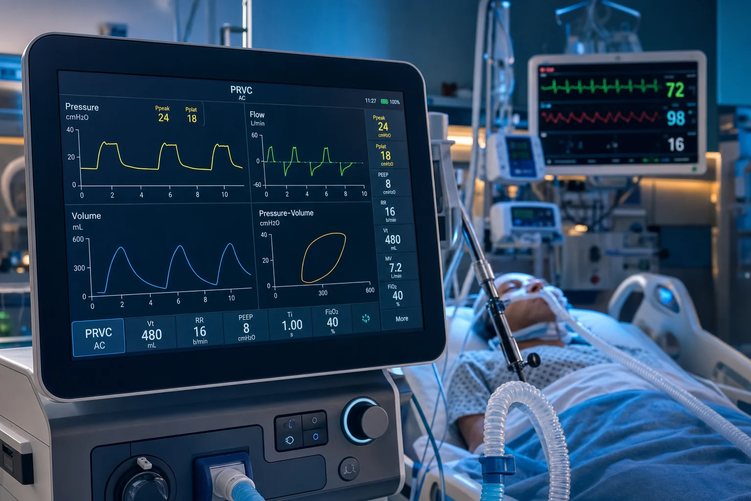
PCV is a pressure-targeted, time-cycled mode. The clinician sets an inspiratory pressure above PEEP, an inspiratory time (or I:E ratio), a respiratory rate, PEEP and FiO2. On each breath the ventilator delivers flow to reach the set pressure and holds it constant for the set inspiratory time. Because pressure is the controlled variable, flow is allowed to vary: it rises rapidly to a peak early in inspiration and then decelerates toward zero as the alveolus fills and the gradient between airway and alveolus shrinks. The decelerating flow pattern is the signature waveform of PCV.[1]
The variable consequence is tidal volume: Vt is the area under the flow-time curve and depends on compliance and resistance, so Vt falls if compliance drops (worsening ARDS, pneumothorax, plugging) or rises if compliance improves (recruitment). Vt must be monitored every breath — a sustained fall is an alarm for worsening mechanics, not a ventilator fault. In PC the peak airway pressure EQUALS the plateau pressure because pressure is held constant with no resistive overshoot — a single pressure reading suffices, unlike volume control where an inspiratory-hold is needed.[1]
Advantages of the decelerating flow pattern: more homogeneous gas distribution in lungs with heterogeneous time constants (fast units fill early, slow units continue to fill during the decelerating tail), a lower peak inspiratory pressure for the same mean airway pressure than constant-flow VC, and improved oxygenation from a higher mean airway pressure at equivalent plateau pressure. Disadvantages: no guaranteed Vt (unsafe if compliance is volatile), and tidal volume must be trended continuously.[2]
Pressure support ventilation (PSV) and CPAP
PSV is a pressure-targeted, flow-cycled, patient-triggered mode designed for spontaneously breathing patients — the backbone of weaning. The clinician sets a pressure support level above PEEP, PEEP, FiO2, a flow trigger and a rise time; the patient sets the rate, the inspiratory time (via flow cycling) and the size of each breath (effort plus support). Inspiration cycles off when inspiratory flow decays to a threshold (typically 25 per cent of peak flow, adjustable). The patient controls everything except the magnitude of support.[9]
CPAP (continuous positive airway pressure) is the lowest-support extreme of the same family: a single continuous pressure throughout the respiratory cycle with no added inspiratory support — every breath is the patient own, with PEEP recruiting alveoli and reducing the work imposed by PEEP-deprived lung. CPAP tests the patient intrinsic ability to ventilate and oxygenate; it is the basis of the T-piece / CPAP spontaneous breathing trial.[9]
PSV advantages: maximal synchrony (patient-triggered and cycled), preserved respiratory-muscle conditioning, comfortable, and the natural mode for weaning. The classic weaning trials (Brochard, Esteban) established that PSV and T-piece weaning outperform SIMV for the duration of weaning and the rate of successful extubation.[9][10] Risks: support set too low = fatigue and failed trials; too high = over-assistance, respiratory-muscle atrophy, delayed weaning, and asynchrony (delayed cycling, double triggering). Flow cycling mistimes in obstructive disease (premature cycling from early flow decay) and in circuit leak.[6]
Synchronized intermittent mandatory ventilation (SIMV)
SIMV guarantees a set number of mandatory (machine) breaths per minute while allowing the patient to breathe spontaneously between them; crucially, the ventilator synchronises each mandatory breath to a patient effort within a timing window, so a mandatory breath is not delivered on top of a spontaneous breath (avoiding breath-stacking). Spontaneous breaths between mandatory breaths may be unsupported, pressure-supported, or both.[1]
SIMV was historically popular as a weaning mode (progressively reduce the mandatory rate to wean). Modern evidence has overturned this: both the Brochard and Esteban trials showed SIMV weans patients slower and with more failures than pressure-support or T-piece weaning, because the intermittent mandatory breaths impose variable load on the diaphragm and the patient must still work during the mandatory cycle. SIMV is now rarely used as a weaning strategy; it survives mainly as an initial controlled-support mode or combined with pressure support.[9][10]
Dual-control modes: PRVC, VC+ (adaptive pressure ventilation), AutoFlow
Dual-control modes attempt to deliver the best of volume and pressure control: a guaranteed tidal volume (volume-targeted) delivered with a decelerating, pressure-limited breath (pressure-controlled). They are the modern default on most ICU ventilators.[1]
- PRVC (pressure-regulated volume control): the operator sets a target Vt, RR, inspiratory time, PEEP, FiO2, and an upper pressure alarm limit. The ventilator delivers a test breath, measures compliance, then delivers each subsequent breath in pressure-control form (constant pressure, decelerating flow) at the lowest pressure needed to achieve the target Vt. If Vt drifts off target (compliance or resistance change), the ventilator automatically adjusts the inspiratory pressure breath-by-breath (typically in 1-3 cmH2O steps) to keep Vt on target — but never exceeds the set pressure limit.[1]
- VC+ / APV (adaptive pressure ventilation): the Hamilton name for the same dual-control principle — volume-targeted, pressure-limited, adaptive. The ventilator continuously recalculates the pressure required for the target Vt and adapts.
- AutoFlow (Drager): the volume-control variant that adds decelerating flow and adaptive pressure to a conventional VC breath.
The key advantage: guaranteed Vt with the decelerating-flow benefits (homogeneous distribution, lower peak pressure), and automatic adaptation to changing compliance without clinician intervention. The key danger: if compliance suddenly worsens, the ventilator will increase pressure up to the limit to chase the Vt — potentially delivering injurious pressures. The pressure alarm limit (and plateau/driving pressure) must therefore be set tight and monitored; "set it and forget it" is unsafe.[2]
Adaptive support ventilation (ASV)
ASV is a closed-loop mode: the operator enters only the patient height and sex (to compute predicted body weight) and a target minute ventilation (as a percentage of predicted, typically 100 per cent). Using the measured compliance and resistance each breath, the ventilator computes the rate-tidal-volume combination that delivers the target minute ventilation at the minimum work of breathing (the Otis time-constant optimum), while respecting lung-protection limits (Vt within roughly 4-8 mL/kg PBW, plateau under a safety limit). As the patient breathes spontaneously, ASV automatically reduces mandatory support and shifts toward pressure support; if apnoea occurs, it automatically delivers full controlled ventilation.[5][7]
ASV advantages: a single control to set, automatic weaning as the patient recovers (reduced clinician workload), and built-in lung protection. An RCT in difficult-to-wean patients (Kirakli 2015) found ASV shortened weaning time vs a physician-driven protocol in a selected COPD weaning unit.[7] Limitations: it optimises for work of breathing, not necessarily for oxygenation or driving pressure, and requires an intact feedback loop; it is not a substitute for clinician judgement in severe ARDS.
Neurally adjusted ventilatory assist (NAVA)
NAVA delivers support proportional to the electrical activity of the diaphragm (EAdi), measured by an array of electrodes on a specialised nasogastric tube positioned at the gastro-oesophageal junction opposite the crura. The EAdi is a direct, real-time readout of the neural respiratory drive (brainstem to phrenic nerve to diaphragm), independent of airflow and airway pressure. The clinician sets a NAVA gain (cmH2O per microvolt of EAdi); on each breath, support rises and falls in exact proportion to the neural signal — the patient governs every aspect of every breath.[5]
Because trigger and cycling are neural (not pneumatic), NAVA virtually eliminates the common pneumatic asynchronies: ineffective triggering (missed small efforts), auto-triggering (false breaths from leaks), and delayed cycling. It is the most physiological mode available and is uniquely effective for non-invasive ventilation with large mask leaks, where pneumatic triggers fail.[5][6]
Requirements and limits: an intact respiratory drive and neuromuscular pathway (it fails in high cervical cord injury, phrenic nerve injury, severe neuromuscular disease, or deep sedation), a correctly positioned EAdi catheter (verified by a characteristic waveform), and the cost/complexity of the catheter. A backup pneumatic mode is mandatory in case of catheter displacement.[5]
Proportional assist ventilation (PAV+)
PAV+ amplifies patient effort in proportion to the instantaneous flow and volume the patient generates. The ventilator continuously measures compliance and resistance (from a brief inspiratory-hold or flow-interrupter manoeuvre), computes the patient pressure output via the equation of motion, and delivers a clinician-set percentage of the load (the gain, e.g. 50 per cent means the ventilator does half the elastic and resistive work, the patient does half). A small patient effort yields small support; a large effort yields large support — the patient controls rate, Vt, flow and inspiratory time.[5]
Like NAVA, PAV+ gives near-physiological synchrony and patient-governed breathing. Unlike NAVA, it requires no specialised catheter (it works from standard flow/pressure sensing) but needs a reliable estimate of elastance and resistance (PAV+ uses an iterative auto-tune to estimate them). Its main hazard is "run-away": if elastance is underestimated, the gain can exceed 100 per cent and the ventilator output reinforces itself, causing pressure overshoot and breath-stacking — modern PAV+ has an auto-limit that caps pressure and warns the clinician.[5]
Airway pressure release ventilation (APRV) and Bilevel
APRV applies two levels of CPAP — a high pressure (P-high, typically 25-35 cmH2O) held for a long T-high (commonly 4-6 seconds), with a brief release to a low pressure (P-low, 0-5 cmH2O) for a short T-low (0.4-0.8 seconds). The patient breathes spontaneously throughout — at P-high and during release. The sustained P-high maintains alveolar recruitment and a high mean airway pressure (oxygenation, an open-lung strategy); the release clears CO2; the spontaneous breaths preserve venous return, reduce sedation, and protect the diaphragm.[8]
Bilevel (BiVent, Duo-PAP) is the generalised two-level pressure mode with a more conventional ratio and set mandatory breaths — comfortable and synchronised for patients with some drive who still need a controlled background. APRV is the extreme inverse-ratio form.[8]
Settings and titration: start P-high near the previous plateau pressure; set P-low to 0-5; set the release frequency (10-14/min) and titrate T-low so expiratory flow returns to about 50-75 per cent of peak (a partial release, not a full expiration — full expiration derecruits the lung). Monitor the expiratory flow waveform — it is the single most important adjustment.[8]
Avoid APRV in obstructive disease (asthma, COPD): the sustained high pressure and brief releases worsen dynamic hyperinflation and air-trapping — the opposite of the long-expiration strategy these patients need. A systematic review and meta-analysis found APRV improves oxygenation in ARDS without a clear mortality benefit over conventional lung-protective ventilation; it is not first-line.[8]
High-frequency oscillatory ventilation (HFOV)
HFOV uses an oscillating piston/membrane (the diaphragm in the oscillator circuit) to deliver very small tidal volumes (often less than anatomic dead space, 1-3 mL/kg) at very high frequencies (3-15 Hz, i.e. 180-900 breaths/min) around a sustained high mean airway pressure. Gas exchange occurs by unconventional mechanisms (bulk flow, Taylor dispersion, pendelluft, molecular diffusion, coaxial flow) rather than conventional bulk convection. The high mean airway pressure keeps the lung recruited (open-lung oxygenation), while the tiny tidal volumes minimise volutrauma.[2]
Settings: mean airway pressure (mPaw, ~25-35 cmH2O — the recruitment/oxygenation variable), power/amplitude (delta-P, the oscillation magnitude — the CO2 clearance variable), frequency (Hz — lower frequency = larger effective tidal volume and better CO2 clearance), inspiratory time percentage, and FiO2. A common pitfall is using too high a frequency, which shrinks tidal volume and causes CO2 retention.[3]
The evidence turned HFOV from promising to near-abandoned in adult ARDS: two large multicentre RCTs published in the same 2013 NEJM issue (OSCILLATE and OSCAR) failed to show benefit. OSCILLATE (Ferguson) was stopped early because HFOV increased in-hospital mortality (47 per cent vs 35 per cent) — driven by heavy sedation, paralysis, and haemodynamic compromise from the high intrathoracic pressure. OSCAR (Young) found no mortality difference. HFOV is now reserved for select rescue scenarios (severe refractory hypoxaemia failing proning and optimised conventional ventilation) and is not standard for adult ARDS.[3][4]
Master comparison: all advanced modes at a glance
Volume control (VC)
Volume-targeted, flow-limited, time-cycled
- Trigger: time or patient; Cycle: set inspiratory time (volume delivered at set flow)
- Target/control: guaranteed Vt; pressure VARIES with compliance
- Flow pattern: constant (square); peak pressure HIGHER than PC for the same Vt
- Advantage: guaranteed Vt — safe when compliance is volatile; easy to monitor
- Disadvantage: high peak/plateau pressure in low-compliance lung (barotrauma risk if not lung-protective)
Pressure control (PCV)
Pressure-targeted, time-cycled
- Trigger: time or patient; Cycle: set inspiratory time
- Target/control: guaranteed inspiratory pressure; Vt VARIES with compliance
- Flow pattern: DECELERATING (high initial then falls) — the signature waveform
- Advantage: homogeneous gas distribution, lower peak pressure, higher mean airway pressure
- Disadvantage: no guaranteed Vt — dangerous if compliance drops; must trend Vt each breath
PSV / CPAP
Pressure support, patient-triggered, flow-cycled
- Trigger: patient (flow); Cycle: flow decay to threshold (~25% peak) — patient sets inspiratory time
- Target/control: set pressure support above PEEP; patient sets rate, Vt, flow
- CPAP = single continuous pressure, no inspiratory support (T-piece equivalent for the SBT)
- Advantage: maximal synchrony; the backbone of weaning; preserves muscle conditioning
- Disadvantage: no guaranteed backup if patient apnoeic; over/under-support causes asynchrony; flow cycling mistimes in leak/obstruction
SIMV
Mandatory synchronised breaths + spontaneous
- Trigger: patient within sync window, else time; Cycle: mandatory = time, spontaneous = flow
- Target/control: set mandatory rate and Vt/pressure; spontaneous breaths supported or not
- Synchronises mandatory breath to patient effort to avoid breath-stacking
- Advantage: guaranteed minimum minute ventilation with spontaneous contribution
- Disadvantage: SLOWER weaning and more failures than PSV/T-piece (Brochard, Esteban) — not a weaning mode
PRVC / VC+ / AutoFlow (dual control)
Volume-targeted, pressure-limited, adaptive
- Trigger: time or patient; Cycle: set inspiratory time
- Target/control: guaranteed Vt at the LOWEST pressure needed; pressure auto-adjusts breath-to-breath
- Delivers a decelerating-flow, constant-pressure breath; adapts to changing compliance
- Advantage: combines guaranteed Vt with PC gas distribution; automatic adaptation — the modern default
- Disadvantage: will CHASE the Vt by raising pressure if compliance worsens — set and monitor a pressure limit
ASV (adaptive support)
Closed-loop, target minute ventilation
- Trigger: patient or auto; Cycle: automatic
- Target/control: operator sets target minute ventilation (% predicted); ventilator chooses rate:Vt
- Computes the minimum-work-of-breathing pattern (Otis) within lung-protection limits; auto-weans to PSV
- Advantage: single control; automatic weaning; built-in lung protection and apnoea backup
- Disadvantage: optimises WOB not oxygenation; not a substitute for clinician judgement in severe ARDS
NAVA
EAdi-proportional neural assist
- Trigger and cycle: NEURAL (diaphragm EMG) — independent of airflow and pressure
- Target/control: gain set in cmH2O per microvolt EAdi; support proportional to neural drive
- Requires a specialised NG tube with electrodes at the crural level
- Advantage: most physiological synchrony; eliminates ineffective/auto-triggering; best for leaky NIV
- Disadvantage: needs intact drive/phrenic/diaphragm; catheter cost and position; needs backup pneumatic mode
PAV+
Proportional to patient effort
- Trigger and cycle: patient; support proportional to measured flow and volume
- Target/control: gain set as % of elastic + resistive load (e.g. 50%)
- Computes patient pressure output via the equation of motion from measured elastance/resistance
- Advantage: near-physiological synchrony; patient governs every breath; no special catheter
- Disadvantage: run-away risk if elastance underestimated (auto-limit guards this); needs reliable mechanics
APRV / Bilevel
Two-level CPAP, spontaneous throughout
- Trigger: time (release) + patient; Cycle: time (release to P-low for a short T-low)
- Target/control: P-high 25-35, P-low 0-5, T-high long, T-low short (partial release)
- High mean airway pressure recruits; release clears CO2; spontaneous breaths preserve venous return
- Advantage: oxygenation (open lung), less sedation, preserved diaphragm and haemodynamics
- Disadvantage: AVOID in COPD/asthma (dynamic hyperinflation); oxygenation benefit, not a mortality benefit
HFOV
Oscillator, very high rate, tiny Vt
- Mechanism: oscillating diaphragm at 3-15 Hz, Vt 1-3 mL/kg around a high mean airway pressure
- Target/control: mPaw (oxygenation), delta-P power (CO2), frequency Hz, I-time %
- Gas exchange by unconventional mechanisms (dispersion, pendelluft) not bulk convection
- Advantage: recruitment + minimal volutrauma (theoretical lung protection)
- Disadvantage: OSCILLATE showed HIGHER mortality; deep sedation/paralysis, haemodynamic compromise — near-abandoned in adult ARDS
Decelerating (PC) vs constant (VC) flow — the waveform signature
Constant flow (VC)
Square flow-time waveform
- Flow rises instantly to the set value and holds constant until the set inspiratory time elapses
- Pressure rises GRADUALLY throughout inspiration — peak pressure reached at END of inspiration
- Peak airway pressure is HIGHER than PC for the same Vt and inspiratory time
- Gas distribution: preferentially fills fast (short time-constant) units; slow units underfilled
- Identify on the screen: flat-topped flow curve; rising-ramp pressure curve
Decelerating flow (PC / PRVC)
Descending-ramp flow-time waveform
- Flow spikes to a peak early, then DECELERATES toward zero as the alveolus fills
- Pressure rises rapidly to the set value and stays CONSTANT (flat-top pressure curve)
- Peak airway pressure EQUALS plateau pressure (the defining feature of pressure control)
- Gas distribution: slow (long time-constant) units continue to fill during the decelerating tail — more homogeneous
- Identify on the screen: descending flow curve; square-top pressure curve
How dual-control modes adapt — breath by breath
PRVC / adaptive pressure ventilation — the adaptive loop
Operator sets a target Vt and a pressure limit
Enter target Vt (typically 6 mL/kg PBW), RR, inspiratory time, PEEP, FiO2, and an upper pressure alarm limit (e.g. 40 cmH2O). The ventilator will not exceed this limit.
The ventilator delivers a test breath and measures compliance
A first pressure-control breath (or an inspiratory hold) lets the ventilator calculate the dynamic compliance (Vt divided by the pressure above PEEP). This is the baseline it works from.
It sets the working pressure just enough for the target Vt
The ventilator computes the inspiratory pressure that, delivered as a constant-pressure decelerating-flow breath, will yield the target Vt at the measured compliance. It uses the lowest pressure that achieves this.
Each breath is delivered in pressure-control form
Every subsequent breath is a constant-pressure, decelerating-flow breath — giving the homogeneous gas distribution and lower peak pressure of PC, while delivering the target Vt.
Vt is measured every breath and the pressure auto-adjusts
If the measured Vt drifts off target (compliance or resistance changed), the ventilator adjusts the inspiratory pressure up or down in small steps (typically 1-3 cmH2O) to return Vt to target — continuously, breath-by-breath, with no clinician input.
The pressure limit is the safety net
If compliance collapses, the ventilator will raise pressure to chase the Vt — up to the set limit. At the limit it caps pressure and the Vt falls below target (an alarm fires). This is why the pressure limit and plateau/driving pressure MUST be set and watched: a dual-control mode can silently deliver injurious pressures if the limit is too high. "Set it and forget it" is unsafe.<Cite id="1" />
How NAVA works
NAVA — from neural drive to proportional support
Place the EAdi catheter
A nasogastric tube carrying a ring of electrodes is passed into the stomach and withdrawn to the gastro-oesophageal junction, where the electrodes sit opposite the diaphragmatic crura. Correct position is confirmed by a characteristic phasic EAdi waveform (a peak with each inspiration) over a smaller ECG trace.
The EAdi is measured continuously
The electrical activity of the diaphragm (EAdi, in microvolts) is a direct real-time readout of the neural respiratory drive — brainstem to phrenic nerve to diaphragm — independent of airflow, airway pressure, or circuit leak. It rises with each inspiratory effort.
Inspiration is triggered by the rise in EAdi
Instead of a flow or pressure drop at the airway, the ventilator triggers on the rising EAdi — the neural signal itself. This eliminates ineffective triggering (small efforts that fail to trigger) and auto-triggering (false breaths from leaks or circuit motion).<Cite id="5" />
Support is proportional to EAdi
The clinician sets a NAVA gain (cmH2O per microvolt of EAdi). On each breath, airway pressure rises in exact proportion to the EAdi — the harder the diaphragm fires, the more support is delivered. The patient governs rate, Vt, flow and inspiratory time.
Cycling is neural
Inspiration cycles off when the EAdi falls (the neural signal to stop), so cycling is intrinsically synchronous with the patient own neural timing — no delayed cycling, no double triggering from mistimed cycling.
A backup mode protects catheter failure
If the EAdi signal is lost (catheter displaced) or the patient apnoeic, a backup pressure-support or PC mode takes over. This mandatory backup is why NAVA is safe despite its dependence on a neural signal.<Cite id="5" />
How PAV+ works
PAV+ — proportional load-sharing
The ventilator measures elastance and resistance
Via a brief inspiratory hold or an iterative manoeuvre, the ventilator estimates the patient respiratory elastance (the elastic load — how stiff the lung and chest wall are) and resistance (the resistive load — airway resistance). These define the equation of motion.
The clinician sets a gain (% assist)
The gain is the percentage of the total work the ventilator will provide (e.g. 50 per cent). At 50 per cent, the ventilator does half the elastic and half the resistive work; the patient does the other half. The gain is the only real setting.
Support tracks instantaneous patient effort
On each breath, the ventilator reads the instantaneous flow and volume the patient generates and delivers pressure proportional to them — a small effort yields small support, a large effort yields large support. The patient sets rate, Vt, flow and inspiratory time.
Run-away is the hazard — the auto-limit guards it
If elastance is underestimated, the gain can exceed 100 per cent: the ventilator output reinforces itself, causing pressure overshoot and breath-stacking (run-away). Modern PAV+ continuously auto-tunes the gain and has an auto-limit that caps pressure and warns the clinician if the assist approaches run-away.<Cite id="5" />
How ASV works
ASV — closed-loop minimum-work targeting
Operator sets the patient and a target minute ventilation
Enter height and sex (to compute predicted body weight) and a target minute ventilation as a percentage of predicted (typically 100 per cent). This is the primary control — everything else is automatic.
The ventilator tests lung mechanics each breath
From the pressure, flow and volume traces it continuously measures compliance and resistance, and detects the patient spontaneous respiratory drive.
It computes the minimum-work rate:Vt pattern
Using the measured mechanics, ASV solves for the respiratory rate and tidal volume that achieve the target minute ventilation at the MINIMUM work of breathing (the Otis optimum based on the expiratory time constant), while keeping Vt within lung-protection bounds (roughly 4-8 mL/kg PBW) and plateau under a safety limit.<Cite id="7" />
It shifts automatically with the patient
As the patient breathes spontaneously, ASV reduces mandatory support and behaves like pressure support; if the patient apnoeas, it automatically delivers full controlled ventilation. The mode continuously adapts without clinician intervention — a built-in weaning and apnoea strategy in one.<Cite id="7" />
HFOV — setup, titration and weaning
HFOV — the practical setup
Set mean airway pressure (mPaw) from the recruitment goal
Start mPaw about 3-5 cmH2O above the mean airway pressure of the prior conventional mode (commonly 25-35 cmH2O). mPaw is the OXYGENATION variable — it recruits and holds the lung open. Titrate to oxygenation (SpO2/PaO2), not to an arbitrary number.
Set the power/amplitude (delta-P) for CO2 clearance
The oscillation magnitude (delta-P) drives CO2 removal. Start at a moderate setting and titrate to PaCO2/pH — a "wiggle" of the chest wall down to the umbilicus is a traditional bedside marker of adequate amplitude.
Set frequency, then bias flow and FiO2
Frequency (Hz) inversely affects tidal volume: a LOWER frequency gives a larger effective Vt and BETTER CO2 clearance. A common error is setting frequency too high (which shrinks Vt and causes CO2 retention). Typical adult starting frequency 5-6 Hz. Bias flow sets the fresh-gas supply; FiO2 is titrated to oxygenation.<Cite id="3" />
Expect heavy sedation and often paralysis — the cost of HFOV
Patients rarely tolerate the oscillator; deep sedation (and frequently neuromuscular blockade) is required. This, plus the high intrathoracic pressure, is why OSCILLATE found HARM: haemodynamic compromise, reduced venous return, and the risks of prolonged paralysis.<Cite id="3" />
Wean to conventional ventilation when oxygenation improves
When the underlying cause improves and FiO2 can be weaned with acceptable mPaw, transition back to conventional lung-protective ventilation — do not linger on HFOV. Reserve HFOV for the rescue window in refractory hypoxaemia, not for routine ARDS.<Cite id="4" />
Waveform-based troubleshooting at the bedside
Reading the ventilator screen — five patterns and what they mean
Scalloped inspiratory flow curve
In pressure support/PC, the inspiratory flow curve should be a smooth descending ramp. If it is CONCAVE (scalloped), the patient is pulling harder than the ventilator delivers — FLOW STARVATION (the patient is "fighting the ventilator"). Fix by increasing rise time/pressure support, lengthening inspiratory time, or switching to a more responsive mode.<Cite id="6" />
Expiratory flow not returning to baseline
The expiratory flow curve should reach zero before the next breath. If it is still above zero at end-expiration, there is INCOMPLETE EXHALATION = AIR TRAPPING (intrinsic/auto-PEEP). Seen in COPD, asthma, high RR, or too-short expiratory time. Manage by reducing RR, shortening inspiratory time, applying external PEEP about 80% of auto-PEEP.<Cite id="6" />
Double triggering (breath stacking)
Two breaths delivered with little or no exhalation between them — the patient triggers a second breath before the first has cycled off. Causes: inspiratory time too short for neural time (delayed cycling), high drive, low support. Consequence: stacked Vt, high transpulmonary pressure, VILI risk. Fix by lengthening inspiratory time or raising support/cycling threshold.<Cite id="6" />
Ineffective triggering (missed efforts)
On the flow/pressure traces you see patient effort (a dip in pressure, a flow blip) that does NOT trigger a breath. Causes: high auto-PEEP (the trigger threshold is not met), over-sedation, weak drive, tube leak. Common in COPD. Fix by reducing auto-PEEP, lowering trigger sensitivity, or raising external PEEP.<Cite id="6" />
Auto-triggering (false breaths)
Breaths triggered without patient effort — seen as a regular trigger pattern with no preceding effort. Causes: circuit leak, water in the circuit, cardiac oscillation, too-sensitive trigger. Fix by reducing trigger sensitivity, removing leaks and water.<Cite id="6" />
Choosing the mode — match the mode to the patient
Matching the advanced mode to the clinical situation
Standard lung-protective VC or PRVC — for almost all patients
For the great majority of ventilated patients, conventional volume control or PRVC with lung-protective settings (Vt 6 mL/kg PBW, plateau <30, driving pressure <15) is the standard and the evidence base. No advanced mode improves mortality over this.<Cite id="2" />
PRVC — when compliance is changing and you want guaranteed Vt with PC distribution
Dual control is the modern default: it gives the homogeneous gas distribution of pressure control while guaranteeing the tidal volume, and adapts automatically. Set a tight pressure limit and watch plateau/driving pressure.
PSV / CPAP — for weaning and any patient with a reliable drive
Once the patient has a drive and is ready to breathe, pressure support (and CPAP/T-piece for the spontaneous breathing trial) is the evidence-based weaning strategy — faster and more successful than SIMV.<Cite id="9" /><Cite id="10" />
APRV — for recruitable ARDS with an intact drive, as a rescue
In moderate-severe ARDS with refractory hypoxaemia on conventional ventilation and an intact respiratory drive, APRV is an open-lung strategy that may improve oxygenation. AVOID in obstructive disease. Use selectively, not as first-line (no mortality benefit).<Cite id="8" />
NAVA — for refractory asynchrony or leaky NIV
When a patient fights conventional ventilation with ineffective triggering, double triggering, or auto-triggering that cannot be tuned, NAVA synchronises to the neural drive. It is also the best mode for non-invasive ventilation with large mask leaks where pneumatic triggers fail.<Cite id="5" /><Cite id="6" />
PAV+ — for synchrony without a special catheter
When you want proportional, patient-governed support and synchrony but do not have (or need) the NAVA catheter, PAV+ delivers effort-proportional support from standard sensing — set the gain and let the patient breathe.
ASV — for automatic weaning and low clinician workload
In selected patients (e.g. cardiac surgical, COPD weaning units), ASV automates the wean and reduces ventilator adjustments. Confirm an RCT benefit is plausible for your patient; do not use as a substitute for judgement in severe ARDS.<Cite id="7" />
HFOV — reserve, do not routine
Given OSCILLATE/OSCAR, HFOV is reserved for rare rescue scenarios in refractory hypoxaemia failing optimised conventional ventilation and proning — and only with careful attention to sedation, paralysis and haemodynamics. It is not standard for adult ARDS.<Cite id="3" /><Cite id="4" />
Evidence — the landmark trials
OSCILLATE (Ferguson 2013)
NEJM 2013
Multicentre RCT, 548 adults with moderate-severe ARDS (P/F <200) — HFOV vs conventional low-Vt ventilation
Key finding
STOPPED EARLY for harm. In-hospital mortality HFOV 47% vs control 35% (RR 1.33, p=0.005). More sedation, paralysis, vasopressors and haemodynamic compromise with HFOV.
Practice change
HFOV is NOT standard for adult ARDS — demonstrated harm from the high intrathoracic pressure and heavy sedation it requires
OSCAR (Young 2013)
NEJM 2013
Multicentre RCT, 795 adults with acute respiratory distress syndrome — HFOV vs conventional ventilation
Key finding
No significant difference in 30-day mortality (HFOV ~41% vs control ~41%). No benefit, and the companion OSCILLATE trial showed harm.
Practice change
Reinforced that HFOV offers no mortality benefit in adult ARDS; use restricted to rare rescue scenarios
Brochard 1994 — weaning modes
Am J Respir Crit Care Med 1994
Multicentre RCT — pressure support vs SIMV vs T-piece weaning in difficult-to-wean patients
Key finding
Pressure support weaned patients FASTER and with FEWER failures than SIMV or T-piece. SIMV was the slowest weaning method.
Practice change
Established pressure support as the preferred weaning mode; SIMV weaning largely abandoned
Esteban 1995 — four weaning methods
NEJM 1995
Multicentre RCT — once-daily T-piece vs pressure support vs SIMV vs intermittent mandatory ventilation in 546 patients
Key finding
Once-daily T-piece and pressure support weaned faster and more successfully than SIMV/IMV. SIMV again the least effective weaning strategy.
Practice change
Confirmed SIMV is inferior for weaning; T-piece and pressure support are the weaning standards
Kirakli 2015 — ASV for weaning
Chest 2015
RCT comparing adaptive support ventilation (ASV) vs a physician-driven weaning protocol in difficult-to-wean COPD patients
Key finding
ASV shortened the weaning time compared with the physician-driven protocol in this selected weaning-unit population.
Practice change
ASV is a feasible closed-loop weaning mode in selected patients; supports automatic-weaning strategies
Patel 2026 — APRV meta-analysis
J Intensive Care Med 2026
Systematic review and meta-analysis of APRV in ARDS — oxygenation, ventilation and mortality vs conventional ventilation
Key finding
APRV improved oxygenation (PaO2/FiO2, oxygenation index) but showed NO clear mortality benefit over conventional lung-protective ventilation.
Practice change
Confirms APRV as a physiological (oxygenation) rescue, not a mortality-improving intervention — use selectively
More clinical pearls
Red flags (additional)
References
- [1]Chatburn RL, El-Khatib M, Mireles-Cabodevila E A taxonomy for mechanical ventilation: 10 fundamental maxims Respir Care, 2014.PMID 25118309
- [2]Slutsky AS, Ranieri VM Ventilator-induced lung injury N Engl J Med, 2013.PMID 24283226
- [3]Ferguson ND, Cook DJ, Guyatt GH, et al. High-frequency oscillation in early acute respiratory distress syndrome N Engl J Med, 2013.PMID 23339639
- [4]Young D, Lamb SE, Shah S, et al. High-frequency oscillation for acute respiratory distress syndrome N Engl J Med, 2013.PMID 23339638
- [5]Vaporidi K, Akoumianaki E, Georgopoulos D NAVA and PAV+ for lung and diaphragm protection Curr Opin Crit Care, 2020.PMID 31738231
- [6]Mirabella L, Cinnella G, Costa R Patient-Ventilator Asynchronies: Clinical Implications and Practical Solutions Respir Care, 2020.PMID 32665426
- [7]Kirakli C, Naz I, Ediboglu O A randomized controlled trial comparing the ventilation duration between adaptive support ventilation and pressure assist/control ventilation in medical patients in the ICU Chest, 2015.PMID 25742308
- [8]Patel R, Thompson J, et al. Safety, Efficacy, and Clinical Outcomes of APRV in ARDS: A Systematic Review and Meta-Analysis J Intensive Care Med, 2026.PMID 42033378
- [9]Brochard L, Rauss A, Benito S, et al. Comparison of three methods of gradual withdrawal from ventilatory support during weaning from mechanical ventilation Am J Respir Crit Care Med, 1994.PMID 7921460
- [10]Esteban A, Frutos F, Tobin MJ, et al. A comparison of four methods of weaning patients from mechanical ventilation. Spanish Lung Failure Collaborative Group N Engl J Med, 1995.PMID 7823995