ICU · Respiratory / ventilation
Airway Pressure Release Ventilation (APRV) and Advanced Ventilation Modes — Comprehensive ICU Management (APRV, Bilevel, PAV, NAVA, ASV)
Also known as Airway pressure release ventilation · APRV · Bilevel ventilation · BiVent · Duo-PAP · Proportional assist ventilation · PAV · Neurally adjusted ventilatory assist · NAVA · Electrical activity of the diaphragm · EAdi · Adaptive support ventilation · ASV · Open lung ventilation · Release ventilation · Advanced ventilator modes
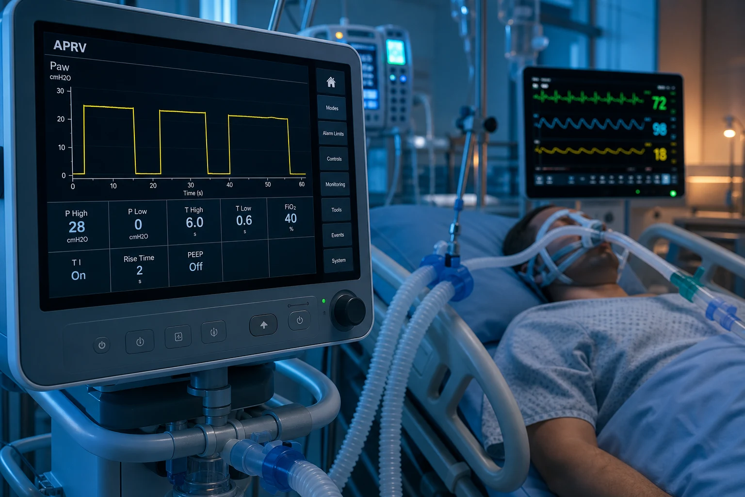
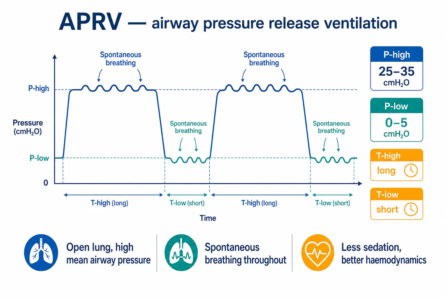
Advanced modes of mechanical ventilation move control away from clinician-set, fixed breaths toward modes that either sustain alveolar recruitment (APRV, bilevel) or proportionalise support to the patient's own effort and neural drive (PAV, NAVA, ASV). Airway pressure release ventilation (APRV) is a pressure-controlled, time-cycled mode that applies a continuous high CPAP (Phigh, typically 25-35 cmH2O) held for a long time (Thigh) with brief release periods to a low pressure (Plow 0-5 cmH2O) for a short time (Tlow 0.4-0.8 s), allowing spontaneous breathing throughout the cycle. The high mean airway pressure maintains alveolar recruitment and oxygenation (an open-lung strategy), the brief release clears CO2, and the preserved spontaneous breathing reduces sedation, preserves venous return, and protects the diaphragm. Bilevel (BiVent, Duo-PAP) is the generalised two-level mode with set mandatory breaths and a more conventional ratio, giving more control than APRV. Proportional assist ventilation (PAV) amplifies the patient's own inspiratory effort — flow and volume are delivered in proportion to patient demand, requiring an intact respiratory drive. Neurally adjusted ventilatory assist (NAVA) delivers support in proportion to the electrical activity of the diaphragm (EAdi), measured via a specialised nasogastric tube — the most physiological mode, because it directly reads neural respiratory drive and uses it to trigger, cycle, and titrate support. Adaptive support ventilation (ASV) automatically adjusts pressure and rate from moment to moment based on the patient's lung mechanics and respiratory drive, targeting the breathing pattern of minimum work of breathing. Clinically, APRV is used as an open-lung strategy in ARDS with an intact drive, NAVA for the difficult-to-ventilate or dyssynchronous patient, and ASV for stable weaning. The critical, repeatedly-tested evidence point is that NO advanced mode has shown a mortality benefit over conventional volume-controlled or pressure-controlled ventilation in ARDS — the PATIENT and the underlying disease, not the mode, determine outcome — but advanced modes may reduce sedation, dyssynchrony, and delirium.
On this page & tools
Your progress
Saved locally on this device.
7 MCQs with explanations
Target exams
Red flags
What "advanced modes" means — the conceptual shift
Conventional ventilation (volume-controlled, VC; pressure-controlled, PC; pressure support, PSV) is clinician-set: the operator fixes the tidal volume or pressure, the rate, and the trigger, and the ventilator delivers that pattern regardless of what the patient wants. Advanced modes hand some of that control back to the patient or to an automatic controller, in two distinct directions: [1]
-
Recruitment-and-spontaneous-breathing modes (APRV, bilevel). The ventilator holds the lung open at a high mean airway pressure (an open-lung strategy) while permitting spontaneous breathing throughout the respiratory cycle. The win is physiological: a recruited lung with better oxygenation, preserved venous return, a diaphragm that keeps working, and less sedation. [1]
-
Proportional-assist modes (PAV, NAVA) and auto-titration (ASV). The ventilator tracks the patient's own effort and drive and delivers support proportional to it (PAV — proportional to flow and volume; NAVA — proportional to diaphragmatic electrical activity) or automatically searches for the optimal pattern (ASV — minimum work of breathing). The win is synchrony: the patient governs the size and timing of every breath, so the ventilator follows the patient rather than the patient fighting the ventilator. [1]
The fellowship-level unifying message is that these modes do not change the laws of lung protection. Lung-protective ventilation (low tidal volume 4-8 mL/kg predicted body weight, plateau pressure <30 cmH2O, driving pressure <15 cmH2O, adequate PEEP) remains the standard proven to reduce mortality in ARDS (the ARMA trial, 2000).[5][6] Advanced modes are tools that may help deliver lung protection with less sedation and better synchrony — they are not a substitute for it, and none has been shown to save lives on top of it.[2]
The mode taxonomy at a glance
APRV
Recruitment + spontaneous breathing
- Continuous high CPAP (Phigh 25-35 cmH2O) held for a long Thigh, with brief releases to Plow 0-5 cmH2O for a short Tlow 0.4-0.8 s
- Patient breathes spontaneously THROUGHOUT — at Phigh and during release
- High mean airway pressure maintains alveolar recruitment (oxygenation); Vt comes from the release phase
- Time-triggered, time-cycled, pressure-controlled; inverse I:E ratio
Bilevel (BiVent, Duo-PAP)
Controlled two-level variant
- Two levels of CPAP (Phigh, Plow) but with SET mandatory breaths and a more conventional I:E ratio than APRV
- Longer release times than APRV — more controlled ventilation, less reliance on spontaneous breathing
- Pressure-supported spontaneous breaths permitted at both levels
- A comfortable, partially supported mode between full control and pressure support
PAV
Proportional to patient effort
- Ventilator AMPLIFIES the patient OWN inspiratory effort — flow and volume delivered in proportion to patient demand
- Gain (the percentage amplification) is set; the patient determines rate, Vt, and flow
- Requires an intact respiratory drive — useless in the paralysed or central-depressed patient
- Runaway protection needed (if the gain exceeds elastance/resistance, support can overshoot)
NAVA
Proportional to diaphragm EAdi
- Support delivered in proportion to the ELECTRICAL ACTIVITY OF THE DIAPHRAGM (EAdi), measured by a specialised nasogastric catheter
- EAdi triggers, cycles, and sets the magnitude of support — the most physiological mode
- Directly measures neural respiratory drive, eliminating ineffective triggering and auto-triggering
- Requires correct EAdi catheter position and an intact neuromuscular pathway
ASV
Auto-titration
- Automatically adjusts pressure control level AND mandatory rate based on the patient lung mechanics and respiratory drive
- Targets the breathing pattern of MINIMUM WORK OF BREATHING (Otis equation / Mead model)
- Operator sets only a target minute ventilation; the ventilator finds the optimal rate:Vt combination
- Used for stable ventilation and weaning — auto-adapts as the patient wakes and breathes more
Airway pressure release ventilation (APRV) — mechanism in depth
APRV is a pressure-controlled, time-triggered, time-cycled mode that alternates between two levels of CPAP: a high pressure (Phigh) held for a long time (Thigh, commonly 4-6 s), and a brief release to a low pressure (Plow) for a short time (Tlow, commonly 0.4-0.8 s). The defining, exam-critical feature is that the patient can breathe spontaneously throughout the entire cycle — at Phigh and during the release.[1]
How APRV works — the pressure-time story
Phigh holds the lung open
Phigh (25-35 cmH2O) is applied and HELD for a long Thigh (4-6 s). The sustained high pressure recruits collapsed alveoli and maintains them open, producing a HIGH MEAN AIRWAY PRESSURE — the source of APRV oxygenation benefit. This is an open-lung strategy: keep the lung inflated most of the time.
Spontaneous breathing occurs at Phigh
During the long Thigh, the patient breathes spontaneously around the CPAP level. Each spontaneous breath is unsupported or pressure-supported at the clinician setting. The diaphragm keeps working, venous return is preserved (less intrathoracic pressure than controlled ventilation), and sedation can be lighter.
The release to Plow clears CO2
Periodically the pressure is released to Plow (0-5 cmH2O) for a SHORT Tlow (0.4-0.8 s). The sudden drop in airway pressure produces a large exhalation — this is where the bulk of CO2-laden gas leaves, and where the tidal volume is generated. Ventilation (CO2 clearance) comes from the release phase.
Tlow is short to prevent derecruitment
The release is deliberately brief so the alveoli do NOT fully collapse before Phigh is restored. Titrate Tlow so the expiratory flow returns to 50-75 per cent of its peak (a partial release), not to baseline. A Tlow too long lets the lung collapse; a Tlow too short traps gas.
Phigh restored, cycle repeats
Pressure returns to Phigh, re-recruiting any alveoli that derecruited during the release, and the cycle repeats at the set frequency. The I:E ratio is inverse (Thigh much longer than Tlow). Mean airway pressure stays high throughout — recruitment is maintained.
Typical APRV settings — the numbers to know
APRV — typical starting settings
The release time — the single most important APRV setting
The Tlow (release time) is the setting that makes or breaks APRV. It is set deliberately SHORT so that exhalation is incomplete — the lung partially empties, CO2 is cleared, but the alveoli do not have time to fully collapse before Phigh is restored. The practical titration target is the expiratory flow waveform: set Tlow so that expiratory flow returns to 50-75 per cent of its peak value by the end of the release (a "partial release"), not to zero/baseline. [1]
[1]APRV — advantages and disadvantages
Advantages
Why it works
- Sustained high mean airway pressure RECRUITS and holds alveoli open — improved oxygenation in refractory hypoxaemia (an open-lung strategy)
- Spontaneous breathing throughout PRESERVES venous return and cardiac output (less mean intrathoracic pressure than deep control ventilation)
- Diaphragm keeps contracting — less diaphragm atrophy and dysfunction (VIDD)
- LIGHTER SEDATION (the patient is breathing), and often no need for paralysis — less delirium, earlier mobilisation
- Improves ventilation-perfusion matching by ventilating dependent (dorsal) lung regions that spontaneous breathing recruits (Putensen 1999)
Disadvantages
Why it is not routine
- INSUFFICIENT EVIDENCE — no mortality benefit over conventional lung-protective ventilation in ARDS (systematic review/meta-analysis)
- AIR-TRAPPING and dynamic hyperinflation if Tlow is too short — AVOID in obstructive disease (asthma, COPD)
- Tidal volume is NOT guaranteed — Vt comes from the release and depends on compliance; risk of injurious Vt if compliance is high
- Mean airway pressure is high — potential for haemodynamic compromise in hypovolaemia or right-heart failure
- Asynchrony is possible (double-triggering, ineffective efforts); requires a spontaneously breathing patient
- Monitoring plateau and driving pressure is harder (no fixed inspiratory hold); expertise and waveform vigilance required
Spontaneous breathing during APRV — the Putensen mechanism
The landmark physiological rationale for APRV came from Putensen et al. (1999), who showed that spontaneous breathing during ventilatory support improves ventilation-perfusion distributions in ARDS.[1] In their randomised controlled trial using computed tomography, spontaneous breathing during APRV (versus full controlled ventilation at the same mean airway pressure) re-opened and ventilated dependent, dorsally-collapsed lung regions — the atelectatic zones that conventional ventilation struggles to recruit. The mechanism is the diaphragm's own contraction: the dependent part of the diaphragm (innervated by the phrenic nerve) moves preferentially during spontaneous effort, expanding the dependent lung where blood flow is greatest. The result is improved V/Q matching, better oxygenation, and improved venous return and splanchnic perfusion — all at the same applied airway pressure.
This is the answer to the classic fellowship question: why does APRV improve oxygenation more than pressure-controlled ventilation at the same mean airway pressure? Because the patient's own diaphragmatic contraction recruits the dependent lung that passive inflation cannot reach.[1]
Bilevel (BiVent, Duo-PAP) — the controlled two-level variant
Bilevel (variously named BiVent, Duo-PAP, Bilevel, or PCV+ depending on the ventilator) is the generalised two-level pressure mode of which APRV is the extreme, inverse-ratio form. The ventilator alternates between Phigh and Plow, but with a more conventional inspiratory-to-expiratory ratio (not the extreme long-Thigh-short-Tlow of APRV), and crucially provides set mandatory breaths — a guaranteed minimum minute ventilation regardless of patient effort.[5]
APRV
The extreme form
- Very long Thigh, very short Tlow — extreme inverse ratio
- No set mandatory breaths — ventilation relies on the release phase + spontaneous breathing
- Requires an intact respiratory drive for reliable CO2 clearance
- Maximal mean airway pressure for maximal recruitment
Bilevel / Duo-PAP
The controlled form
- More conventional Phigh/Plow timing — longer release (Tlow) times than APRV
- SET mandatory breaths guarantee a minimum minute ventilation
- More controlled ventilation — less reliant on the patient drive
- Pressure-supported spontaneous breaths permitted at BOTH Phigh and Plow
- A safer, more controlled step between full control and APRV
Clinically, bilevel is used as a comfortable, partially supported mode for the patient with some respiratory drive who still needs a controlled background — a step between full control and pressure support. APRV is the more aggressive recruitment tool reserved for refractory hypoxaemia with an intact drive. [1]
Proportional assist ventilation (PAV) — amplifying patient effort
Proportional assist ventilation (PAV) is a fundamentally different philosophy: the ventilator does not deliver a set volume or pressure — it amplifies the patient's own inspiratory effort. The patient determines the rate, the tidal volume, and the inspiratory flow; the ventilator measures the patient's instantaneous effort (via flow and volume signals) and adds a clinician-set proportion of support on top. [1]
How PAV works
The ventilator reads patient effort
PAV continuously measures the flow and volume the patient generates, which (via the equation of motion) reflect the patient instantaneous pressure output — how hard the diaphragm is pulling.
A gain is set
The clinician sets the gain — the percentage by which the ventilator amplifies that effort (e.g. 50 per cent means the ventilator does half the work, the patient does half). A higher gain means more assist for the same patient effort.
Support is proportional
On each breath, the more the patient pulls, the more the ventilator pushes — flow- and volume-proportional. A small patient effort yields small support; a large effort yields large support. The patient governs every breath.
Runaway protection
Modern PAV+ measures the patient elastance and resistance breath-by-breath (a brief inspiratory-hold manoeuvre) and limits the gain so that support never exceeds the load — preventing runaway (support overshooting if the gain is set above the patient own mechanics, which can cause pressure to spiral).
Neurally adjusted ventilatory assist (NAVA) — the most physiological mode
NAVA is the most physiological mode available, because it delivers support in direct proportion to the electrical activity of the diaphragm (EAdi) — the body's own neural respiratory drive signal. A specialised nasogastric tube equipped with electrode array is positioned at the level of the diaphragmatic crura (verified by a characteristic EAdi waveform and an ECG-like tracing), and the EAdi is used to trigger, cycle, and titrate every breath.[3][4]
How NAVA works
The EAdi catheter is placed
A nasogastric tube with an array of electrodes is passed into the stomach and pulled back to the gastro-oesophageal junction, where the electrodes sit opposite the diaphragmatic crura. Correct position is confirmed by a distinct EAdi waveform (a phasic signal with each inspiration) overlying a smaller ECG trace.
EAdi is measured continuously
The electrical activity of the diaphragm (EAdi, in microvolts) is a direct, real-time measure of the neural drive from the respiratory centre via the phrenic nerve to the diaphragm. It rises with each inspiratory effort and is independent of airflow or airway pressure.
Support is triggered by EAdi
Inspiration is triggered not by a flow or pressure drop at the airway (which can miss small efforts or misfire in leak), but by the rise in EAdi — the neural signal itself. This eliminates ineffective triggering (missed breaths) and auto-triggering (false breaths from leaks or circuit motion).
Support is proportional to EAdi
A clinician-set gain (the NAVA level, in cmH2O per microvolt of EAdi) means the pressure delivered is directly proportional to neural drive. The harder the patient drives, the more support; the neural centre, not the ventilator, governs the breath size.
Cycling is also neural
The breath cycles off (exhalation begins) when the EAdi falls to a set fraction of its peak — the neural signal that the patient is ending the breath. Cycling therefore tracks the patient own timing, not a set inspiratory time, eliminating delayed or premature cycling.
Why NAVA is "most physiological" — and its killer feature
The central advantage of NAVA is that it bypasses the pneumatic trigger entirely and reads the neural trigger directly. Conventional flow/pressure triggers are confounded by circuit leaks, auto-PEEP, and the threshold load of the endotracheal tube — all of which cause ineffective triggering (the patient makes an effort the ventilator does not detect) or auto-triggering (the ventilator fires without a patient effort). In the difficult-to-ventilate, dyssynchronous, or heavily loaded patient, NAVA reads the diaphragm's command signal before any air has moved, so: [1]
- Triggering is near-perfect — even very small efforts are detected, eliminating wasted work.
- Support is matched to demand — the patient gets exactly what the brain is asking for, breath by breath.
- Cycling is synchronous — the breath ends when the patient ends it.
- It is leak-tolerant — because the trigger is neural, NAVA works through a leaky circuit or a non-invasive interface (NIV-NAVA) where flow triggers fail.[4]
NAVA — requirements and pitfalls
Requirements
What NAVA needs to work
- An intact respiratory DRIVE — the brainstem centre, phrenic nerve, neuromuscular junction, and diaphragm must all function
- A correctly POSITIONED EAdi catheter at the crural level — verified by the characteristic waveform
- A patient who is not paralysed — neuromuscular blockers abolish the EAdi (paralysed diaphragm = no signal)
- A back-up mode (apnoea ventilation) for safety if the EAdi signal is lost
Pitfalls
What can go wrong
- Catheter MALPOSITION — a displaced catheter reads oesophageal peristalsis or ECG noise rather than diaphragmatic activity; support becomes erratic or fails
- The NAVA level (gain) set too high can over-assist and reduce the EAdi (the patient stops driving because support is excessive) — a self-limiting but undesirable effect
- Requires specific hardware (EAdi catheter and compatible ventilator — Servo-i/Getinge)
- An absolute contra-indication: phrenic nerve injury, neuromuscular disease, or deep sedation/paralysis abolish the signal
Adaptive support ventilation (ASV) — auto-titration to minimum work of breathing
ASV is an automated closed-loop mode: the operator sets a target minute ventilation (a percentage of the patient's predicted minute ventilation) and the ventilator automatically adjusts the pressure-control level AND the mandatory respiratory rate breath-by-breath, using the patient's measured lung mechanics (compliance and resistance) and any spontaneous drive, to find the breathing pattern that minimises the work of breathing. [1]
The underlying theory is the Otis equation and the Mead minimum-work model: for any given alveolar minute ventilation, there is an optimal combination of respiratory rate and tidal volume that minimises the total mechanical work of breathing (the sum of elastic work, which favours large slow breaths, and resistive work, which favours small fast breaths). ASV continuously solves for that optimum and steers the pattern toward it. [1]
How ASV works
Operator sets a target minute ventilation
The clinician enters a target minute ventilation (typically as a percentage, e.g. 100 per cent, of the patient predicted minute ventilation based on body weight and sex). This is the ONLY primary setting.
The ventilator tests lung mechanics
On each breath the ventilator measures the patient compliance and resistance (from the pressure, flow, and volume traces) and the spontaneous respiratory drive.
It computes the optimal pattern
Using the measured mechanics, ASV calculates the rate:tidal-volume combination that achieves the target minute ventilation at MINIMUM WORK OF BREATHING — the Otis optimum. It also respects lung-protection limits (Vt within 4-8 mL/kg predicted body weight, plateau < safety limit).
It adjusts pressure and rate
The pressure-control level and the mandatory rate are adjusted automatically toward the optimal pattern. If the patient breathes spontaneously above the target, mandatory breaths drop away; if the patient apnoeas, mandatory pressure-controlled breaths take over at the computed rate and pressure.
It auto-weans
As the patient wakes and increases spontaneous effort, ASV progressively reduces the mandatory component and the pressure, effectively auto-weaning toward pressure support. This makes ASV well suited to stable ventilation and weaning in patients with a recovering drive.
Comparing the proportional and auto modes
PAV
Proportional to effort
- Amplifies patient effort in proportion to measured flow and volume
- Gain is operator-set (%); the patient determines rate, Vt, flow
- Pneumatic (flow/volume) trigger — susceptible to leak and threshold load
- Requires intact drive; no support without patient effort
NAVA
Proportional to EAdi
- Support proportional to diaphragmatic electrical activity (EAdi)
- Neural trigger — leak-tolerant, near-perfect synchrony
- Requires correctly positioned EAdi catheter and intact neuromuscular pathway
- Most physiological; best for dyssynchrony and NIV with leak
ASV
Auto-titration
- Automatically sets rate and pressure to minimum work of breathing
- Operator sets only a target minute ventilation
- Works whether the patient is breathing or apnoeic (mandatory backup)
- Best for stable ventilation and structured weaning
Clinical applications — which mode, when
The exam answer to "which advanced mode for which patient" rests on matching the mode's strength to the patient's problem: [1]
Matching the mode to the patient
APRV — for ARDS with refractory hypoxaemia and an intact drive
APRV is an open-lung strategy for the recruitable, stiff lung of moderate-severe ARDS where oxygenation is failing on conventional ventilation. The patient MUST have an intact respiratory drive (APRV relies on spontaneous breathing for its physiological benefit). Use it to improve oxygenation as a bridge while the underlying cause (pneumonia, sepsis) is treated — NOT as a mortality-improving intervention.<Cite id="1" /><Cite id="2" />
Bilevel — for the patient needing controlled background + spontaneous contribution
When you want the recruitment and synchrony benefits of a two-level mode but need a guaranteed minimum minute ventilation (the patient is tiring or drive is marginal), bilevel/Duo-PAP with its set mandatory breaths is safer than pure APRV.
NAVA — for the dyssynchronous or difficult-to-trigger patient
When a patient fights conventional ventilation — ineffective triggering, double-triggering, asynchrony that cannot be tuned — NAVA synchronises support to the neural drive. Also the best mode for non-invasive ventilation with large leaks (NIV-NAVA), where pneumatic triggers fail.<Cite id="3" /><Cite id="4" />
PAV — for the triggering patient who wants to control their own breaths
For the patient with a stable, intact drive who is uncomfortable on fixed pressure support, PAV proportionalises support to effort and can improve comfort and synchrony. Requires reliable drive and modern runaway-protected PAV+.
ASV — for stable ventilation and structured weaning
ASV auto-adapts as the patient recovers drive, progressively reducing mandatory breaths and pressure — a built-in weaning trajectory. Use for patients with a recovering drive who need a smooth, hands-off path from controlled ventilation to liberation.
NEVER — in the paralysed or central-depressed patient (PAV, NAVA)
PAV and NAVA both require an intact respiratory drive. In the paralysed patient (neuromuscular blockers abolish the signal) or the patient with central drive depression (opiates, brainstem injury, deep sedation), they deliver no support. Use a controlled mode (VC, PC) instead.
APRV titration and weaning
Setting up and weaning APRV is a practical skill tested at fellowship level. The titration aims to recruit the lung (oxygenation) while avoiding air-trapping and haemodynamic compromise; weaning is a graded descent of Phigh. [1]
Setting and titrating APRV
Set Phigh from the plateau pressure
Start Phigh at approximately the plateau pressure of the previous conventional mode (typically 25-35 cmH2O), respecting lung-protection (keep Phigh such that the release Vt does not exceed 6 mL/kg predicted body weight and the transalveolar pressure is safe). Do not exceed ~30-35 cmH2O without a clear rationale.
Set Plow to 0 (or 0-5)
Plow is commonly set to 0 cmH2O (CPAP zero) to maximise the pressure gradient for release ventilation. Some clinicians set Plow at 0-5 to provide a small CPAP floor.
Set the release frequency and Tlow
A release frequency of 10-14/min with a Tlow of 0.4-0.8 s is typical. TITRATE Tlow so the expiratory flow returns to 50-75 per cent of peak (partial release). Check the expiratory flow waveform — this is the single most important APRV adjustment.
Titrate to oxygenation and Vt
Adjust Phigh up (if oxygenation inadequate and plateau permits) or down (if plateau too high); adjust Tlow (if air-trapping/dynamic hyperinflation develops, shorten; if derecruitment, lengthen). Target SpO2 88-95 per cent, release Vt 4-6 mL/kg predicted body weight.
Confirm spontaneous breathing and lighten sedation
APRV REQUIRES spontaneous breathing — titrate sedation to maintain drive (RASS -1 to 0), and avoid paralysis unless specifically indicated. The physiological benefit is lost if the patient is paralysed.
Wean by descending Phigh
As the patient recovers, wean by GRADUALLY REDUCING Phigh (e.g. by 2-3 cmH2O steps) toward Plow, while lengthening Thigh and monitoring tolerance. When Phigh approaches Plow (around 8-12 cmH2O), transition to pressure support or a T-piece/SBT. Do NOT convert abruptly to a conventional mode — the lung may derecruit.
The evidence — no mortality benefit, but real physiological gains
This is the most frequently tested fact about advanced modes: no advanced ventilation mode — APRV, bilevel, PAV, NAVA, or ASV — has demonstrated a mortality benefit over conventional volume- or pressure-controlled lung-protective ventilation in ARDS.[2][5] The mortality benefit in ARDS comes from lung-protective ventilation (low tidal volume 4-6 mL/kg predicted body weight, plateau <30 cmH2O), demonstrated by the ARMA trial (NEJM 2000), and from prone positioning in severe disease (PROSEVA, NEJM 2013).[5]
The systematic review and meta-analysis of APRV in ARDS (Patel et al., J Intensive Care Med 2026) synthesised the available trials and found that APRV improved oxygenation (oxygenation index, PaO2/FiO2) compared with conventional ventilation, but showed no clear mortality benefit.[2] Earlier physiological work (Putensen 1999) established the mechanism — spontaneous breathing during APRV recruits dependent lung and improves V/Q matching — but improved oxygenation has never translated into a survival signal.[1]
Advanced modes — what the evidence shows
For NAVA, the Brander/Sinderby titration study (Chest 2009) demonstrated that NAVA could be safely implemented and titrated in critically ill adults, delivering support proportional to EAdi with improved synchrony, but no subsequent large trial has shown a survival advantage.[3] The narrative review by Tian et al. (J Thorac Dis 2025) summarises the broad clinical application of NAVA — improved patient-ventilator synchrony, leak-tolerance for NIV, and utility in the dyssynchronous patient — without mortality benefit.[4]
For ultra-protective ventilation, Terragni et al. (Anesthesiology 2009) showed that reducing tidal volume below 6 mL/kg predicted body weight (to ~3-4 mL/kg) further reduced lung inflammation (a surrogate of ventilator-induced lung injury) when combined with extracorporeal CO2 removal (ECCO2R) to maintain normocapnia.[6] This underlines the principle that lung protection is driven by tidal volume and transpulmonary pressure, not by the named mode — and that the most injurious variable (excess stretch) is what the patient and disease impose, not which brand of ventilator mode is selected.
Evidence and landmark trials
Putensen (APRV physiology)
Am J Respir Crit Care Med 1999
Randomised controlled CT study — spontaneous breathing during APRV vs controlled ventilation at matched mean airway pressure in ARDS
Key finding
Spontaneous breathing during APRV recruited and ventilated dependent (dorsal) lung regions, improving ventilation-perfusion distributions and oxygenation at the same applied airway pressure
Practice change
Established the physiological rationale for APRV — diaphragmatic contraction recruits the dependent lung that passive inflation cannot reach
Patel (APRV meta-analysis)
J Intensive Care Med 2026
Systematic review and meta-analysis of APRV in ARDS — oxygenation, ventilation, and mortality vs conventional ventilation
Key finding
APRV improved oxygenation (PaO2/FiO2, oxygenation index) but showed NO clear mortality benefit over conventional lung-protective ventilation
Practice change
Confirms APRV as a physiological (oxygenation) tool, not a mortality-improving intervention; use selectively in refractory hypoxaemia
Brander/Sinderby (NAVA)
Chest 2009
Physiological implementation study — titration and feasibility of NAVA (EAdi-proportional support) in critically ill adults
Key finding
NAVA could be safely implemented and titrated, delivering support proportional to diaphragmatic electrical activity with improved patient-ventilator synchrony
Practice change
Established NAVA as a feasible, synchrony-improving mode for the difficult-to-ventilate or dyssynchronous patient
Terragni (ultra-low Vt)
Anesthesiology 2009
Physiological study — tidal volume reduced below 6 mL/kg PBW (to ~3-4 mL/kg) with ECCO2R to maintain normocapnia in ARDS
Key finding
Ultra-low tidal volume further reduced lung inflammatory mediators (surrogate of VILI) compared with standard 6 mL/kg lung protection
Practice change
Demonstrates lung protection is driven by tidal volume and transpulmonary pressure — the injurious variable, not the mode
Prognosis
The prognosis of a patient on an advanced mode is the prognosis of the underlying disease, not the mode. A patient ventilated with APRV for severe ARDS has the mortality of severe ARDS (roughly 30-40 per cent, higher in the most severe), regardless of the mode. What advanced modes can change is the trajectory of recovery: better oxygenation buys time for treatable causes (antibiotics for pneumonia, source control for sepsis); lighter sedation and preserved spontaneous breathing reduce delirium, ICU-acquired weakness, and duration of ventilation; improved synchrony reduces dyssynchrony-related discomfort and the work of breathing. [1]
Outcomes — what the mode does and does not change
The patient who benefits most from an advanced mode is the one with a reversible cause of respiratory failure, an intact respiratory drive, and moderate-severe but recruitable lung disease — the patient in whom a physiological improvement (oxygenation, synchrony) can be converted into a clinical improvement (liberation from the ventilator, survival) as the underlying disease resolves. The patient with irreversible, end-stage lung injury or a devastated neurological drive will not be saved by the mode. [1]
Clinical pearls
Red flags
Exam practice — SAQ
APRV and advanced modes — written SAQ
10 minutes · 10 marks
A critically ill adult is admitted to ICU with aprv and advanced modes. Address the examiner points below with numbers and thresholds.
References
- [1]Putensen C, Mutz NJ, Putensen-Himmer G, Zinserling J. Spontaneous breathing during ventilatory support improves ventilation-perfusion distributions in patients with acute respiratory distress syndrome Am J Respir Crit Care Med, 1999.PMID 10194172
- [2]Patel R, Thompson J, Panchal V, Patel N, Patel V, Iribarren J. Safety, Efficacy, and Clinical Outcomes of APRV in ARDS: A Systematic Review and Meta-Analysis J Intensive Care Med, 2026.PMID 42033378
- [3]Brander L, Leong-Poi H, Beck J, Brunet F, Hutchison SJ, Slutsky AS, Sinderby C. Titration and implementation of neurally adjusted ventilatory assist in critically ill patients Chest, 2009.PMID 19017889
- [4]Tian X, Alizadeh M, Qi H, Shang Y. Application of neurally adjusted ventilatory assist (NAVA): a narrative review J Thorac Dis, 2025.PMID 41229799
- [5]Sklar MC, Patel BK, Beitler JR, Piraino T, Goligher EC. Optimal Ventilator Strategies in Acute Respiratory Distress Syndrome Semin Respir Crit Care Med, 2019.PMID 31060090
- [6]Terragni PP, Del Sorbo L, Mascia L, Urbino R, Martin EL, Birocco A, Faggiano C, Quintel M, Gattinoni L, Ranieri VM. Tidal volume lower than 6 ml/kg enhances lung protection: role of extracorporeal carbon dioxide removal Anesthesiology, 2009.PMID 19741487