ICU · Respiratory / ventilation
APRV, Bilevel & Airway-Pressure Release Ventilation
Also known as APRV · Airway pressure release ventilation · Bilevel · BiVent · Duo-PAP · Open lung ventilation · P-high · P-low · Release ventilation · Inverse ratio ventilation · T-high · T-low · Continuous positive airway pressure with release · BiPhasic positive airway pressure
Airway pressure release ventilation (APRV) is a pressure-controlled, time-cycled mode that applies two levels of CPAP — a high pressure (P-high, 25-35 cmH2O) held for a long time (T-high, 4-6 s) and a brief release to a low pressure (P-low, 0-5 cmH2O) for a short T-low (0.2-0.8 s) — allowing spontaneous breathing throughout the cycle. The high mean airway pressure maintains alveolar recruitment and oxygenation (an open-lung strategy), while the intermittent release clears CO2 and the spontaneous breaths preserve venous return and reduce sedation. The defining inverse I:E ratio (commonly 8:1 to 10:1) keeps the lung inflated most of the time and derecruits only partially on each brief release. APRV is used selectively in moderate-severe ARDS as a rescue or an alternative to conventional ventilation, and in refractory hypoxaemia; systematic reviews and a COVID-era RCT show improved oxygenation without a clear mortality benefit, so it is not the standard first-line. Contraindications are obstructive airway disease (CO2 retention, air-trapping) and profound shock (the elevated intrathoracic pressure). Bilevel (BiVent/Duo-PAP) is the generalised two-level mode with a more conventional ratio and set mandatory breaths; APRV is its extreme, inverse-ratio, spontaneous-breathing form.
On this page & tools
Your progress
Saved locally on this device.
Target exams
Overview & definition
Airway pressure release ventilation (APRV) is a pressure-controlled, time-triggered, time-cycled mode that alternates between two levels of CPAP: a high pressure (P-high) held for most of the cycle (a long T-high), and a brief release to a low pressure (P-low) (a short T-low) for ventilation. The defining feature is that the patient can breathe spontaneously throughout the cycle — at both P-high and P-low.[1][1]
Bilevel (BiVent, Duo-PAP) is the generalised two-level pressure mode with a more conventional inspiratory-to-expiratory ratio; APRV is the extreme, inverse-ratio form (a very long T-high and a very short T-low).[1]
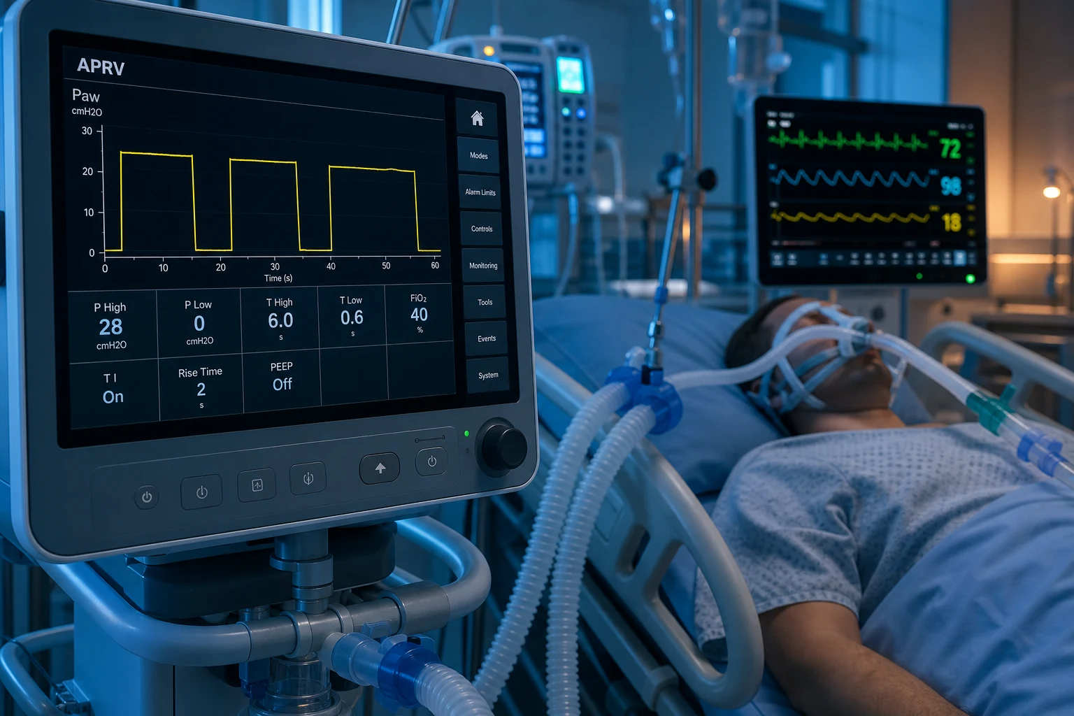
How APRV works
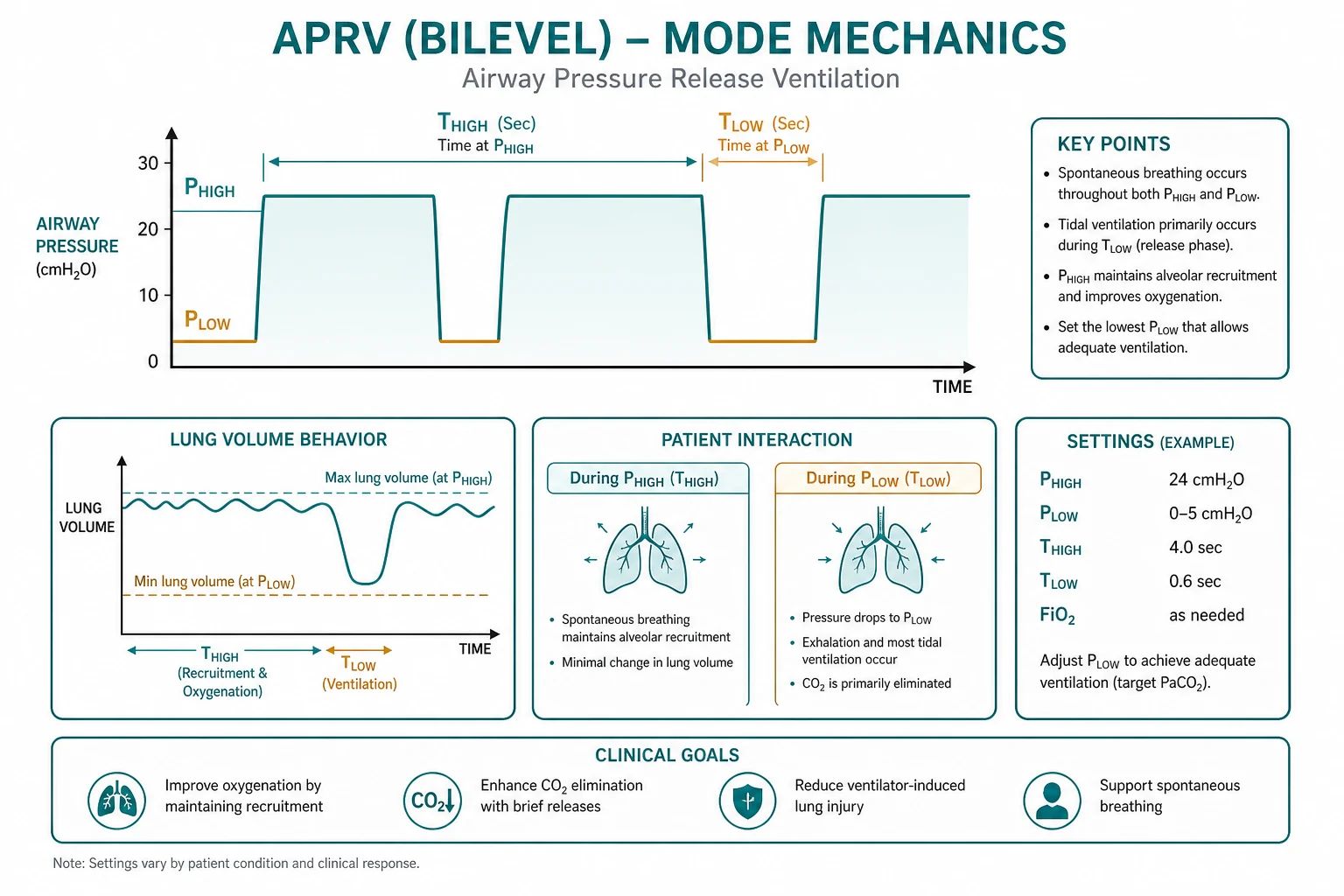
- P-high (high pressure, 25-35 cmH2O) is held for a long T-high (commonly 4-6 seconds), maintaining alveolar recruitment and a high mean airway pressure for oxygenation (an open-lung strategy).[1]
- P-low (release pressure, often 0-5 cmH2O) is held for a short T-low (about 0.2-0.8 seconds). The release drops the alveolar pressure, allowing CO2-rich gas to escape and fresh gas to enter on the next P-high — this is the "ventilation" (CO2 clearance) of APRV.[1]
- The T-low is deliberately short so that the alveoli do not fully derecruit on the release — set so the expiratory flow returns to about 50-75 per cent of its peak (a partial release) rather than to baseline, to prevent alveolar collapse.[1]
- Spontaneous breathing occurs throughout — the patient takes unsupported breaths at both levels. These spontaneous breaths improve venous return and cardiac output (the negative intrathoracic pressure), reduce the need for sedation and paralysis, and reduce diaphragm disuse atrophy.[1][2]
APRV mechanics in depth — the four settings and the inverse ratio
APRV is governed by exactly four operator-set variables, and the fellowship candidate must be able to state each with its typical range and its physiological purpose. The mode is fundamentally a continuous high CPAP (P-high) interrupted by brief releases (to P-low); the spontaneous breaths that occur throughout are the patient's own, unsupported (or lightly pressure-supported) breaths superimposed on the CPAP floor.[1][5]
APRV — the four settings to know
- P-high (25-35 cmH2O). The upper CPAP level. It is the lever for recruitment and oxygenation — a sustained high airway pressure that holds alveoli open and raises mean airway pressure. It is set from the plateau pressure of the preceding conventional mode, respecting lung protection (the transalveolar pressure must remain safe; do not exceed ~30-35 cmH2O without rationale).[1][5]
- T-high (4-6 s). The duration P-high is held on each cycle. Because T-high is long relative to T-low, the lung spends most of the cycle inflated — this is what produces the inverse I:E ratio (commonly 8:1 to 10:1) and the high mean airway pressure that underpins the open-lung concept.[1]
- P-low (0-5 cmH2O). The lower CPAP level, the release pressure. It is commonly set to 0 cmH2O (true CPAP zero) to maximise the pressure gradient driving CO2 out during the release; some clinicians leave a small floor of 0-5 cmH2O. Unlike PEEP in conventional ventilation, P-low is NOT the recruitment variable in APRV — P-high is.[1]
- T-low (0.2-0.8 s). The release duration — the single most important and most error-prone setting. It is set deliberately short so exhalation is incomplete: the lung partially empties (clearing CO2) but does not have time to fully deflate before P-high is restored. The titration target is the expiratory flow waveform: set T-low so flow returns to 50-75 per cent of peak (a partial release), not to baseline.[1][5]
The release rate (the number of releases per minute) is the frequency at which P-high is released to P-low; a typical starting pattern is 10-14 releases per minute with a T-low of 0.5-0.8 s. The tidal volume is not set — it is generated by the release and equals the volume emptied from the lung during T-low (governed by compliance, resistance, and the P-high–P-low gradient).[1]
APRV — the open-lung concept
Why it is built this way
- Inverse I:E ratio (8:1 to 10:1) — the lung is inflated for MOST of the cycle (long T-high, short T-low)
- Mean airway pressure is HIGH and sustained — recruits and holds alveoli open (oxygenation)
- Brief releases clear CO2 without letting the lung fully deflate — partial release prevents atelectrauma
- Spontaneous breathing throughout — the diaphragm keeps working, venous return preserved
- The lung is "open" for the majority of the respiratory cycle — the opposite of the cyclic open-close of conventional ventilation
Conventional volume/pressure control
What APRV is being contrasted with
- Conventional I:E ratio (1:2) — lung inflates and deflates fully each breath
- Mean airway pressure lower — recruitment depends on PEEP alone
- Cyclic opening and closing of unstable alveoli — atelectrauma if PEEP too low
- Patient-triggered breaths are machine-cycled; deep sedation and often paralysis needed for synchrony
- Tidal volume is guaranteed (VC) or pressure-limited (PC); CO2 clearance is by each mechanical breath
Why APRV works — the physiological rationale
The rationale for APRV rests on three linked mechanisms, each of which addresses a specific failure of conventional ventilation in ARDS.[2][5]
Why APRV works — three mechanisms
Continuous alveolar recruitment (the open lung)
ARDS collapses dependent (dorsal) alveoli that conventional ventilation re-opens and re-collapses with every breath — atelectrauma. APRV holds the lung at a sustained high pressure (P-high) for most of the cycle, recruiting and STABILISING alveoli so they stay open. The high mean airway pressure is the source of the oxygenation benefit: more open, ventilated alveoli means less shunt and a higher PaO2/FiO2.
Spontaneous breathing recruits the dependent lung (Putensen)
Putensen et al. (1999) showed, using CT, that spontaneous breathing during APRV re-opened and ventilated the atelectatic DEPENDENT lung that passive inflation could not reach. The mechanism is the diaphragm: its dependent portion (phrenic-nerve-driven) moves preferentially during spontaneous effort, expanding the dorsal lung where blood flow is greatest. The result is improved V/Q matching and oxygenation at the SAME applied airway pressure as full control ventilation.<Cite id="2" />
Improved V/Q matching and venous return
Because the spontaneous breaths are generated by NEGATIVE intrathoracic pressure (not positive-pressure inflation), venous return and cardiac output are preserved, splanchnic perfusion is better, and the ventilation is distributed preferentially to well-perfused dependent regions — improving V/Q matching rather than overdistending the non-dependent lung that positive pressure preferentially ventilates.
Reduced need for sedation and NMBAs
A patient who can breathe spontaneously throughout the cycle does not need to be deeply sedated or paralysed to achieve synchrony. Lighter sedation (RASS -1 to 0), less or no neuromuscular blockade, earlier mobilisation, less delirium, and reduced ICU-acquired weakness and ventilator-induced diaphragm dysfunction (VIDD) are all downstream benefits. This is the practical advantage examiners reward: APRV trades a fixed tidal volume for a spontaneously breathing, less-sedated patient.
The answer to the classic fellowship question — why does APRV improve oxygenation more than pressure-controlled ventilation at the same mean airway pressure? — is the Putensen mechanism: the patient's own diaphragmatic contraction recruits the dependent lung that passive inflation cannot reach.[2]
Advantages
- Oxygenation — the sustained high mean airway pressure recruits and holds open alveoli, improving oxygenation in refractory hypoxaemia.
- Spontaneous breathing — less sedation and paralysis, better haemodynamics (preserved venous return), and less diaphragm atrophy than fully controlled ventilation.[2]
- Lung-protective — the high mean pressure with limited peak pressure and maintained recruitment reduces the cyclic collapse-reopening of atelectrauma.[1][1]
Disadvantages and risks
- Air-trapping and dynamic hyperinflation — if the T-low is too long, the lung derecruits; if too short, gas traps; careful T-low titration is needed. APRV is generally avoided in obstructive disease (asthma, COPD), where it worsens air-trapping.[1]
- Hypercapnia — the short release limits CO2 clearance; a permissive-hypercapnia approach is accepted. The Ibarra-Estradá RCT reported transient severe hypercapnia (PaCO2 ≥55 with pH <7.15) in 42 per cent of APRV patients versus 15 per cent on low-Vt ventilation.[6]
- Asynchrony — the release can fall during a spontaneous inspiratory effort.
- Less familiar — APRV requires expertise to set and wean.[1]
- Tidal volume is not guaranteed — Vt is generated by the release and depends on compliance; an injurious Vt is possible in a highly compliant lung. Lung protection (release Vt ~4-6 mL/kg PBW) still applies.[5]
Indications — when to reach for APRV
APRV is a rescue or alternative strategy, not a first-line mode. The standard of care for ARDS remains lung-protective ventilation (low tidal volume, low plateau/driving pressure, adequate PEEP) with proning in severe disease.[3][4][5] APRV is considered when:
- Moderate-severe ARDS where oxygenation is failing on optimised conventional ventilation, as an open-lung alternative that may reduce the depth of sedation and the need for paralysis.[1][5]
- Refractory hypoxaemia as a bridge while a reversible cause (pneumonia, sepsis) is treated, or while proning/ECMO is being arranged.
- A recruited lung with an intact drive — the patient who benefits most is one with recruitable, stiff lung disease AND a preserved respiratory drive, because APRV's physiological advantage (spontaneous breathing recruiting the dependent lung) is lost in the paralysed patient.[2]
- To permit lighter sedation / wean paralysis — when deep sedation or NMBAs are themselves the problem (delirium, weakness), APRV may allow spontaneous breathing and reduce exposure.[1]
Indications
APRV may help
- Moderate-severe ARDS as a rescue/alternative to conventional ventilation
- Refractory hypoxaemia unresponsive to optimised PEEP and FiO2
- A recruitable, stiff lung WITH an intact respiratory drive
- Need to reduce sedation or wean neuromuscular blockade while keeping the lung open
- Bridge to recovery, proning, or ECMO while the cause is treated
Contraindications
APRV may harm
- Obstructive airway disease (asthma, COPD) — CO2 retention and air-trapping
- Profound shock / severe hypovolaemia — elevated intrathoracic pressure drops venous return
- Right-heart failure / severe pulmonary hypertension — high intrathoracic pressure worsens RV afterload
- No respiratory drive (deep sedation, paralysis, brainstem injury) — loses the spontaneous-breathing benefit
- Large bronchopleural fistula or unrescued barotrauma — high mean airway pressure worsens air leak
Contraindications — when to avoid APRV
The two exam-favourite contraindications both turn on the elevated intrathoracic pressure and the short release:[1][5]
- Obstructive airway disease (asthma, COPD). The defining problem in obstruction is slow expiration. APRV's brief releases (T-low 0.2-0.8 s) do not allow time for full exhalation through obstructed airways, so gas traps, intrinsic PEEP and dynamic hyperinflation rise, and CO2 is retained — the opposite of the long-expiration strategy these patients need. This is the single most important contraindication.[1]
- Profound shock / severe hypovolaemia. The sustained high mean airway pressure of APRV raises intrathoracic pressure, which reduces venous return and can depress cardiac output in the under-filled or vasoplexic patient. APRV is best started after reasonable resuscitation; profound shock is a relative contraindication until filling and vasoactive support are addressed.
- Right-heart failure / severe pulmonary hypertension. High intrathoracic pressure raises pulmonary vascular resistance and right-ventricular afterload, which can precipitate or worsen acute cor pulmonale.
- No respiratory drive. If the patient is deeply sedated, paralysed, or has a depressed central drive (opiates, brainstem injury), APRV loses its spontaneous-breathing advantage and degenerates into inverse-ratio pressure control — there are simpler ways to ventilate a paralysed patient.[2]
- Uncontrolled air leak / bronchopleural fistula. The high mean airway pressure can worsen a large air leak; APRV is relatively contraindicated until the leak is controlled.
Setting up APRV — initial settings and the first hour
Setting up APRV is a practical, examinable skill. The aim on initiation is to recruit the lung (oxygenation) while avoiding air-trapping and haemodynamic compromise. A consistent starting recipe and prompt titration of T-low are what separate a safe APRV run from a dangerous one.[1][5]
Setting up APRV — a practical sequence
Set P-high from the plateau pressure
Start P-high at approximately the PLATEAU PRESSURE of the previous conventional mode — typically 25-30 cmH2O (range 25-35). Respect lung protection: keep the release tidal volume within 4-6 mL/kg predicted body weight and do not exceed ~30-35 cmH2O without a clear rationale. P-high is the recruitment/oxygenation lever.
Set P-low to 0
P-low is commonly set to 0 cmH2O (CPAP zero) to maximise the pressure gradient driving CO2 out during the release. A small floor of 0-5 cmH2O is acceptable. P-low is NOT the recruitment variable in APRV — P-high is.
Set T-high 4-6 s
A long T-high (commonly 4-6 s) keeps the lung inflated for most of the cycle and produces the inverse I:E ratio. Most ventilators set the release FREQUENCY rather than T-high directly — a release rate of ~10-14/min with T-high 4-6 s is typical.
Set T-low 0.5-0.8 s (then titrate)
Start T-low at 0.5-0.8 s and IMMEDIATELY titrate to the expiratory flow waveform: the release should end when expiratory flow has fallen to 50-75 per cent of its peak (a partial release). This is the single most important APRV adjustment — get it right in the first hour.
Confirm spontaneous breathing and lighten sedation
APRV REQUIRES spontaneous breathing for its physiological benefit. Reduce sedation toward RASS -1 to 0, and stop or avoid neuromuscular blockade unless specifically indicated. A paralysed APRV patient is simply on inverse-ratio pressure control and has lost the point of the mode.
Reassess oxygenation, ventilation, and haemodynamics
Target SpO2 88-95 per cent and release Vt 4-6 mL/kg PBW. Accept permissive hypercapnia (pH >7.15-7.20) for ventilation. Check blood pressure and that cardiac output is preserved — if shock deepens, lower P-high and reassess filling. Re-check an ABG at 30-60 minutes.
APRV — a safe starting recipe
Titrating APRV over the first day
- If oxygenation is inadequate and the plateau permits, raise P-high in 2-3 cmH2O steps (the recruitment lever), or lengthen T-high (raises mean airway pressure). Ensure FiO2 is weaned as oxygenation improves.
- If CO2 is too high (pH too low), increase the release rate (shorter T-high) or, cautiously, lengthen T-low — but watch for derecruitment if T-low is too long.
- If air-trapping develops (rising intrinsic PEEP, falling blood pressure, expiratory flow not returning toward baseline), SHORTEN T-low and reduce P-high.[1]
Weaning from APRV — a graded descent of P-high
Weaning from APRV is a gradual reduction of P-high toward P-low as the lung recovers, followed by a transition to a conventional spontaneous mode. The principle is to maintain recruitment while progressively handing the work of breathing to the patient.[1][5]
Weaning from APRV — the descent
Confirm the patient is ready
Readiness: resolving ARDS (rising PaO2/FiO2, falling FiO2 requirement, improving compliance), minimal vasopressors, intact respiratory drive with a sustainable spontaneous rate (<30/min), and adequate cough. Do not begin the descent in an unstable patient.
Drop P-high by 2-3 cmH2O at a time
Reduce P-high in steps of 2-3 cmH2O (e.g. 30 → 27 → 24), assessing oxygenation, respiratory rate, and comfort after each step. The aim is to narrow the P-high–P-low gap progressively while keeping the lung recruited. If oxygenation falls sharply, the lung is not yet ready — return to the prior P-high.
Extend T-high as P-high falls
Lengthening T-high (or reducing the release rate) raises the mean airway pressure and helps preserve recruitment as P-high drops. The transition is a smooth convergence of the two pressure levels.
Transition to PSV / CPAP when P-high approaches P-low
When P-high is approaching P-low (around 8-12 cmH2O) and the patient is breathing comfortably with good oxygenation on low FiO2, switch to pressure support ventilation (PSV) or CPAP/pressure-support for a spontaneous breathing trial. Do NOT convert abruptly from full APRV — the lung may derecruit.
Proceed to extubation via an SBT
A successful SBT on low pressure support (e.g. 5-7 cmH2O) with low FiO2, a sustainable rate, and adequate cough leads to extubation per the unit weaning protocol. APRV weaning is one path among several; the endpoint is a successful spontaneous breathing trial, not a particular mode name.
The two cardinal errors in APRV weaning are (1) dropping P-high too fast (derecruitment and a failed transition) and (2) converting abruptly to a conventional mode (loss of the recruited lung). The graded descent with progressive T-high extension avoids both.[1]
Evidence and indications
A systematic review and meta-analysis of APRV in ARDS found that it improved oxygenation without a clear mortality benefit over conventional lung-protective ventilation.[1] APRV is therefore not the standard first-line for ARDS (which remains low-tidal-volume ventilation with PEEP and proning in severe disease) — it is used selectively, as an open-lung strategy in refractory hypoxaemia, or to reduce sedation and paralysis in a spontaneously breathing but recruited lung.[1][1]
The mortality benefit in ARDS comes from interventions APRV does not replace: lung-protective ventilation (the ARMA trial, NEJM 2000 — tidal volume 6 mL/kg PBW reduced mortality from 40 to 31 per cent)[3] and prone positioning in severe disease (PROSEVA, NEJM 2013 — 28-day mortality 16 vs 33 per cent with ≥16 h/day proning in PaO2/FiO2 <150).[4] APRV is a tool layered on top of these fundamentals, not a substitute.[5]
Evidence — landmark studies
Patel (APRV meta-analysis)
J Intensive Care Med 2026
Systematic review and meta-analysis of APRV in ARDS — oxygenation, ventilation, and mortality vs conventional ventilation
Key finding
APRV improved oxygenation (PaO2/FiO2, oxygenation index) but showed NO clear mortality benefit over conventional lung-protective ventilation
Practice change
Confirms APRV as a physiological (oxygenation) tool, not a mortality-improving intervention; use selectively in refractory hypoxaemia
Putensen (APRV physiology)
Am J Respir Crit Care Med 1999
Randomised CT study — spontaneous breathing during APRV vs controlled ventilation at matched mean airway pressure in ARDS
Key finding
Spontaneous breathing during APRV recruited and ventilated dependent (dorsal) lung regions, improving V/Q distributions and oxygenation at the same applied airway pressure
Practice change
Established the physiological rationale for APRV — diaphragmatic contraction recruits the dependent lung passive inflation cannot reach
Ibarra-Estrada (APRV in COVID-19)
Crit Care Med 2022
Single-centre RCT, 90 intubated COVID-19 ARDS patients — APRV vs low-tidal-volume ventilation within 48 h of intubation
Key finding
APRV gave higher PaO2/FiO2 and compliance in week 1 but MORE transient severe hypercapnia (PaCO2 ≥55 with pH <7.15: 42% vs 15%); no difference in ventilator-free days; mortality 78% vs 60% (p=0.07, NS)
Practice change
Modern RCT evidence that APRV improves oxygenation but does not improve outcomes and carries a hypercapnia cost
ARMA / ARDSNet (low tidal volume)
N Engl J Med 2000
Multicentre RCT — tidal volume 6 mL/kg vs 12 mL/kg PBW in acute lung injury/ARDS
Key finding
Low tidal volume REDUCED mortality (31% vs 40%) — the foundation of lung-protective ventilation that APRV does not replace
Practice change
Established low tidal volume as the mortality-reducing standard for ARDS — applies to APRV (release Vt 4-6 mL/kg) as to any mode
Bilevel (BiVent, Duo-PAP)
Bilevel is the generalised two-level pressure mode: it alternates between P-high and P-low with a more conventional inspiratory-to-expiratory ratio (not the extreme inverse ratio of APRV), and allows pressure-supported spontaneous breaths at both levels. It is used as a comfortable, partially supported mode for patients with some respiratory drive who still need a controlled background — a step between full control and pressure support.[1]
BiLevel vs APRV — the distinction made explicit
The two modes are frequently confused because both alternate between two CPAP levels and both permit spontaneous breathing. The distinction is one of degree and control, not of kind, and it matters for how the mode is used:[1][5]
APRV
The extreme, spontaneous form
- Very long T-high, very short T-low — extreme INVERSE ratio (8:1 to 10:1)
- P-low commonly 0 cmH2O; mean airway pressure is HIGH (maximal recruitment)
- NO set mandatory breaths — ventilation relies on the release phase PLUS spontaneous breathing
- Requires an intact respiratory drive for reliable CO2 clearance and its physiological benefit
- Maximal recruitment tool; reserved for refractory hypoxaemia with an intact drive
Bilevel (BiVent, Duo-PAP)
The controlled form
- More conventional T-high/T-low timing — NOT an extreme inverse ratio
- P-low usually ≥5 cmH2O (a genuine PEEP floor); mean airway pressure lower than APRV
- SET mandatory breaths guarantee a minimum minute ventilation regardless of patient effort
- Less reliant on spontaneous drive — safer when drive is marginal or tiring
- Pressure-supported spontaneous breaths permitted at BOTH levels; a comfortable step toward PSV
The practical rule: APRV is recruitment-driven and spontaneous-breathing-dependent; bilevel is control-driven and drive-tolerant. Reach for APRV when the priority is maximal recruitment in a patient who can breathe; reach for bilevel when you want a two-level mode but need a guaranteed minute ventilation or the patient's drive is unreliable.[5]
[1]Exam practice
SAQ — Setting up APRV in moderate-severe ARDS
10 minutes · 10 marks
A 48-year-old man (height 178 cm, PBW 73 kg) is intubated for severe pneumococcal pneumonia with ARDS. On volume control: Vt 440 mL (6 mL/kg PBW), RR 28, PEEP 14, FiO2 0.85. Plateau pressure 29 cmH2O. ABG: pH 7.30, PaCO2 48, PaO2 61, HCO3 24. P/F = 72 (severe ARDS). He remains hypoxaemic despite optimised lung-protective ventilation and a conservative fluid strategy. He is triggering the ventilator and lightly sedated (RASS -1). You elect to convert to APRV (airway pressure release ventilation).
SAQ — Liberation from APRV
10 minutes · 10 marks
A 52-year-old woman has been on APRV for 5 days for severe ARDS from H1N1 influenza. Current settings: P-high 28 cmH2O, P-low 0, T-high 5 s, T-low 0.6 s, FiO2 0.35. ABG: pH 7.40, PaCO2 42, PaO2 88, HCO3 26. She is triggering well, breathing at a spontaneous rate of 22/min, RASS -1, on no vasopressors, with improving compliance and a productive cough. P/F ratio is now 251. She is ready to be liberated from APRV.
Exam-exhaustive clinical pearls
Red flags
References
- [1]Patel R, Thompson J, et al. Safety, Efficacy, and Clinical Outcomes of APRV in ARDS: A Systematic Review and Meta-Analysis J Intensive Care Med, 2026.PMID 42033378
- [2]Putensen C, Mutz NJ, Putensen-Himmer G, Zinserling J. Spontaneous breathing during ventilatory support improves ventilation-perfusion distributions in patients with acute respiratory distress syndrome Am J Respir Crit Care Med, 1999.PMID 10194172
- [3]The Acute Respiratory Distress Syndrome Network (ARDSNet). Ventilation with lower tidal volumes as compared with traditional tidal volumes for acute lung injury and the acute respiratory distress syndrome N Engl J Med, 2000.PMID 10793162
- [4]Guérin C, Reignier J, Richard JC, et al.; PROSEVA Trial Investigators. Prone positioning in severe acute respiratory distress syndrome N Engl J Med, 2013.PMID 23688302
- [5]Sklar MC, Patel BK, Beitler JR, Piraino T, Goligher EC. Optimal Ventilator Strategies in Acute Respiratory Distress Syndrome Semin Respir Crit Care Med, 2019.PMID 31060090
- [6]Ibarra-Estrada MÁ, García-Salas Y, Mireles-Cabodevila E, et al. Use of Airway Pressure Release Ventilation in Patients With Acute Respiratory Failure Due to COVID-19: Results of a Single-Center Randomized Controlled Trial Crit Care Med, 2022.PMID 34593706