ICU · Respiratory / acid-base
Systematic Arterial Blood Gas Interpretation
Also known as Arterial blood gas · ABG interpretation · Acid-base analysis · Anion gap · Winter's formula · Delta ratio · High anion gap metabolic acidosis · HAGMA · Normal anion gap metabolic acidosis · NAGMA · Compensation
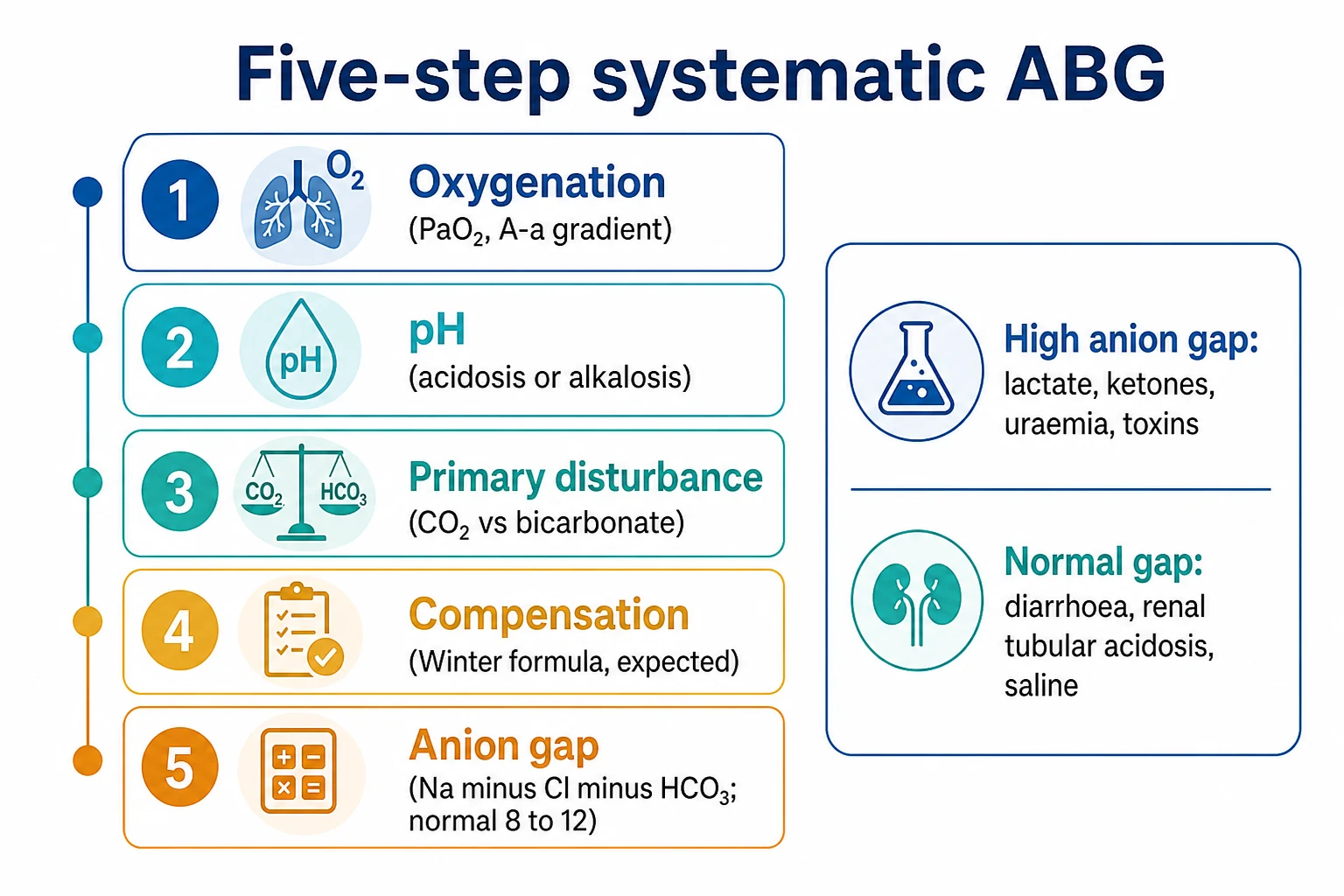
A systematic six-step approach to the arterial blood gas: (1) assess oxygenation (PaO2 with FiO2, the P/F ratio, the A-a gradient); (2) read the pH (acidaemia or alkalaemia); (3) identify the primary disturbance (CO2 versus bicarbonate); (4) check the compensation (Winter's formula for metabolic acidosis; the 1-per-10 and 4-per-10 rules for respiratory disorders); (5) calculate the anion gap (albumin-corrected) with the delta ratio for mixed metabolic disturbances; and (6) read the base excess, the standard bicarbonate, the SpO2, and the lactate. The anion gap divides metabolic acidosis into a high-anion-gap group (lactate, ketones, uraemia, toxins — GOLD MARK) and a normal-anion-gap group (diarrhoea, renal tubular acidosis, saline). The delta-delta (ΔAG/ΔHCO3) detects a mixed metabolic disorder; the osmolar gap flags toxic alcohols; and the A-a gradient separates hypoventilation from a lung problem.
On this page & tools
Your progress
Saved locally on this device.
Target exams
Overview & definition
The arterial blood gas (ABG) reports oxygenation, acid-base status, and (on most analysers) lactate, electrolytes, and haemoglobin. Interpreting it systematically — in the same order every time — avoids missing a mixed disturbance. The six steps are: (1) oxygenation, (2) pH, (3) the primary disturbance, (4) compensation, (5) the anion gap (albumin-corrected) with the delta-delta, and (6) the base excess, the standard bicarbonate, the SpO2, and the lactate.[1][1][3]
The normal values (at sea level, on room air): pH 7.35-7.45, PaCO2 35-45 mmHg, PaO2 80-100 mmHg, bicarbonate 22-26 mmol/L, base excess minus 2 to plus 2 mmol/L, standard bicarbonate 22-26 mmol/L, chloride 98-106 mmol/L, sodium 135-145 mmol/L, lactate under 2 mmol/L, and the anion gap 8-12 mmol/L (or, more precisely, 3 if corrected for albumin). The "six-step" framework is the construct used here and in the major texts; older sources describe five steps (oxygenation through the anion gap) and fold the base excess and lactate into the earlier steps. Either sequence works provided the anion gap, the delta ratio, the base excess, and the lactate are all examined on every ABG.[1][3]
Normal ABG values at sea level (room air) — the reference ranges to memorise
| Variable | Normal range | Significance of an abnormal value |
|---|---|---|
| pH | 7.35-7.45 | <7.35 acidaemia; >7.45 alkalaemia. Normal pH does NOT exclude a mixed disorder. |
| PaCO2 | 35-45 mmHg (4.7-6.0 kPa) | Respiratory component. High = respiratory acidosis; low = respiratory alkalosis. |
| PaO2 | 80-100 mmHg (10.7-13.3 kPa) | Oxygenation. <60 mmHg (SpO2 <90%) = hypoxaemic respiratory failure. |
| HCO3 (actual) | 22-26 mmol/L | Metabolic component. Reflects the in-vivo bicarbonate at the measured PaCO2. |
| Standard bicarbonate | 22-26 mmol/L | Bicarbonate standardised to PaCO2 40 mmHg — isolates the metabolic component. |
| Base excess (BE) | −2 to +2 mmol/L | The metabolic story independent of PaCO2. Negative = metabolic acidosis; positive = metabolic alkalosis. |
| Lactate | <2.0 mmol/L | 2-4 = mild (hyperlactataemia); >4 = severe (tissue hypoperfusion / sepsis). |
| SpO2 | 95-100% | The haemoglobin oxygen saturation; flat above PaO2 ~60 mmHg (the sigmoid curve). |
| Anion gap (Na−Cl−HCO3) | 8-12 mmol/L | >12 with acidaemia = high-anion-gap metabolic acidosis. |
| A-a gradient | <15 mmHg (young) to <25 mmHg (elderly) | A high gradient = a lung (V/Q) problem; a normal gradient = pure hypoventilation. |
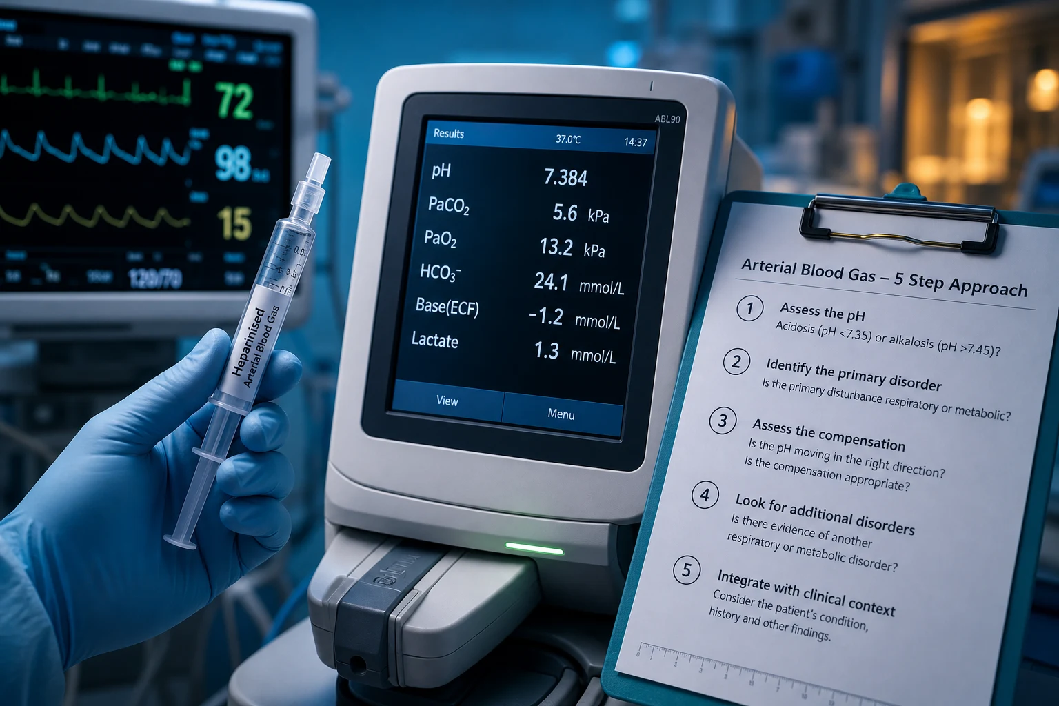
Step 1 — Oxygenation
Assess the PaO2 in the context of the FiO2. A naked PaO2 is meaningless without knowing the inspired oxygen:[1][1]
- A PaO2 under about 60 mmHg (an SpO2 under 90 per cent) is hypoxaemic; below this point the haemoglobin dissociation curve is steep, so small falls in PaO2 produce large falls in saturation.
- Calculate the PaO2/FiO2 ratio (the P/F ratio) — normal over 400; under 300 defines acute hypoxaemic respiratory failure (the Berlin ARDS thresholds: mild under 300, moderate under 200, severe under 100). The P/F ratio corrects for the FiO2 and so allows a single threshold to be applied across patients on different levels of oxygen.
- Calculate the A-a gradient (PAO2 − PaO2) to separate hypoventilation (a normal gradient) from a lung problem (a high gradient). This is the single most discriminating calculation on the ABG for hypoxaemia.
- The alveolar gas equation: PAO2 = FiO2 × (Patm − PH2O) − (PaCO2 / R). At sea level on room air this simplifies to PAO2 = 150 − (PaCO2 / 0.8). The respiratory quotient (R) is about 0.8 on a mixed diet (1.0 on pure carbohydrate). PH2O is 47 mmHg (water vapour pressure at 37°C); Patm is 760 mmHg at sea level and falls with altitude.
- Normal A-a gradient: under about 15 mmHg in a young adult, rising by roughly 3 mmHg per decade (the formula "age/4 + 4" gives a quick bedside upper limit of normal). A PaO2 that is lower than expected for the age means the gradient is widened.
- A normal A-a gradient with hypoxaemia = pure hypoventilation (the CO2 is high and displaces alveolar oxygen — opioid overdose, neuromuscular weakness, obesity-hypoventilation). The lungs themselves are fine.
- A high A-a gradient = a V/Q mismatch, shunt, diffusion impairment, or a low inspired oxygen. The five causes of a high gradient are the "lung" causes of hypoxaemia and dictate the rest of the work-up.
The A-a gradient — separating hypoventilation from a lung problem
| A-a gradient | Mechanism | Typical PaCO2 | Classic causes |
|---|---|---|---|
| Normal (<15-25 mmHg) | Pure hypoventilation — the alveoli are fine but under-ventilated | Raised (CO2 displaces O2) | Opioid, brainstem stroke, neuromuscular weakness, obesity-hypoventilation, COPD with CO2 retention |
| High (>25 mmHg) | V/Q mismatch, shunt, diffusion impairment | Variable | Pneumonia, pulmonary oedema, ARDS, pulmonary embolism, atelectasis, pulmonary fibrosis, R-to-L shunt |
Step 2 — The pH
- pH under 7.35 is acidaemia; over 7.45 is alkalaemia. Normal is 7.35-7.45.
- A normal pH does not exclude a disturbance — a mixed disorder can bring the pH into the normal range. Read on to steps 3-6.[1]
- pH is a log scale: a pH of 7.30 represents a hydrogen-ion concentration about 1.6 times that at 7.40 (50 vs 40 nmol/L). Small pH changes reflect large changes in [H+]. The relationship is approximately linear between pH 7.20 and 7.55: [H+] ≈ 80 − (pH × 10) at the midpoint.
- The Henderson-Hasselbalch equation (pH = 6.1 + log([HCO3] / (0.03 × PaCO2))) ties the three variables together. The pH is determined only by the ratio of bicarbonate to CO2, not by their absolute values — which is why a normal pH with an abnormal PaCO2 and HCO3 always means a compensated or mixed disorder.
Step 3 — The primary disturbance
Match the pH to the PaCO2 and the bicarbonate. The change that matches the pH is the primary disturbance:[1]
| pH | PaCO2 | HCO3 | Primary disturbance |
|---|---|---|---|
| Low (acidaemia) | High | — | Respiratory acidosis |
| Low (acidaemia) | — | Low | Metabolic acidosis |
| High (alkalaemia) | Low | — | Respiratory alkalosis |
| High (alkalaemia) | — | High | Metabolic alkalosis |
If the CO2 and the bicarbonate both move in the same direction away from normal, there is a mixed disturbance (for example, a low pH with a high CO2 and a low bicarbonate — a combined respiratory and metabolic acidosis). [1]
Step 4 — Compensation
The body compensates to bring the pH back toward normal. The expected compensation can be calculated; if the measured value differs from the expected, a second (mixed) disturbance is present.[1][1]
- Metabolic acidosis — Winter's formula: expected PaCO2 = (1.5 x HCO3) + 8, plus or minus 2. If the measured PaCO2 is higher than expected, there is an additional respiratory acidosis; if lower, an additional respiratory alkalosis.
- Metabolic alkalosis: the PaCO2 rises by about 0.7 mmHg for each 1 mmol/L rise in bicarbonate (up to about 55 mmHg).
- Acute respiratory acidosis: the bicarbonate rises by about 1 mmol/L for every 10 mmHg rise in PaCO2.
- Chronic respiratory acidosis: the bicarbonate rises by about 3.5-4 mmol/L for every 10 mmHg (renal compensation over days, as in COPD).
- Acute respiratory alkalosis: the bicarbonate falls by about 2 mmol/L for every 10 mmHg fall in PaCO2.
- Chronic respiratory alkalosis: the bicarbonate falls by about 4-5 mmol/L for every 10 mmHg. [1]
The body compensates to bring the pH back toward (but never fully to) normal. Metabolic disturbances are compensated within minutes by a change in ventilation; respiratory disturbances are compensated within minutes (acute, by intracellular buffering) and over 2-5 days (chronic, by renal bicarbonate reabsorption or excretion). A measured compensation that is outside the predicted range means a second (mixed) primary disorder is also present.[1][3][6]
The six compensation rules — the expected values the examiner wants verbatim
| Primary disorder | Expected compensation | Time-frame | Notes |
|---|---|---|---|
| Metabolic acidosis | Winter's: PaCO2 = (1.5 × HCO3) + 8 ± 2 | Minutes | Most-examined formula. If measured PaCO2 > expected → additional respiratory acidosis (the tiring patient). If < → additional respiratory alkalosis. |
| Metabolic alkalosis | PaCO2 rises 0.7 mmHg per 1 mmol/L rise in HCO3 (cap ~55 mmHg) | Minutes | The least reliable rule — hypoventilation limited by hypoxaemia. Expected PaCO2 ≈ 0.7 × (HCO3 − 24) + 40. |
| Acute respiratory acidosis | HCO3 rises 1 mmol/L per 10 mmHg rise in PaCO2 | Minutes | Buffered by haemoglobin and intracellular proteins. ΔHCO3 = 0.1 × ΔPaCO2. |
| Chronic respiratory acidosis | HCO3 rises 4 (3.5-4) mmol/L per 10 mmHg rise in PaCO2 | 2-5 days | Renal bicarbonate reabsorption (COPD retainers). ΔHCO3 = 0.35 × ΔPaCO2. |
| Acute respiratory alkalosis | HCO3 falls 2 mmol/L per 10 mmHg fall in PaCO2 | Minutes | Tissue buffering. ΔHCO3 = 0.2 × ΔPaCO2. |
| Chronic respiratory alkalosis | HCO3 falls 4-5 mmol/L per 10 mmHg fall in PaCO2 | 2-5 days | Renal bicarbonate excretion (pregnancy, high altitude, sepsis, hepatic failure). ΔHCO3 = 0.5 × ΔPaCO2. |
A useful single check for respiratory disorders: in acute respiratory acidosis the pH falls about 0.08 per 10 mmHg rise in PaCO2; in chronic respiratory acidosis the pH falls only about 0.03 per 10 mmHg (because the kidney has reclaimed bicarbonate). A COPD patient with a PaCO2 of 70 and a pH of 7.32 is therefore chronic (well-compensated); the same PaCO2 with a pH of 7.15 is acute (decompensated). This single comparison ("acute" vs "chronic") is the most common respiratory-acidosis viva question. [1]
Step 5 — The anion gap (for metabolic acidosis)
The anion gap = sodium minus (chloride plus bicarbonate) — Na − (Cl + HCO3). Normal is about 8-12 mmol/L (some sources quote 7-12; the variation reflects differences in measured vs calculated methodology). It divides metabolic acidosis into two groups, depending on whether the lost bicarbonate has been replaced by chloride (normal gap) or by an unmeasured anion (high gap). The anion gap exists because total cations and total anions must be equal, and "unmeasured" anions (proteins, phosphates, sulphates, organic anions) exceed "unmeasured" cations (potassium, calcium, magnesium) by about 10 mmol/L.[1][2]
Correcting the anion gap for albumin (the Figge correction)
Albumin is the dominant contributor to the "unmeasured anion" pool — about 75 per cent of the normal anion gap is albumin. Hypoalbuminaemia falsely lowers the anion gap and can mask a high-gap acidosis. For every 10 g/L the albumin falls below 40 g/L, the anion gap falls by about 2.5 mmol/L (the Figge correction):[2]
Corrected AG = measured AG + 2.5 × (40 − albumin in g/L) [1]
Equivalently, the normal anion gap in hypoalbuminaemia = 2.5 × albumin (so a patient with an albumin of 20 g/L has a normal gap of only 5 mmol/L, and a measured gap of 10 is markedly raised). In the ICU, where albumin is often low, always correct the anion gap — failing to do so is the commonest single reason a high-gap acidosis is missed. The corrected gap should be used in all subsequent delta-ratio calculations. [1]
High anion gap metabolic acidosis (HAGMA)
The bicarbonate has been consumed by an added acid whose anion is unmeasured (lactate, ketones, etc.). The mnemonic GOLD MARK: Glycols (ethylene glycol), Oxoproline, L-lactate, D-lactate, Methanol, Aspirin, Renal failure (uraemia), Ketoacidosis. (The older MUDPILES is similar: methanol, uraemia, DKA, propylene glycol, iron/INH, lactic acidosis, ethylene glycol, salicylates.)[1]
Normal anion gap (hyperchloraemic) metabolic acidosis (NAGMA)
The lost bicarbonate has been replaced by chloride, so the gap stays normal. The causes are bicarbonate loss or failure to excrete acid: diarrhoea (the commonest), renal tubular acidosis, acetazolamide, uretero-enteric fistula, pancreatic drainage, Addison's disease, and hyperchloraemia from large-volume normal saline.[1]
The delta ratio (for mixed metabolic disturbances)
When the anion gap is high, compare the rise in the anion gap to the fall in bicarbonate — this is the delta-delta (ΔAG/ΔHCO3), also called the delta ratio:[1][4][5]
Delta ratio = (measured AG − 12) / (24 − measured HCO3) [1]
(Use the corrected anion gap if albumin is abnormal.) The logic: in a pure high-gap acidosis, every mmol/L of unmeasured anion added displaces one mmol/L of bicarbonate, so the rise in AG should equal the fall in HCO3 — a ratio of about 1. Deviations from 1 reveal a second metabolic disorder. [1]
- Around 1-2 — a pure high-anion-gap acidosis (the unmeasured anion displaces the bicarbonate one-for-one). The classic single number to remember is 1 (lactic acidosis) to 1-2 (ketoacidosis).
- Under 0.4 — a pure normal-anion-gap (hyperchloraemic) acidosis (chloride has risen, the AG is little changed, but bicarbonate has fallen a lot).
- 0.4-0.8 — a mixed high-anion-gap and normal-anion-gap acidosis (e.g. diarrhoea plus sepsis).
- Over 2 — a high-anion-gap acidosis with an additional metabolic alkalosis (the bicarbonate has not fallen as much as expected, because an alkalosis is also present — the classic case is the patient who is both ketoacidotic and vomiting). [1]
The delta ratio (ΔAG/ΔHCO3) — interpreting the four bands
| Delta ratio | Interpretation | Worked example | What it means |
|---|---|---|---|
| <0.4 | Pure normal-anion-gap (hyperchloraemic) acidosis | AG 12, HCO3 14 → ΔAG 0, ΔHCO3 10 → ratio 0 | The chloride has replaced the lost bicarbonate (diarrhoea, RTA, saline). |
| 0.4-0.8 | Mixed HAGMA + NAGMA | AG 18, HCO3 12 → ratio 0.5 | A high-gap process plus a hyperchloraemic process (sepsis + diarrhoea). |
| 1-2 | Pure high-anion-gap metabolic acidosis | AG 28, HCO3 12 → ratio 1.3 | The unmeasured anion has displaced bicarbonate 1:1 (lactate, ketones). |
| >2 | HAGMA + metabolic alkalosis | AG 30, HCO3 22 → ratio 4.5 | Bicarbonate higher than expected → a second alkalotic process (vomiting, diuretics). |
The delta gap (ΔGap) is an alternative expression: ΔGap = (measured AG − 12) − (24 − measured HCO3). A ΔGap of −6 mmol/L means an additional normal-gap acidosis; a ΔGap of +6 mmol/L means an additional metabolic alkalosis. The two methods (ratio and gap) are mathematically interchangeable; use whichever your unit uses. [1]
Step 6 — The base excess, the standard bicarbonate, the SpO2, and the lactate
The last step pulls together the four values that quantify the metabolic story and the severity of the disturbance.[1][1][8]
Base excess (BE) and standard bicarbonate (SBC)
The base excess is the amount of strong acid or base (in mmol/L) that would return a blood sample to pH 7.40 at a PaCO2 of 40 mmHg and 37°C — it is the metabolic component of an acid-base disorder, isolated from the respiratory component. A negative base excess (base deficit, e.g. −10) means metabolic acidosis; a positive base excess (e.g. +8) means metabolic alkalosis. Normal is −2 to +2 mmol/L. The standard bicarbonate is the bicarbonate the sample would have at PaCO2 40 mmHg — it conveys the same information as the base excess (the metabolic component, respiratory component removed) and is used interchangeably with it.[8]
The base excess is most useful for tracking the metabolic component over time in a ventilated patient whose PaCO2 is changing (where the bicarbonate alone is confounded by the CO2). A base excess that becomes more negative over successive ABGs signals a worsening metabolic acidosis even if the pH is held by hyperventilation. It is also the basis of the dose of bicarbonate: the base deficit × 0.3 × body weight (kg) gives the total body deficit in mmol — but this is a maximum estimate and is rarely used to guide therapy, because correcting to a normal base excess overshoots and the reasons for the acidosis matter more than the number.[8]
Base excess interpretation — what the number tells you
| Base excess (mmol/L) | Interpretation | Examples |
|---|---|---|
| −2 to +2 | Normal metabolic component | Healthy reference |
| −2 to −6 | Mild metabolic acidosis | Early sepsis, mild chronic kidney disease |
| −6 to −10 | Moderate metabolic acidosis | Established DKA, moderate AKI |
| < −10 | Severe metabolic acidosis | Severe septic shock, cardiac arrest, methanol poisoning |
| +2 to +6 | Mild metabolic alkalosis | Mild vomiting, diuretic use |
| > +6 | Marked metabolic alkalosis | Severe vomiting, nasogastric suction, chloride depletion |
Two flavours of base excess appear on most analyser printouts. The base excess, blood (BE-b) is calculated from the blood sample alone and is sensitive to changes in PaCO2; the base excess, extracellular fluid (BE-ecf, or "standard base excess") is the preferred variable because it is standardised to an average haemoglobin of about 30-60 g/L and better reflects whole-body (interstitial + intracellular) metabolic state. In the stable ICU patient the two differ little; quote the standard (extracellular) base excess. [1]
SpO2 and the oxyhaemoglobin dissociation curve
The SpO2 reported on the ABG is the directly measured arterial saturation (by co-oximetry), not the pulse-oximeter value. Normal is 95-100 per cent. Because the dissociation curve is sigmoid, the SpO2 is a poor guide to PaO2 in the flat upper portion: a saturation of 97 per cent corresponds to a PaO2 anywhere from 80 to 110 mmHg. Below a PaO2 of 60 mmHg (saturation ~90 per cent) the curve is steep, and small falls in PaO2 produce large falls in saturation — this is the physiological justification for the 90 per cent / 60 mmHg threshold for hypoxaemic respiratory failure. An SpO2 above 90 per cent does NOT exclude hypercarbia; check the PaCO2. [1]
Lactate
The lactate quantifies tissue hypoperfusion and anaerobic metabolism. Normal is under 2 mmol/L. 2-4 mmol/L is "hyperlactataemia" (mild, often from adrenergic stress or beta-agonists); above 4 mmol/L is "lactic acidosis" (severe, typically from tissue hypoperfusion, sepsis, or a mitochondrial toxin). In septic shock the lactate trend (the rate of clearance) is more prognostic than a single value: a lactate that falls by 10 per cent per hour or that normalises within 24 hours is associated with survival (Jansen 2010, lactate-guided resuscitation). A lactate that fails to clear suggests ongoing hypoperfusion, an ischaemic territory (bowel, limb), or mitochondrial dysfunction (metformin, linezolid, antiretrovirals).[1]
Causes of a raised lactate — the Type A / Type B framework (Cohen & Woods)
| Type | Mechanism | Examples |
|---|---|---|
| A — with tissue hypoxia | Anaerobic glycolysis from poor oxygen delivery | Shock (septic, haemorrhagic, cardiogenic), severe hypoxia, mesenteric ischaemia, seizures, shivering |
| B1 — disease, no hypoxia | Impaired lactate clearance or upregulated pyruvate | Renal failure, hepatic failure, malignancy, alkalosis, diabetes |
| B2 — drugs/toxins | Mitochondrial toxin or upregulated glycolysis | Metformin, linezolid, antiretrovirals (NRTIs), propofol infusion syndrome, salicylates, cyanide, beta-agonists, propylene glycol |
| B3 — inborn errors of metabolism | Enzyme defects | Mitochondrial myopathies, GSD, fatty-acid oxidation defects |
Two additional lactate nuances for the exam. D-lactate (produced by gut bacteria, absorbed in blind-loop syndrome or short-bowel) is not measured by the standard L-lactate assay and causes an unexplained high anion gap with a normal L-lactate — request it specifically. A normal lactate does NOT exclude sepsis — about 15-20 per cent of septic shock patients have a normal lactate on presentation; trend it. [1]
The osmolar gap — flagging a toxic alcohol
In any unexplained high-anion-gap metabolic acidosis, calculate the osmolar gap to look for a toxic alcohol (ethylene glycol, methanol, propylene glycol). The measured osmolality minus the calculated osmolality should normally be under 10 mOsm/kg:[9][10][11]
Calculated osmolality = 2 × Na + glucose + urea + ethanol (all in mmol/L) (If ethanol is in mmol/L, add it; the simplified 2-Na + glucose + urea version omits ethanol.) Osmolar gap = measured osmolality − calculated osmolality [1]
An osmolar gap above 10-15 mOsm/kg in the presence of a high anion gap metabolic acidosis strongly suggests a toxic alcohol (the parent alcohol raises the osmolar gap; its metabolites — glycolate, formate — raise the anion gap). The gap is highest early (before the alcohol is metabolised) and falls as the anion gap rises. A normal osmolar gap does NOT exclude toxic-alcohol poisoning once metabolism is advanced — the combination of a high anion gap, a low/normal osmolar gap, and visual symptoms (methanol) or renal failure (ethylene glycol) still demands treatment. Fomepizole (15 mg/kg IV, then 10 mg/kg q12h) blocks alcohol dehydrogenase and should be given empirically while awaiting levels; haemodialysis removes both the parent alcohol and the toxic metabolites.[9][10]
The toxic alcohols — distinguishing the three (plus the surrogate marker)
| Toxin | Osmolar gap | Anion-gap acidosis | Specific organ damage | Urine/findings |
|---|---|---|---|---|
| Methanol | High (early) | High (formate) | Retina / optic nerve (blindness) | No crystals; visual symptoms |
| Ethylene glycol | High (early) | High (glycolate) | Kidney (AKI from calcium oxalate) | Calcium oxalate crystals; Wood's-lamp fluorescence of urine |
| Diethylene glycol | High (early) | High | Kidney + liver + neuro (facial palsy) | Renal failure |
| Propylene glycol | Mildly high | High (with large IV loads) | Lactate (metabolised to lactate) | Common in IV lorazepam/diazepam/phenytoin infusions |
Putting it together
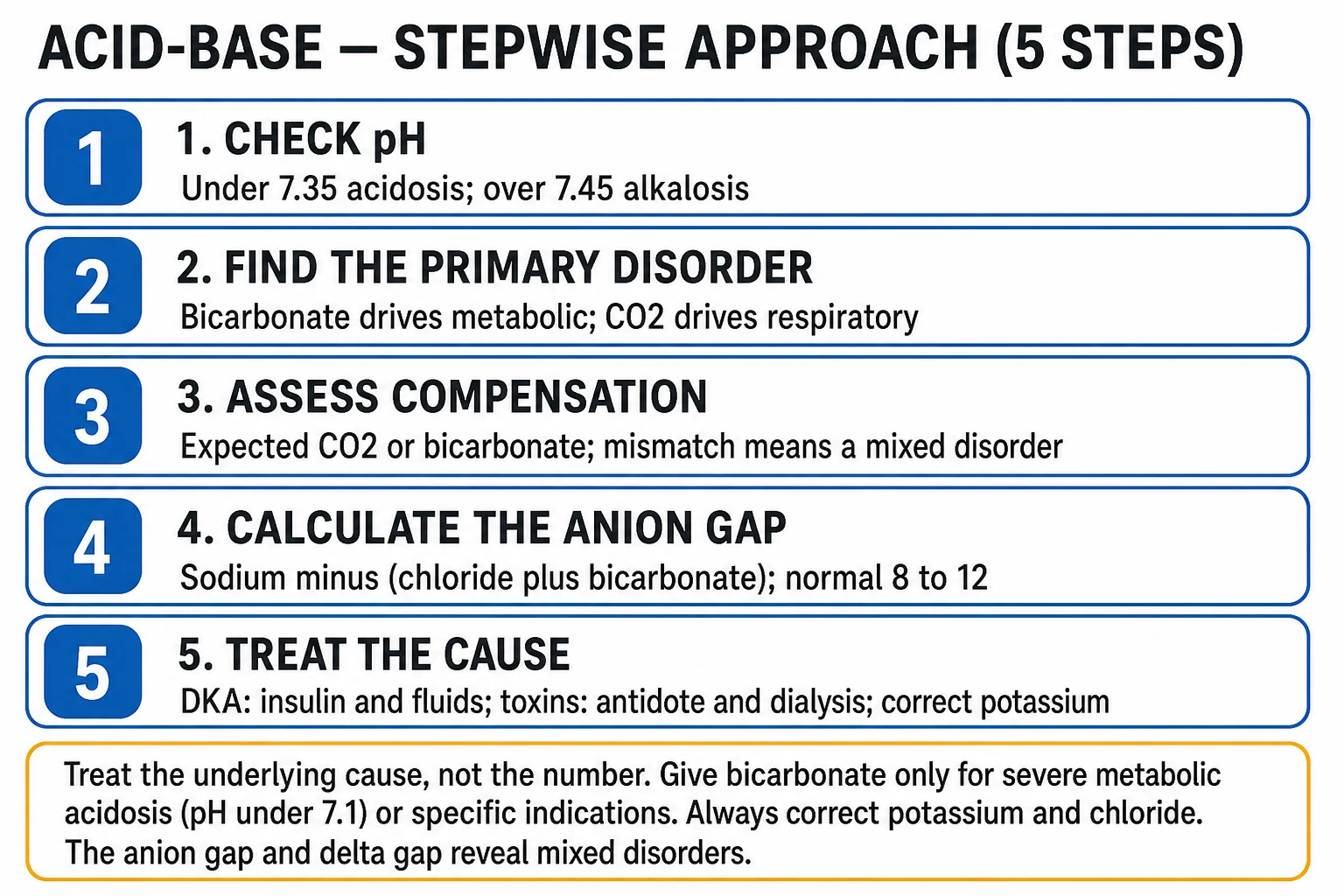
— a worked example [1]
A 60-year-old with sepsis: pH 7.20, PaCO2 28, HCO3 11, PaO2 70 on room air, lactate 6, sodium 140, chloride 100.[1]
- Oxygenation — PaO2 70 (mildly low); A-a gradient raised (the lung is part of the problem).
- pH 7.20 — acidaemia.
- Primary disturbance — low bicarbonate (11) with acidaemia = metabolic acidosis.
- Compensation — Winter's expected PaCO2 = (1.5 x 11) + 8 = 24.5, range 22-26; the measured PaCO2 is 28, slightly higher than expected — an additional mild respiratory acidosis (the patient is tiring).
- Anion gap = 140 - (100 + 11) = 29 (raised). Delta ratio = (29 - 12) / (24 - 11) = 17/13 = about 1.3 — a pure high-anion-gap metabolic acidosis (lactate 6).
- Base excess and lactate — base excess about −16 (severe metabolic acidosis); lactate 6 confirms lactic acidosis from sepsis. The picture is a lactic acidosis (sepsis) with a mild additional respiratory acidosis (early fatigue) — a single ABG that, untreated, will worsen as the patient tires.
Worked example 2 — a mixed disorder (the classic viva trap)
A 22-year-old with DKA and vomiting: pH 7.30, PaCO2 30, HCO3 14, PaO2 95 on room air, lactate 1.5, sodium 140, chloride 95, albumin 40.[3][4]
- Oxygenation — PaO2 95 on room air (normal); A-a gradient = [150 − (30/0.8)] − 95 = 112.5 − 95 = 17.5 (upper normal for a young adult — the lung is not the problem).
- pH 7.30 — acidaemia.
- Primary disturbance — low bicarbonate (14) with acidaemia = metabolic acidosis.
- Compensation — Winter's expected PaCO2 = (1.5 × 14) + 8 = 29, range 27-31; the measured PaCO2 is 30 — appropriately compensated (no additional respiratory disorder).
- Anion gap = 140 − (95 + 14) = 31 (raised). Delta ratio = (31 − 12) / (24 − 14) = 19/10 = 1.9 — at the upper end of the "pure HAGMA" range but suspicious; the bicarbonate has fallen only 10 when the gap has risen 19. This is the delta ratio that betrays a hidden metabolic alkalosis — the patient is both ketoacidotic (raising the gap) and vomiting (raising the bicarbonate back toward normal). The "1-2" range is a guide, not a rule; in this stem the clinical story (vomiting) plus a ratio approaching 2 is the diagnosis.
- Base excess −10 (metabolic acidosis); lactate 1.5 (normal — the acidosis is not lactate-driven). The picture is a ketoacidosis with a superimposed metabolic alkalosis from vomiting — the two opposing processes keep the bicarbonate higher than the gap alone would predict, and the pH only mildly low. Treat both the DKA (fluid, insulin, potassium) and recognise the alkalosis will unmask as the ketosis clears (the chloride will need to be replenished).
Worked example 3 — a respiratory acidosis with acute-on-chronic decompensation
A 68-year-old COPD retainer with a chest infection: pH 7.25, PaCO2 75, HCO3 33, PaO2 52 on 2 L/min nasal spec, lactate 1.2.[1]
- Oxygenation — PaO2 52 on FiO2 ~0.28 (2 L via nasal spec ≈ 28 per cent); P/F ratio = 52/0.28 = 186 (acute hypoxaemic respiratory failure). A-a gradient = [0.28 × 713 − (75/0.8)] − 52 = (199 − 94) − 52 = 53 (mildly raised — a degree of V/Q mismatch from the infection, but the dominant problem is CO2 retention).
- pH 7.25 — acidaemia.
- Primary disturbance — high PaCO2 (75) with acidaemia = respiratory acidosis.
- Compensation — for a chronic respiratory acidosis the expected HCO3 rises 4 per 10 mmHg: ΔPaCO2 = 75 − 40 = 35, so ΔHCO3 = 4 × 3.5 = 14, expected HCO3 = 24 + 14 = 38. The measured HCO3 is only 33 — lower than expected for a purely chronic picture, meaning there is an additional metabolic acidosis (the infection/lactic-poor sepsis, or a developing renal tubular acidosis from the COPD). Equivalently, applying the acute rule (1 per 10) would predict HCO3 ~27.5 — the measured 33 sits between acute and chronic, the classic acute-on-chronic picture.
- Anion gap — assume Na 138, Cl 96: AG = 138 − (96 + 33) = 9 (normal — a pure respiratory acidosis does not raise the gap).
- Base excess +7 (the chronic renal compensation shows as a positive base excess). The picture is an acute-on-chronic respiratory acidosis (a COPD retainer who has acutely decompensated with infection). The pH of 7.25 (rather than 7.34 for a fully compensated chronic PaCO2 of 75) signals the acute component. Treat the trigger (antibiotics, physiotherapy), optimise ventilation (NIV/bi-level), and correct the oxygen to a target SpO2 88-92 per cent.
The four primary acid-base disorders at a glance
The four primary acid-base disorders — pH, cause, compensation, and the exam one-liner
| Disorder | pH | Primary change | Compensation | Classic causes |
|---|---|---|---|---|
| Metabolic acidosis | Low (acidaemia) | ↓ HCO3 | ↓ PaCO2 (Winter's formula) | Sepsis (lactate), DKA, AKI, diarrhoea, toxins |
| Metabolic alkalosis | High (alkalaemia) | ↑ HCO3 | ↑ PaCO2 (0.7 per 1 HCO3, cap 55) | Vomiting, NG suction, diuretics, antacids, Conn's |
| Respiratory acidosis | Low (acidaemia) | ↑ PaCO2 | ↑ HCO3 (1 acute / 4 chronic per 10 mmHg) | COPD, opioid, neuromuscular, brainstem stroke |
| Respiratory alkalosis | High (alkalaemia) | ↓ PaCO2 | ↓ HCO3 (2 acute / 4-5 chronic per 10 mmHg) | Sepsis, hypoxia, anxiety, pregnancy, salicylates (early), pain |
The systematic six-step approach (a one-page workflow)
The six-step ABG interpretation — the bedside workflow
Step 1 — Oxygenation
Read the PaO2 with the FiO2. Calculate the P/F ratio (PaO2/FiO2; normal >400, ARDS <300). Calculate the A-a gradient = PAO2 − PaO2 = [FiO2 × (Patm − PH2O) − PaCO2/R] − PaO2. At sea level on room air: 150 − PaCO2/0.8 − PaO2. Normal <15 (young) to <25 (elderly). A high gradient = a lung problem; a normal gradient with hypoxaemia = pure hypoventilation.
Step 2 — The pH
pH <7.35 = acidaemia; >7.45 = alkalaemia. A normal pH does NOT exclude a disorder — a mixed disturbance can sit at 7.40. Read on. The pH is determined by the HCO3/PaCO2 ratio (Henderson-Hasselbalch), not by their absolute values.
Step 3 — The primary disturbance
Match the pH to the PaCO2 and the HCO3. Acidaemia + high PaCO2 = respiratory acidosis; acidaemia + low HCO3 = metabolic acidosis; alkalaemia + low PaCO2 = respiratory alkalosis; alkalaemia + high HCO3 = metabolic alkalosis. If PaCO2 and HCO3 move in the SAME direction, the disorder is mixed.
Step 4 — Compensation
Calculate the expected compensation: metabolic acidosis → Winter's (PaCO2 = 1.5 × HCO3 + 8 ± 2); metabolic alkalosis → PaCO2 up 0.7 per HCO3; acute respiratory acidosis → HCO3 up 1 per 10 mmHg; chronic → 4 per 10; acute respiratory alkalosis → HCO3 down 2 per 10; chronic → 4-5 per 10. A value outside the expected = a second primary disorder.
Step 5 — The anion gap (albumin-corrected) with the delta-delta
AG = Na − (Cl + HCO3), normal 8-12. Correct for albumin: add 2.5 × (40 − albumin). A high gap = added acid (GOLD MARK); a normal gap = bicarbonate loss (diarrhoea, RTA, saline). Then the delta ratio = (AG − 12)/(24 − HCO3): <0.4 NAGMA; 1-2 pure HAGMA; >2 HAGMA + metabolic alkalosis.
Step 6 — Base excess, standard bicarbonate, SpO2, lactate
Base excess (−2 to +2): negative = metabolic acidosis, positive = metabolic alkalosis; tracks the metabolic component over time independent of PaCO2. Standard bicarbonate conveys the same. Lactate: <2 normal, 2-4 mild, >4 severe lactic acidosis (sepsis, hypoperfusion). SpO2 is the flat part of the curve above 90% / PaO2 60 mmHg. In any unexplained HAGMA, add the osmolar gap (measured − calculated, normal <10) to flag a toxic alcohol.
The metabolic acidoses compared
High-anion-gap vs normal-anion-gap metabolic acidosis — the two groups
| Feature | High anion gap (HAGMA) | Normal anion gap (NAGMA, hyperchloraemic) |
|---|---|---|
| Mechanism | Added acid with an unmeasured anion (lactate, ketones, toxins) | Bicarbonate loss or failure to excrete acid; chloride replaces the lost bicarbonate |
| Anion gap | >12 (corrected for albumin) | 8-12 |
| Chloride | Normal or low | High (the defining feature) |
| Delta ratio | 1-2 (pure) | <0.4 (pure) |
| Mnemonic / causes | GOLD MARK: Glycols, Oxoproline, L-lactate, D-lactate, Methanol, Aspirin, Renal failure, Ketoacidosis | USED CARP: Ureteroenterostomy, Small bowel fistula/pancreatic drainage, Endocrine (Addison's), Diarrhoea, Carbonic anhydrase inhibitors, Acid infusion, Renal tubular acidosis, Pancreatic transplant; HARDUP for the diarrhoea/RTA cluster |
| Work-up | Lactate, ketones, creatinine, toxic-alcohol screen (osmolar gap), salicylate level, methaemoglobin | Urinary anion gap (positive in RTA, negative in diarrhoea); stool history; drugs (acetazolamide) |
The HAGMA mnemonics — GOLD MARK (modern) vs MUDPILES (legacy)
| GOLD MARK (2008, preferred) | MUDPILES (older) |
|---|---|
| Glycols (ethylene, propylene) | Methanol |
| Oxoproline (5-oxoproline — chronic paracetamol) | Uraemia |
| L-lactate | DKA |
| D-lactate | Propylene glycol |
| Methanol | Iron / INH |
| Aspirin (salicylates) | Lactic acidosis |
| Renal failure (uraemia) | Ethylene glycol |
| Ketoacidosis | Salicylates |
GOLD MARK is the modern mnemonic — it adds D-lactate and oxoproline (both increasingly recognised in ICU) and drops the now-rare iron/INH. Recite GOLD MARK in the viva; mention MUDPILES only as the legacy alternative.[1][9]
When to give bicarbonate in metabolic acidosis
Bicarbonate in metabolic acidosis — when it helps, when it does not
| Scenario | Give bicarbonate? | Rationale |
|---|---|---|
| pH < 7.10-7.15 with haemodynamic instability | Yes (50-100 mmol IV over 30-60 min, target pH ~7.20) | Below pH 7.10 the catecholamine response fails — pressors do not work. The aim is to raise the pH just enough to restore pressor responsiveness, not to normalise it. |
| DKA / ketoacidosis | No (routine) — only if pH <6.9 | Insulin stops ketogenesis; the bicarbonate regenerates as ketones are metabolised. Bicarbonate delays ketone clearance and risks hypokalaemia and cerebral oedema (in children). |
| Lactic acidosis (sepsis/shock) | No (routine) — only if pH <7.10 and unstable | Treat the cause (source control, resuscitation). BICAR-ICU (2018) showed no mortality benefit from bicarbonate even at pH 7.1-7.2 in stable patients. |
| Renal acidosis / uraemia | Yes if pH <7.20 or symptomatic | The kidney cannot regenerate bicarbonate; give to keep HCO3 >18 (or in dialysis, where bicarbonate is in the bath). |
| Toxic alcohol / salicylate / TCA poisoning | Yes (to alkalise) | Urinary alkalinisation for salicylate (pH 7.45-7.55, target urine pH 8); sodium bicarbonate for TCA-induced cardiac toxicity (Na load + alkalaemia overcomes the sodium-channel block). |
The dose of bicarbonate, if needed: base deficit (× 0.3 × weight in kg) gives the total deficit in mmol, but this is a maximum — give half, reassess, and aim for a pH of about 7.20 (not 7.40). Hypertonic sodium bicarbonate (8.4%) causes hypernatraemia and hyperosmolality; use 1.26% or 4.2% where possible, especially in children.[7]
The evidence base for acid-base interpretation and the key references
Berend 2014 (NEJM) — physiological approach
The widely cited single-reference framework for the systematic six-step approach: identify the primary disorder, calculate the expected compensation, classify the metabolic acidosis by the (albumin-corrected) anion gap, and apply the delta ratio to detect mixed disorders. PMID 25295502.
Figge 1998 (Crit Care Med) — albumin correction
Established that the anion gap must be corrected for albumin: the gap falls 2.5 mmol/L for every 10 g/L the albumin is below 40 g/L. Without correction, hypoalbuminaemia masks a high-gap acidosis in ~30% of ICU patients. PMID 9824071.
Rastegar 2007 (JASN) — delta ratio
The definitive derivation and validation of the DeltaAG/DeltaHCO3 ratio for the bedside detection of mixed metabolic acid-base disorders — the four bands (<0.4, 0.4-0.8, 1-2, >2) used today. PMID 17656477.
Wrenn 1990 (Ann Emerg Med) — the delta gap
The original description of the delta gap as an approach to mixed acid-base disorders — the forerunner of the modern delta ratio. PMID 2240729.
Kraut & Madias 2010 (Nat Rev Nephrol) — metabolic acidosis review
Comprehensive review of pathophysiology, diagnosis, and management of metabolic acidosis, including the Type A/B lactate framework and the role of bicarbonate. PMID 20308999.
Fenves & Emmett 2021 (AJKD Core Curriculum) — HAGMA work-up
The contemporary nephrology approach to the high-anion-gap metabolic acidosis: the differential (GOLD MARK), the work-up (lactate, ketones, osmolar gap, toxicology), and the indications for dialysis. PMID 34400023.
Siggaard-Andersen 1995 (Acta Anaesthesiol Scand Suppl) — base excess
The conceptual foundation of the base excess (and standard base excess) as the measure of the non-respiratory (metabolic) acid-base disturbance, independent of PaCO2 — and the comparison with the Stewart strong-ion-difference approach. PMID 8599264.
Common pitfalls
- Reporting only the pH and missing a mixed disturbance — always work through all six steps.
- Using a venous sample and treating the values as arterial (venous pH is about 0.03 lower and the CO2 about 6 mmHg higher).
- Forgetting the lactate in a metabolic acidosis — it is the commonest single cause of a high anion gap in the ICU.
- Missing the delta ratio in a patient with both vomiting (alkalosis) and ketoacidosis (the bicarbonate is higher than the gap alone would predict).
- Failing to correct the anion gap for albumin — a "normal" gap of 12 in a hypoalbuminaemic ICU patient is, after Figge correction, a high gap. This is the commonest reason a high-gap acidosis is missed in critical care.[2]
- Interpreting the PaO2 without the FiO2 — a PaO2 of 90 is fine on room air, alarming on 100% oxygen (P/F ratio 90, severe ARDS). Always state the FiO2 first.
- Stopping at a single ABG — acid-base is dynamic. A trajectory (the base excess getting more negative, the lactate rising) is more informative than a single snapshot. Trend the ABG.
- Treating the number, not the patient — giving bicarbonate for a low pH in DKA (where insulin is the treatment), or chasing a base excess in a stable patient. The cause, not the pH, drives the therapy.[1][2]
SAQ — The mixed metabolic disorder: DKA with vomiting
10 minutes · 10 marks
A 23-year-old woman with type 1 diabetes presents with a 3-day history of polyuria, polydipsia and profuse vomiting. She is drowsy, breathing deeply and rapidly (Kussmaul), and has the smell of ketones. Arterial blood gas on room air: pH 7.28, PaCO₂ 24 mmHg, HCO₃⁻ 11 mmol/L, PaO₂ 96 mmHg, base excess −15. Sodium 132, chloride 92, potassium 5.6, glucose 34, ketones 6.8 mmol/L, albumin 40 g/L, lactate 1.6.
SAQ — Acute-on-chronic respiratory acidosis in COPD
10 minutes · 10 marks
A 69-year-old man with severe COPD (usual PaCO₂ 60 mmHg) is brought in with a chest infection, increasing drowsiness and a coarse tremor. He was given high-flow oxygen in the ambulance. Arterial blood gas on FiO₂ 0.4: pH 7.22, PaCO₂ 85 mmHg, HCO₃⁻ 34 mmol/L, PaO₂ 70 mmHg, base excess +9, lactate 1.4. He is drowsy but rousable, using accessory muscles, with a respiratory rate of 28.
Red flags
References
- [1]Sanagustín MN, Osredkar J Blood gas analysis: Clinical applications, interpretation and future directions (Review) Med Int (Lond), 2026.PMID 41473681
- [2]Figge J, Jabor A, Kazda A, Fencl V Anion gap and hypoalbuminemia Crit Care Med, 1998.PMID 9824071
- [3]Berend K, de Vries APJ, Gans ROB Physiological approach to assessment of acid-base disturbances N Engl J Med, 2014.PMID 25295502
- [4]Rastegar A Use of the DeltaAG/DeltaHCO3- ratio in the diagnosis of mixed acid-base disorders J Am Soc Nephrol, 2007.PMID 17656477
- [5]Wrenn K The delta (delta) gap: an approach to mixed acid-base disorders Ann Emerg Med, 1990.PMID 2240729
- [6]Kraut JA, Madias NE Approach to patients with acid-base disorders Respir Care, 2001.PMID 11262558
- [7]Kraut JA, Madias NE Metabolic acidosis: pathophysiology, diagnosis and management Nat Rev Nephrol, 2010.PMID 20308999
- [8]Siggaard-Andersen O, Fogh-Andersen N Base excess or buffer base (strong ion difference) as measure of a non-respiratory acid-base disturbance Acta Anaesthesiol Scand Suppl, 1995.PMID 8599264
- [9]Fenves AZ, Emmett M Approach to Patients With High Anion Gap Metabolic Acidosis: Core Curriculum 2021 Am J Kidney Dis, 2021.PMID 34400023
- [10]Ross JA, Borek HA, Holstege CP Toxic Alcohols Crit Care Clin, 2021.PMID 34053711
- [11]Mullins ME, Kraut JA The Role of the Nephrologist in Management of Poisoning and Intoxication: Core Curriculum 2022 Am J Kidney Dis, 2022.PMID 34895948