ICU · Respiratory
Acute severe community-acquired pneumonia: aspiration pneumonia update
Also known as Aspiration pneumonia · Aspiration pneumonitis update · Chemical pneumonitis vs bacterial pneumonia · Antibiotic stewardship in aspiration
Aspiration events in ICU patients can cause two distinct entities: aspiration PNEUMONITIS (sterile chemical injury from acidic gastric contents — acute onset, sterile, NO routine antibiotics) or aspiration PNEUMONIA (bacterial infection from aspirated oropharyngeal flora — develops over 24-48h, needs antibiotics covering anaerobes). This topic provides an update on the critical distinction and management. Key update: do NOT routinely give antibiotics for aspiration pneumonitis. Give only if infection develops (fever 48h, purulent sputum, progressive infiltrates). Anaerobic coverage is often over-prescribed — piperacillin-tazobactam or clindamycin + ceftriaxone covers both aerobes and anaerobes for confirmed aspiration pneumonia.
On this page & tools
Your progress
Saved locally on this device.
Target exams
Red flags
SAQ — Witnessed aspiration under anaesthesia: pneumonitis vs pneumonia
10 minutes · 10 marks
A previously well 28-year-old woman undergoes an emergency caesarean section under general anaesthesia for fetal distress. On induction she regurgitates and aspirates a large volume of gastric contents. She is rapidly intubated and suctioned. Two hours later in recovery she is tachypnoeic (RR 32), SpO₂ 89% on room air, with diffuse bilateral wheeze and frothy pink sputum. Chest X-ray shows bilateral perihilar infiltrates. Temperature 37.1°C, WCC 9.8, CRP 18, procalcitonin 0.08.
SAQ — Aspiration pneumonia with cavitation in an elderly patient
10 minutes · 10 marks
An 82-year-old nursing-home resident with advanced dementia and poor dentition is admitted with a 3-day history of low-grade fever, productive cough and increasing confusion. He is cachectic with foul-smelling breath. Chest X-ray shows a dense consolidation with a 4 cm cavity and air-fluid level in the posterior segment of the right upper lobe. Blood pressure 95/60, RR 26, SpO₂ 92% on room air, WCC 18.4, CRP 240, procalcitonin 2.8, creatinine 145. Blood cultures grow Peptostreptococcus.
Clinical pearls
Red flags
Aspiration pneumonitis vs aspiration pneumonia: the core distinction
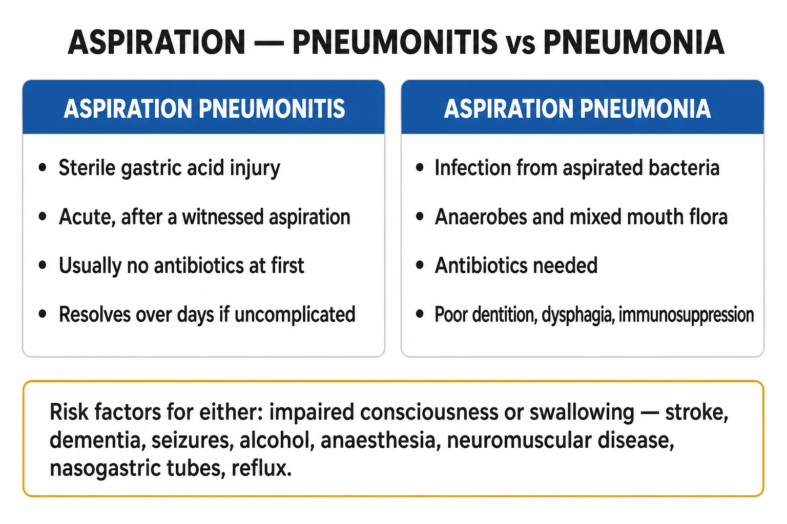
A single aspiration event can produce two entirely different syndromes, and the difference determines whether antibiotics are given. Confusing them is one of the commonest antibiotic-stewardship errors in the ICU.[3]
Aspiration pneumonitis (Mendelson syndrome)
Aspiration of sterile, acidic gastric contents (pH < 2.5) causes an acute chemical burn of the bronchial and alveolar epithelium. It is sterile at onset — the injuring agent is the acid, not bacteria. Onset is within minutes to hours, classically after anaesthesia, seizure, intoxication, or resuscitation. Features include abrupt dyspnoea, bronchospasm, frothy or pink sputum, hypoxaemia that is often out of proportion to the initial chest X-ray, and bilateral infiltrates that develop over hours. Roughly 10 per cent of massive aspirations progress to ARDS, and a minority develop secondary bacterial infection over the following days.[3][4]
Management is supportive: airway protection, supplemental oxygen, NIV or invasive ventilation for respiratory failure, and judicious fluids. No routine antibiotics, no corticosteroids, no bronchoalveolar lavage. Antibiotics are added only if infection declares itself (fever beyond 48 hours, purulent sputum, rising inflammatory markers, progressive infiltrates).[3]
Aspiration pneumonia
Aspiration of bacteria-laden oropharyngeal or gastric secretions causes a bacterial pneumonia. It is more indolent, evolving over 24 to 48 hours (or insidiously over days in elderly or demented patients with repeated small aspirations). It arises in patients with impaired swallow or chronic aspiration: stroke, dementia, Parkinson's disease, neuromuscular weakness, poor dentition, prolonged intubation. The infiltrate localises to dependent segments and may cavitate (anaerobes) or progress to abscess or empyema.[3]
Management requires antibiotics covering both aerobes and anaerobes.[2]
Aspiration pneumonitis vs aspiration pneumonia — the exam answer side by side
| Feature | Aspiration pneumonitis (Mendelson) | Aspiration pneumonia |
|---|---|---|
| Mechanism | Chemical injury — sterile gastric acid | Bacterial infection — oropharyngeal flora |
| pH of aspirate | < 2.5 (acidic) | Any (usually colonised, near-neutral) |
| Onset | Minutes to hours | 24 to 48 hours (or insidious) |
| Typical setting | Anaesthesia, post-ictal, intoxication, resuscitation | Stroke, dementia, neuromuscular, poor dentition |
| Inoculum | Sterile gastric acid | Bacteria-laden secretions |
| Fever / purulent sputum | Absent initially | Present |
| Chest X-ray | Bilateral diffuse infiltrates ± ARDS | Focal dependent-segment consolidation ± cavitation |
| Dependent localisation | Less localised | Posterior upper lobe + apical lower lobe (supine); basal lower lobe (upright) |
| Anaerobes | — | Peptostreptococcus, Fusobacterium, Prevotella, Bacteroides |
| Procalcitonin | Low (< 0.1) | Elevated (> 0.25) |
| Antibiotics | NO (unless infection develops) | YES — aerobes + anaerobes |
| Corticosteroids | NO | NO |
| Bronchoalveolar lavage | NO | NO |
| Duration of therapy | Supportive | 7 days (longer if abscess or empyema) |
Why the distinction matters
Up to a third of inpatients labelled "aspiration pneumonia" actually have sterile pneumonitis for which antibiotics add nothing and cause harm — Clostridioides difficile colitis, antimicrobial resistance, adverse drug events, and cost.[9] A 48 to 72 hour observation window with procalcitonin and CRP trends and repeat imaging is the stewardship-friendly approach in equivocal cases.[9]
Risk factors for aspiration
All risk factors converge on two mechanisms: impaired airway protection and increased bacterial inoculum.[2]
Reduced consciousness / protective reflexes
- Decreased GCS — stroke (especially brainstem or large MCA territory), post-ictal state, traumatic head injury, metabolic encephalopathy (hepatic, uraemic, hyperosmolar)
- Anaesthesia and procedural sedation — especially emergency or non-fasted cases; the setting of Mendelson's original 1946 description
- Intoxication — alcohol, opioids, benzodiazepines
- ICU sedation — minimised by daily sedation interruption (SAT) and spontaneous awakening trials [1]
Neurological and swallowing impairment
- Acute stroke — dysphagia in up to 50 per cent; pneumonia risk is highest in the first 72 hours, and a formal swallow screen prevents pneumonia[10]
- Dementia, Parkinson's disease, motor neuron disease
- Bulbar weakness — myasthenia gravis, Guillain-Barré syndrome, multiple sclerosis
- Post-extubation dysphagia — common after more than 48 hours of intubation (cuff-related injury, recurrent laryngeal nerve neuropraxia)
Mechanical and anatomical
- Mechanical ventilation — micro-aspiration around the cuff, especially with low cuff pressure, patient-ventilator dys-synchrony, or reintubation
- Gastrointestinal obstruction, recent upper GI surgery
- Oesophageal disorders — achalasia, Zenker's diverticulum, stricture, scleroderma, severe GERD
- Feeding tubes — an NG tube splints the lower oesophageal sphincter and causes stasis; PEG does NOT eliminate aspiration (oropharyngeal secretions are still aspirated)[2]
- Tracheostomy, vocal cord palsy
Increased bacterial load / colonisation
- Poor dentition and periodontal disease — the reservoir for anaerobes
- Poor oral hygiene — addressed by chlorhexidine oral care
- Proton-pump inhibitor use — raises gastric pH and permits gastric colonisation with Gram-negatives
- Chronic or recurrent micro-aspiration — elderly, nursing-home residents [1]
Aspiration risk factors grouped by mechanism — what each does and how to counter it
| Mechanism | Representative risk factors | Modifiable counter-measure |
|---|---|---|
| Impaired consciousness | ↓GCS, post-ictal, intoxication, sedation, encephalopathy | Minimise sedation (SAT/SBT); intubate if GCS < 8; sit upright |
| Swallow failure | Stroke, dementia, Parkinson's, MG/GBS/MND, post-extubation | Formal swallow screen (SLT); NPO until cleared; texture-modified diet[10] |
| Airway not protected | Intubation, low cuff pressure, reintubation, tracheostomy | Cuff pressure 20-30 cmH₂O; subglottic suction ETT; minimise reintubation |
| GI regurgitation | Obstruction, achalasia, Zenker's, ileus, GERD | Post-pyloric feeding; prokinetics; treat obstruction |
| High bacterial inoculum | Poor dentition, poor oral hygiene, PPI use, nursing home | Chlorhexidine oral care; toothbrushing; review PPI need |
Pathophysiology
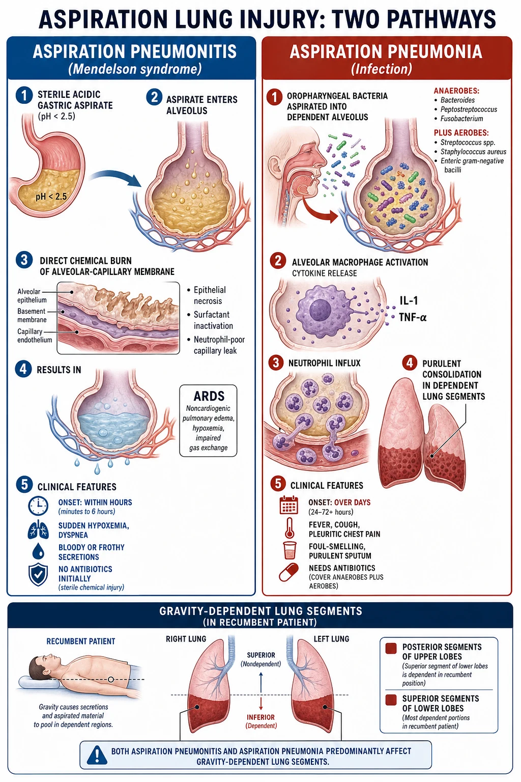
Pneumonitis — the Mendelson cascade
- Acid reaches the alveoli and spreads throughout the bronchial tree within 12 to 18 seconds.[4]
- A pH below 2.5 causes immediate coagulative necrosis of bronchial and alveolar epithelium.
- Alveolar-capillary membrane disruption within minutes allows a protein-rich exudate to flood the alveoli — non-cardiogenic pulmonary oedema.
- Neutrophil infiltration (1 to 4 hours) releases reactive oxygen species and proteases that amplify injury — this is acute inflammation, not infection.
- Surfactant inactivation causes atelectasis and a fall in compliance.
- V/Q mismatch and intrapulmonary shunt produce hypoxaemia, often disproportionate to the initial radiographic findings.
- By 24 to 36 hours, roughly 10 per cent progress to ARDS; a subset develop secondary bacterial infection over subsequent days.[4]
The injuring agent is the acid, which is precisely why antibiotics, corticosteroids, and lavage do not alter the acute chemical injury.[3]
Pneumonia — bacterial overgrowth in dependent lung
- An aspirated bacterial inoculum overwhelms mucociliary clearance and alveolar macrophages in dependent, poorly ventilated segments.
- Anaerobic, low-oxygen-tension micro-environments (atelectasis, necrotic tissue) favour anaerobe proliferation.
- Necrotising anaerobes (Fusobacterium, Bacteroides) drive tissue destruction → cavitation, lung abscess, necrotising pneumonia, empyema.
- Dental plaque (biofilm) is the reservoir for anaerobes — which is why dentition and oral hygiene are the key modifiable risk factors.[8]
Causative organisms
Aspiration pneumonia — the classic anaerobic + aerobic mix
Anaerobes (originate from gingival crevices and dental plaque; need anaerobic transport and are rarely recovered from expectorated sputum):
- Peptostreptococcus species — the single most common anaerobe
- Fusobacterium nucleatum — necrotising
- Prevotella, Bacteroides (including the B. fragilis group in abscess)
- Porphyromonas, Actinomyces (chronic, indolent infection) [1]
Aerobes:
- Streptococcus pneumoniae, Haemophilus influenzae — community-acquired
- Staphylococcus aureus (including MRSA)
- Enteric Gram-negative bacilli — Klebsiella, E. coli, Pseudomonas — more prominent in healthcare-associated aspiration, alcoholism, and structural lung disease[8]
High-yield exam pearl: Edentulous patients have markedly fewer anaerobes (no gingival crevices), so anaerobic coverage can often be omitted, and aspiration in an edentulous patient is more likely aerobic (S. pneumoniae, H. influenzae, Gram-negative bacilli).[8]
Aspiration pneumonitis
Sterile — no organism. If secondarily infected, expect the same oropharyngeal flora as above. [1]
Organisms by syndrome and setting — what to cover
| Setting | Predominant organisms | Empirical cover |
|---|---|---|
| Community aspiration, dentate | Anaerobes + S. pneumoniae, H. influenzae | Amoxil-clav OR clindamycin ± respiratory FQ |
| Community aspiration, edentulous | Aerobes (S. pneumo, H. flu, GNB) — few anaerobes | Ceftriaxone ± metronidazole (anaerobe cover optional) |
| Severe / hospitalised | Anaerobes + aerobes ± GNB | Pip-tazo OR clindamycin + ceftriaxone |
| Healthcare-associated / MDR risk | Anaerobes + Pseudomonas, MRSA, ESBL | Meropenem/cefepime + vanc/linezolid + metronidazole |
| Lung abscess / empyema | Anaerobes (necrotising) | Drain + prolonged anaerobe cover (4-6 weeks) |
| Pneumonitis (sterile) | None | None (antibiotics only if infection develops) |
Clinical features and radiology
- Onset — pneumonitis within hours; pneumonia over 24 to 48 hours (or insidious in the elderly)
- Dyspnoea, tachypnoea, tachycardia; fever, purulent sputum and leucocytosis favour infection
- Crackles, bronchial breathing, decreased breath sounds over dependent zones
- Hypoxaemia — classically out of proportion to the chest X-ray early in pneumonitis [1]
Radiographic distribution — gravity dictates the site
| Patient position | Involved segments |
|---|---|
| Supine / recumbent (most ICU patients) | Posterior segment of the upper lobes + apical segment of the lower lobes |
| Upright / ambulant | Basal segments of the lower lobes |
| Right > Left | The right main bronchus is wider, shorter and more vertical |
Imaging patterns
- Pneumonitis — bilateral patchy or perihilar infiltrates within hours; may evolve into an ARDS-pattern of diffuse alveolar and interstitial opacities
- Pneumonia — focal consolidation in dependent segments; ± cavitation or abscess (anaerobes), effusion or empyema, necrotising pneumonia
- CT chest — more sensitive than chest X-ray for cavitation, abscess, empyema, necrosis, and an aspirated foreign body [1]
Investigations
- Bloods — FBC, EUC, LFTs, CRP, procalcitonin (low in pneumonitis, high in bacterial infection)[9]
- ABG/VBG — quantify hypoxaemia, the A-a gradient, and acid-base status
- Chest X-ray on presentation and serially; CT chest if cavitation, abscess, empyema is suspected or for poor-resolution cases
- Blood cultures before antibiotics (in pneumonia or sepsis)
- Sputum / endotracheal aspirate Gram stain and culture — aerobic; anaerobic culture of expectorated sputum is unhelpful because of contamination by oral flora
- Invasive sampling (bronchoscopic BAL, protected specimen brush) — for severe, healthcare-associated, immunocompromised, or non-responding cases
- Bronchoscopy — for retrieval of large particulate / food / foreign body aspiration; not for lavage of acidic liquid
- Percutaneous or bronchoscopic aspirate of an abscess for anaerobes in complicated cases
Management
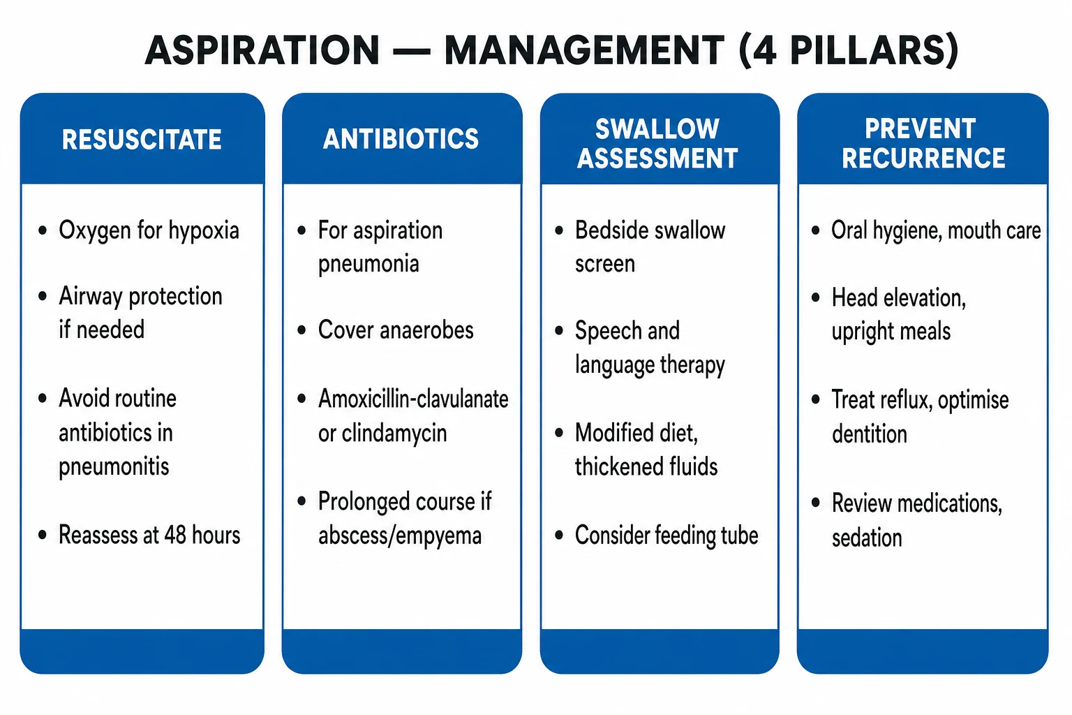
1. Immediate airway and respiratory support
- Airway — clear and protect the airway; suction the oropharynx; intubate if GCS < 8, unable to protect the airway, or severe hypoxaemia
- Oxygen — target SpO₂ 92-96 per cent (88-92 per cent if COPD/hypercapnia risk)
- Ventilatory support — NIV may avoid intubation in moderate pneumonitis (each intubation raises VAP risk); invasive ventilation for refractory hypoxaemia or ARDS
- Positioning — head of bed elevated; lateral with the affected side down to localise the process
- Fluids — judicious; capillary leak favours cautious resuscitation, but hypovolaemia worsens perfusion [1]
2. The antibiotic decision
Aspiration pneumonitis → NO empirical antibiotics. Reassess at 48 to 72 hours and start only if infection develops (fever, purulent sputum, leucocytosis, progressive infiltrates), or if there is high baseline suspicion of bacterial co-infection (sepsis, immunocompromise).[3]
Aspiration pneumonia → antibiotics covering aerobes + anaerobes:[2][7]
- Community-acquired, mild to moderate — amoxicillin-clavulanate OR clindamycin (± a respiratory fluoroquinolone)
- Severe / hospitalised — piperacillin-tazobactam OR clindamycin + ceftriaxone (or cefepime + metronidazole)
- Healthcare-associated / MDR risk — extended-spectrum antipseudomonal β-lactam (pip-tazo, cefepime, meropenem) ± MRSA cover (vancomycin or linezolid)
- Abscess / empyema — drain + prolonged anaerobic cover (4-6 weeks)[8]
3. Therapies NOT recommended
- Corticosteroids — no benefit and possible harm across multiple RCTs and meta-analyses[3]
- Bronchoalveolar lavage to neutralise or remove acid — distributes the injury more widely and worsens outcomes[3]
- Empirical antibiotics for sterile chemical pneumonitis[9]
4. Supportive and adjunctive
- Chest physiotherapy — postural drainage, percussion, breathing exercises once the patient is stable
- Bronchodilators for bronchospasm
- VAP prevention bundle (see below)
- DVT and stress-ulcer prophylaxis per ICU protocol
- Early enteral nutrition via NG or NJ with swallow reassessment before any oral intake [1]
Aspiration event in the ICU — the management algorithm
Immediate (0-10 min): airway and oxygen
Clear and suction the oropharynx; intubate if GCS < 8 or airway unprotected; oxygen to SpO2 92-96 per cent (88-92 per cent if COPD). Position head up, affected side down.
Assess: pneumonitis vs pneumonia
Timing (hours = pneumonitis; 24-48 h = pneumonia), setting (anaesthesia/intoxication vs stroke/dementia), purulence of sputum, fever, CXR pattern, procalcitonin (low vs high).
Decision: antibiotics or not
If pneumonitis → supportive care, NO antibiotics, observe 48-72 h. If pneumonia → antibiotics covering aerobes + anaerobes (pip-tazo OR clindamycin + ceftriaxone); send blood + sputum cultures first.
Supportive care
NIV/invasive ventilation for respiratory failure; judicious fluids; chest physiotherapy once stable; bronchodilators for wheeze; DVT and stress-ulcer prophylaxis; early enteral nutrition.
Reassess at 48-72 h
For pneumonitis: start antibiotics only if fever, purulent sputum, rising inflammatory markers, or progressive infiltrates appear. For pneumonia: narrow to culture results; duration 7 days (longer if abscess or empyema).
Prevent recurrence (the bundle)
HOB 30-45 degrees; oral care with chlorhexidine; subglottic suction ETT; cuff pressure 20-30 cmH2O; minimise sedation; swallow assessment before oral intake; review PPI and feeding route.
Antibiotic regimens for aspiration pneumonia by severity and setting
| Setting | First-line regimen | Alternative | Notes |
|---|---|---|---|
| Community, mild (oral) | Amoxicillin-clavulanate 875/125 mg PO BD | Clindamycin 300 mg PO QID ± moxifloxacin | Covers aerobes + anaerobes; edentulous may drop anaerobe cover |
| Community, severe (IV) | Clindamycin 600 mg IV QID + ceftriaxone 2 g IV OD | Piperacillin-tazobactam 4.5 g IV TDS | Classic anaerobe + aerobic combination |
| Hospitalised, broad cover | Piperacillin-tazobactam 4.5 g IV TDS | Cefepime + metronidazole; meropenem | Single agent covers anaerobes + GNB |
| Healthcare-associated / MDR | Meropenem 1 g IV TDS + vancomycin (if MRSA) | Cefepime + metronidazole + linezolid | Add MRSA and Pseudomonas cover |
| Abscess / empyema | Drainage + prolonged anaerobe cover (4-6 wk) | Clindamycin or amox-clav IV→PO | Drainage is essential; antibiotics alone fail |
Aspiration prophylaxis bundle (prevention)
The most effective "treatment" for aspiration pneumonia is prevention. The following evidence-based measures reduce aspiration in ICU and other high-risk patients.[6]
- Head of bed elevation 30 to 45 degrees — unless contraindicated; the strongest single intervention (Drakulovic 1999 showed a near-halving of VAP)[6]
- Oral care with chlorhexidine (0.12 per cent or 0.2 per cent) every 4 to 6 hours — reduces oropharyngeal bacterial load and pneumonia risk[2]
- Subglottic suctioning endotracheal tube (continuous or intermittent) — removes pooled contaminated secretions above the cuff[6]
- Cuff pressure management 20 to 30 cmH₂O — check every 8 to 12 hours; avoid under-inflation (leak and micro-aspiration) and over-inflation (mucosal ischaemia)[6]
- Minimise sedation — daily sedation interruption, spontaneous awakening trials, spontaneous breathing trials; the awake patient protects their own airway
- Avoid unnecessary reintubation — each intubation or reintubation event multiplies VAP and aspiration risk
- Swallow assessment (speech-language therapist) before any oral intake in stroke and post-extubation patients[10]
- Good oral hygiene — toothbrushing and denture care, not chlorhexidine alone
- Enteral feeding safeguards — jejunal rather than gastric feeding if aspiration risk is high; prokinetics; small-bore tube; post-pyloric placement
- Review stress-ulcer prophylaxis — balance the risk (a PPI raises gastric pH and promotes Gram-negative gastric colonisation) against benefit (indicated only in high-risk patients: mechanically ventilated > 48 h, coagulopathy, major burns)
The aspiration prophylaxis bundle — measure, what it does, strength of evidence
| Measure | What it does | Evidence |
|---|---|---|
| HOB 30-45° | Gravity opposes reflux and pooling above the cuff | Strong (Drakulovic 1999 RCT) |
| Oral care / chlorhexidine | Lowers oropharyngeal bacterial load | Moderate (reduces VAP in cardiac surgery) |
| Subglottic suction ETT | Removes contaminated secretions above cuff | Strong (multiple RCTs) |
| Cuff pressure 20-30 cmH₂O | Prevents leak around the cuff | Moderate |
| Daily sedation interruption | Awakened patient protects own airway | Strong (SAT/SBT trials) |
| Minimise reintubation | Each intubation event multiplies risk | Strong (observational) |
| Formal swallow screen | Detects dysphagia before oral intake | Strong in stroke (Hinchey 2005) |
Special situations
Acute stroke
Up to 50 per cent of acute stroke patients have dysphagia, and aspiration pneumonia is the commonest medical complication. A formal dysphagia screen within 4 hours of admission and keeping the patient NPO until swallow is cleared reduce pneumonia significantly.[10] Texture-modified diets and fluid thickeners are used per SLT assessment, recognising that thickened fluids themselves may increase dehydration and are not uniformly protective.
Post-extubation dysphagia
Common after more than 48 hours of intubation — cuff-related laryngeal injury and recurrent laryngeal nerve neuropraxia cause transient swallow impairment. Screen before restarting oral intake; the majority recover within days to weeks. [1]
Anaesthesia
The setting of Mendelson's original description (obstetric anaesthesia, 1946). Modern practice — fasting, rapid sequence induction with cricoid pressure, securing the cuff before ventilation, and avoiding positive-pressure mask ventilation in the at-risk patient — has made aspiration during anaesthesia uncommon but it remains a leading cause of anaesthesia-related morbidity when it occurs. [1]
Pregnancy
Historically a leading cause of maternal anaesthetic death. The combination of increased intra-abdominal pressure, delayed gastric emptying, and reduced lower oesophageal sphincter tone makes the parturient high-risk; RSI with a cuffed tube is standard. [1]
Complications
- ARDS — roughly 10 per cent of massive aspiration pneumonitis progresses to ARDS with its attendant mortality
- Lung abscess and necrotising pneumonia — typically anaerobic (Fusobacterium, Bacteroides); may need percutaneous or surgical drainage plus prolonged antibiotics
- Empyema — requires chest-tube drainage ± surgical decortication
- Secondary bacterial pneumonia — supervenes on a fraction of pneumonitis cases
- Death — hospitalised aspiration pneumonia mortality is 20 to 30 per cent, higher in the elderly and nursing-home residents [1]
Prognosis
- Pneumonitis — most cases resolve within 24 to 72 hours with supportive care; mortality rises sharply if ARDS develops
- Aspiration pneumonia — hospitalised mortality 20 to 30 per cent; higher in the elderly, nursing-home residents, and those with recurrent aspiration or multiple comorbidities
- Recurrence — the patient who aspirates once is at high risk of repeating; address the underlying cause (swallow, dentition, feeding route, sedation) [1]
Evidence and trials
Aspiration pneumonitis and pneumonia — the key evidence
Mendelson 1946 (Am J Obstet Gynecol): the original description of acid aspiration in obstetric anaesthesia — the eponymous syndrome. Established the chemical-injury paradigm and the rationale for fasting and airway protection.[3] Marik 2001 (NEJM) — the seminal review: crystallised the pneumonitis vs pneumonia distinction. Concluded that routine antibiotics are NOT indicated for aspiration pneumonitis, corticosteroids are unhelpful, and anaerobic coverage is over-prescribed. The single most-cited reference on the topic.[3] Raghavendran 2011 (Crit Care Med) — pathophysiology review: detailed the acid-injury cascade (epithelial necrosis, neutrophil-mediated amplification, surfactant loss, ARDS) and confirmed that the acute injury is chemical and sterile.[4] Singh 2000 (AJRCCM): the classic antibiotic-stewardship study in ICU patients with pulmonary infiltrates — clinical and procalcitonin-guided stopping rules safely shortened antibiotic courses, underpinning the observation window for aspiration pneumonitis.[9] Klompas 2014 (ICHE) — VAP prevention update: synthesised the evidence for the VAP bundle (HOB elevation, oral care, subglottic suction, cuff pressure, sedation minimisation).[6] Drakulovic 1999 (Lancet): the RCT showing that a semi-recumbent position (45°) vs supine halved the incidence of nosocomial pneumonia in mechanically ventilated patients — the evidence base for HOB 30-45°.[6] Hinchey 2005 (Stroke): a large registry study showing that hospitals with a formal dysphagia screening protocol had significantly lower post-stroke pneumonia rates — the basis for universal early swallow screening.[10] El-Solh 2003 (AJRCCM): microbiology of severe aspiration pneumonia in institutionalised elderly — confirmed the anaerobic + aerobic mix and the lower anaerobe burden in edentulous patients.[8]
Additional clinical pearls — exam-exhaustive
[1]Red flags — additional
References
- [1]Martin-Loeches I, Torres A. Severe community-acquired pneumonia Eur Respir Rev, 2022.PMID 36517046
- [2]DiBardino DM, Underwood EL. Notum palmitoleoyl-protein carboxylesterase regulates Fas cell surface death receptor-mediated apoptosis via the Wnt signaling pathway in colon adenocarcinoma Bioengineered, 2021.PMID 34402722
- [3]Marik PE. Management development of clinicians in Indonesia Med Teach, 2001.PMID 11260753
- [4]Raghavendran K, Nemzek J, Napolitano LM, Knight PR. Changes in health-related quality of life and factors predicting long-term outcomes in older adults admitted to intensive care units Crit Care Med, 2011.PMID 21263318
- [5]Metlay JP, Waterer GW, Long AC, et al. Diagnosis and Treatment of Adults with Community-acquired Pneumonia. An Official Clinical Practice Guideline of the American Thoracic Society and Infectious Diseases Society of America Am J Respir Crit Care Med, 2019.PMID 31573350
- [6]Klompas M, Branson R, Eichenwald EC, et al. Infectious risk moments: a novel, human factors-informed approach to infection prevention Infect Control Hosp Epidemiol, 2014.PMID 25026623
- [7]Mandell LA, Wunderink RG, Anzueto A, et al. Infectious Diseases Society of America/American Thoracic Society consensus guidelines on the management of community-acquired pneumonia in adults Clin Infect Dis, 2007.PMID 17278083
- [8]El-Solh AA, Pietrantoni C, Bhat A, et al. Effect of (+/-)-epibatidine, a nicotinic agonist, on the central pathways controlling voiding function in the rat Am J Physiol Regul Integr Comp Physiol, 2003.PMID 12689850
- [9]Singh N, Rogers P, Atwood CW, Wagner MM, Yu VL. Long-term follow-up of hexamethylene diisocyanate-, diphenylmethane diisocyanate-, and toluene diisocyanate-induced asthma Am J Respir Crit Care Med, 2000.PMID 10934080
- [10]Hinchey JA, Shephard T, Furie K, Smith D, Wang D, Tonn S. Cerebral vein and dural sinus thrombosis in elderly patients Stroke, 2005.PMID 16100024