ICU · Respiratory
Atypical pneumonias in ICU: Legionella, Mycoplasma, Chlamydia
Also known as Atypical pneumonia · Legionnaires disease · Legionella pneumophila · Mycoplasma pneumoniae · Chlamydia psittaci · Psittacosis
Atypical pneumonias: caused by atypical pathogens (Legionella, Mycoplasma, Chlamydia psittaci, Chlamydia pneumoniae, Coxiella burnetii). 'Atypical' because: (1) different clinical features (dry cough, headache, myalgia, prominent extrapulmonary symptoms). (2) Not visible on Gram stain (intracellular, cell-wall deficient). (3) Do not respond to beta-lactams (need macrolides, tetracyclines, fluoroquinolones). MYCOPLASMA PNEUMONIAE: 1 atypical worldwide, young adults, 'walking pneumonia', cold agglutinins (autoimmune haemolysis), macrolide-resistant strains emerging. LEGIONELLA PNEUMOPHILA: most severe — Pontiac fever (mild self-limiting flu-like illness) vs Legionnaires disease (severe pneumonia with GI/neurological symptoms, SIADH, urinary antigen). CHLAMYDIA PSITTACI: bird exposure (psittacosis). CHLAMYDIA PNEUMONIAE: person-to-person, common cause of CAP, linked to atherosclerosis. COXIELLA BURNETII: Q fever (cattle/sheep exposure), hepatitis, endocarditis. Treatment: macrolide (azithromycin, clarithromycin) OR doxycycline OR fluoroquinolone (levofloxacin, moxifloxacin).
On this page & tools
Your progress
Saved locally on this device.
Target exams
Red flags
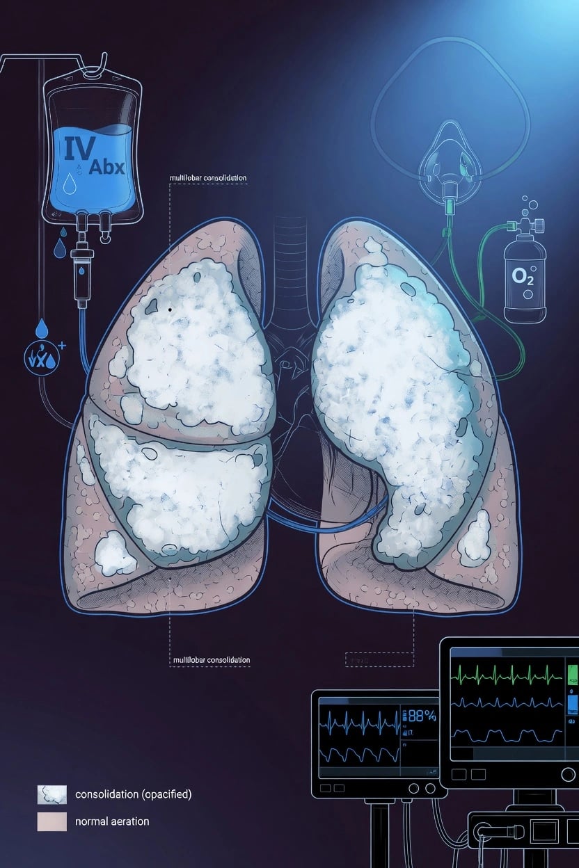
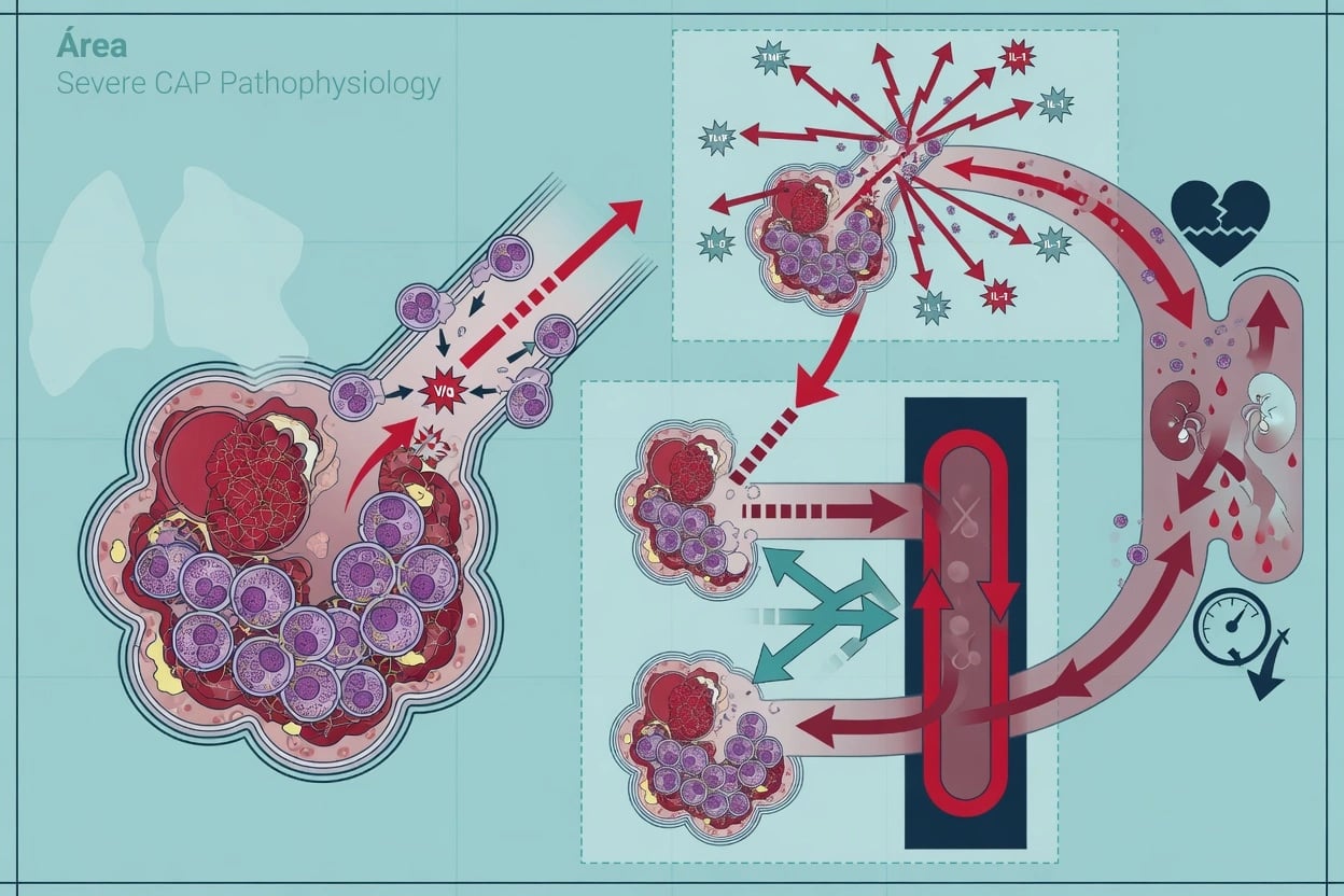
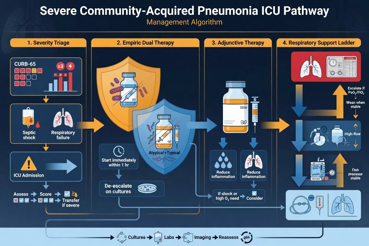
Atypical pneumonia pathogens
| Feature | Legionella pneumophila | Mycoplasma pneumoniae | Chlamydia psittaci | Coxiella burnetii |
|---|---|---|---|---|
| Severity | SEVERE (ICU common) | Mild-moderate ('walking pneumonia') | Variable | Usually mild (Q fever) |
| Source | Water (cooling towers, spas, showers) | Person-to-person (droplet) | Birds (parrots, pigeons — psittacosis) | Animals (cattle, sheep — Q fever) |
| Age | >50, smokers, immunocompromised | Young adults (5-20) | Bird owners, pet shop workers | Farmers, veterinarians |
| Clinical clues | GI (diarrhoea, nausea), neurological (confusion), SIADH/hyponatraemia, relative bradycardia, high fever | Dry cough, prominent extrapulmonary (haemolysis, rash, neurological, arthritis) | Dry cough, headache, splenomegaly, hepatitis | Fever, hepatitis, endocarditis |
| Diagnosis | Urine antigen (L. pneumophila serogroup 1), PCR, serology | PCR (throat), serology | Serology (4-fold rise) | Serology |
| Treatment | Levofloxacin OR azithromycin | Azithromycin OR doxycycline | Doxycycline | Doxycycline |
| Mortality | 5-30% (higher if untreated) | <1% | 1-5% (untreated 20%) | 1-2% |
Empiric antibiotic strategy for severe CAP (covering atypicals)
- Severe CAP (ICU) — cover BOTH typical (S. pneumoniae) AND atypical pathogens
- Beta-lactam + macrolide: ceftriaxone 2g IV OD + azithromycin 500mg IV OD (PREFERRED — covers typicals + atypicals)
- OR Beta-lactam + fluoroquinolone: ceftriaxone + levofloxacin/moxifloxacin (if macrolide allergy)
- OR fluoroquinolone monotherapy (moxifloxacin) — covers both typicals + atypicals (for non-severe only)
- Add atypical cover EARLY — beta-lactams ALONE do NOT cover atypicals (Legionella, Mycoplasma, Chlamydia)
- Narrow when pathogen identified — if Legionella confirmed: levofloxacin or azithromycin monotherapy
- Duration: 5-7 days (CAP), 7-14 days (Legionella — longer, immunocompromised)
SAQ — Legionnaires\u2019 disease in severe community-acquired pneumonia
10 minutes · 10 marks
A 62-year-old male smoker returns from a cruise with a 4-day history of high fever (39.4°C), dry cough, confusion and profuse watery diarrhoea. On examination he is delirious, pulse 88 in sinus, BP 96/60, RR 30, SpO₂ 90% on room air with focal right lower lobe crackles. Bloods: Na⁺ 126, K⁺ 3.2, creatinine 150, ALT 110, CK 1200, WCC 14. Chest X-ray shows a dense right lower lobe consolidation. He is admitted to ICU for severe CAP.
SAQ — Mycoplasma cold-agglutinin haemolytic anaemia
10 minutes · 10 marks
A 19-year-old university student presents with a 10-day dry cough, low-grade fever and malaise. On the day of admission he notices dark urine and blue-tinged fingers after walking to hospital in the cold. Examination: pale, mildly jaundiced, acrocyanosis of the fingers, extensive bilateral crackles. Haemoglobin 68 g/L, reticulocytes 9%, unconjugated bilirubin 65, LDH 1200, haptoglobin undetectable. Chest X-ray shows patchy bilateral lower-lobe infiltrates far more extensive than the clinical picture suggests.
Clinical pearls
Red flags
Prognosis
2019 ATS/IDSA CAP guidelines (Metlay 2019)
Updated guidelines for community-acquired pneumonia:
- Empiric atypical cover: recommended for all severe CAP (beta-lactam + macrolide OR fluoroquinolone)
- Routine atypical testing: NOT recommended (unless severe, outbreak, specific suspicion)
- Legionella urine antigen: recommended in severe CAP (especially if SIADH, neurological, summer-fall)
- Sputum culture: recommended for severe CAP, healthcare-associated risk factors [1]
Legionella mortality: 5-30% overall. Severe (ICU): 15-30%. Reduced with early appropriate antibiotics. Mycoplasma mortality: <1% (usually mild). Rare severe cases in elderly/immunocompromised. Psittacosis mortality: 1-5% treated, 20% untreated. Q fever mortality: 1-2% (higher with endocarditis — 25%).
Pathogen deep-dive
Mycoplasma pneumoniae — the #1 atypical pathogen
Mycoplasma pneumoniae is the COMMONEST atypical pathogen worldwide and the leading cause of community-acquired pneumonia (CAP) in older children and young adults (5-35 years). It is a cell-wall-deficient bacterium (smallest free-living organism, ~0.1-0.3 µm) — the absence of a cell wall explains BOTH why it is invisible on Gram stain AND why beta-lactams (which target peptidoglycan cross-linking) are useless. Only three drug classes work: macrolides (50S ribosome), tetracyclines (30S), and fluoroquinolones (DNA gyrase/topoisomerase IV). [1]
Classic presentation is 'walking pneumonia': an indolent illness (1-3 week prodrome) with dry cough, sore throat, low-grade fever, malaise and headache — the patient looks surprisingly well despite radiographic infiltrates that are often more extensive than the examination suggests. Chest X-ray typically shows patchy, unilateral, lower-lobe bronchopneumonia; pleural effusion is usually small when present. The white cell count is frequently normal or only mildly raised. [1]
Extrapulmonary disease is a hallmark and occurs in up to 25% — driven by immune-mediated mechanisms (molecular mimicry, autoantibodies) and direct organism spread: [1]
- Haematological: cold agglutinin autoimmune haemolytic anaemia (IgM anti-I antibodies).
- Dermatological: erythema multiforme, Stevens-Johnson syndrome (especially in children/young adults).
- Neurological: aseptic meningitis, meningoencephalitis, transverse myelitis, cerebellar ataxia, Guillain-Barré syndrome (including the anti-GQ1b antibody variant).
- Cardiac: pericarditis, myocarditis (rare but a leading cause of cardiovascular death in young patients with Mycoplasma).
- Musculoskeletal: polyarthritis, myalgia.
- Renal/ GI: glomerulonephritis, pancreatitis, hepatitis. [1]
Diagnosis: respiratory PCR (throat swab, nasopharyngeal aspirate or sputum) is now first-line — rapid, sensitive, and available early. Serology requires paired sera (4-fold rise in IgM/ complement-fixing antibody 2-4 weeks apart), so it confirms retrospectively. Cold agglutinin titre (>1:64) supports the diagnosis but is non-specific. Culture is slow and needs special media (Eaton's PPLO agar) — rarely performed.[3][6]
Legionella — the severe atypical
Legionella is a Gram-negative intracellular bacillus that thrives in warm aqueous environments (20-45°C). It causes a SPECTRUM: at one end the mild, self-limiting Pontiac fever (flu-like illness, no pneumonia, 95%+ attack rate, full recovery in ~1 week) and at the other Legionnaires' disease — a severe, sometimes fatal CAP that disproportionately reaches the ICU. Infection is by inhalation of contaminated water aerosols from cooling towers, evaporative condensers, spa pools/ hot tubs, showers, mist machines and decorative fountains; there is NO person-to-person spread. Risk factors for severe disease: age >50, smoking, chronic lung disease, immunosuppression (transplant, steroids, anti-TNF), and male sex.[1][2]
Clinical clues pointing to Legionnaires' disease (the "Winawer-Clancy" / Cunha constellation): high fever (>39°C), relative bradycardia, dry cough, prominent GI symptoms (diarrhoea, nausea, abdominal pain ~50%), neurological features (confusion, lethargy, headache ~30%), SIADH with hyponatraemia (Na <130), deranged liver enzymes, mild AKI and sometimes rhabdomyolysis. The chest X-ray classically shows a unilateral lower-lobe patchy consolidation that progresses rapidly and may cavitate (especially in the immunocompromised). Mortality ranges 5-10% in treated immunocompetent cases to 25-30% in ICU/ immunosuppressed patients.[1]
Diagnosis: the cornerstone is the Legionella urinary antigen test — a rapid (~1 hour), highly specific (>95%) immunochromatographic assay that detects soluble lipopolysaccharide antigen of L. pneumophila serogroup 1 (responsible for ~80-90% of community cases). Sensitivity ~70-90%; antigen is excreted from day 1 and persists for weeks, but the assay will MISS non-serogroup-1 and non-pneumophila species (e.g. L. longbeachae from potting mix — common in Australia/ New Zealand). Newer combined assays (e.g. ImmuView) detect both L. pneumophila sg1 and L. longbeachae. Confirm with respiratory PCR and culture on buffered charcoal yeast extract (BCYE) agar (3-10 days, allows molecular typing for outbreak investigation), and paired serology (4-fold rise).[10][2]
Chlamydia psittaci — psittacosis (ornithosis)
An obligate intracellular bacterium acquired by inhalation of aerosolised dried droppings, nasal secretions or feather dust from infected birds — psittacines (parrots, cockatiels, budgerigars), pigeons, poultry (turkeys, ducks), and poultry workers, bird breeders and pet-shop owners are at risk. Presents after a 5-14 day incubation with abrupt high fever, severe headache (often out of proportion), dry cough, myalgia, and a characteristic splenomegaly with a faint Horder's spots rash. Hepatitis (raised transaminases), myocarditis, endocarditis and encephalitis are recognised complications. Chest X-ray shows variable patchy consolidation. Diagnosis is by serology (4-fold rise in microimmunofluorescence antibody titre, or a single high IgM); PCR is increasingly available. Doxycycline 100 mg PO/IV BD for 2-3 weeks is the treatment of choice. Untreated mortality is ~20%; treated 1-5%. It is a NOTIFIABLE zoonosis in many jurisdictions.[8]
Chlamydia pneumoniae
A human-only (no animal reservoir) obligate intracellular pathogen transmitted person-to-person by respiratory droplets, with an incubation of 3-4 weeks. It is a common — and frequently under-recognised — cause of CAP, sinusitis, pharyngitis and bronchitis, and causes both endemic and epidemic disease in semi-closed populations (military recruits, university dormitories, nursing homes). Clinically it overlaps with Mycoplasma: prolonged dry cough, sore throat, hoarseness (laryngitis is prominent), headache and sinus tenderness. Severity is usually mild-moderate but can cause severe pneumonia in the elderly and immunocompromised. An intriguing epidemiological association exists between C. pneumoniae and atherosclerosis (organism detected in coronary plaques), though causality and a treatment benefit remain unproven. Diagnosis is serology (microimmunofluorescence) or PCR; treatment is a macrolide or doxycycline.[6]
Coxiella burnetii — Q fever
A Gram-negative obligate intracellular bacterium (formerly a rickettsia) with a remarkable spore-like small cell variant that resists desiccation and persists in the environment. The classic reservoirs are cattle, sheep and goats; birth products, faeces, urine and milk are heavily contaminated. Inhalation of infected dust (wind-borne spread — patients may NOT recall direct animal contact), or less often tick vectors or unpasteurised milk, transmits disease. Farmers, abattoir workers and veterinarians are at highest risk; it is a recognised bioterrorism agent (CDC category B). [1]
Q fever has two phases. Acute Q fever: incubation ~2-3 weeks, then high fever, severe headache, myalgia, and a mild atypical pneumonia (often only on imaging) OR a self-limiting granulomatous hepatitis (doughnut granulomas). Chronic Q fever (<5%, developing months-years later) is defined by infection lasting >6 months and most often manifests as culture-negative endocarditis of prosthetic or previously damaged valves — the leading cause of death; vascular infections (aneurysms) and chronic hepatitis also occur. Pregnancy raises the risk of chronic infection and obstetric complications. [1]
Diagnosis: serology with phase-variation antibodies is key — acute disease is dominated by phase II IgM (and rising phase II IgG), while chronic disease shows high-titre phase I IgG (≥1:800 is the diagnostic hallmark of endocarditis). PCR on blood/ tissue is useful early. Doxycycline 100 mg BD for 14 days treats acute disease; chronic Q fever needs doxycycline PLUS hydroxychloroquine for ≥18 months (hydroxychloroquine raises the phagolysosomal pH and overcomes Coxiella's intracellular alkalinisation defence). An effective whole-cell vaccine (Q-Vax) is available and recommended for high-risk occupational groups in Australia.[7]
[1]Distinguishing atypical from typical pneumonia
Typical vs atypical pneumonia — bedside contrast
| Feature | Typical (S. pneumoniae, H. influenzae) | Atypical (Mycoplasma, Legionella, Chlamydia, Coxiella) |
|---|---|---|
| Onset | Abrupt (hours), rigors | Gradual/ subacute (days-weeks) |
| Cough | Purulent (rusty sputum in pneumococcal) | Dry, hacking, scant sputum |
| Fever | High, shaking rigors | Moderate (Legionella high); relative bradycardia |
| Systemic | Localised to chest | Headache, myalgia, GI, neurological prominent |
| Examination | Focal crackles, consolidation signs | Often disproportionate — exam underestimates X-ray |
| Chest X-ray | Lobar consolidation | Patchy, interstitial, lower-lobe, bilateral |
| WCC | Leucocytosis (neutrophils) | Normal/ mildly raised |
| Gram stain | Neutrophils + organism | Neutrophils, NO organism seen |
| Beta-lactam response | Good within 48-72 h | NO response (need macrolide/ doxycycline/ fluoroquinolone) |
Pontiac fever vs Legionnaires' disease (both Legionella)
| Feature | Pontiac fever | Legionnaires' disease |
|---|---|---|
| Clinical syndrome | Acute self-limiting flu-like illness (NO pneumonia) | Severe pneumonia ± multi-organ |
| Attack rate | Very high (~90-95%) | Low (~5%) of those exposed |
| Incubation | 5-66 hours (short) | 2-10 days |
| Chest X-ray | Normal | Patchy/ lobar consolidation, progresses, may cavitate |
| Severity | Mild, no deaths | 5-30% mortality |
| Treatment | Supportive only (antibiotics not required) | Fluoroquinolone or macrolide urgently |
| Host | Any age, healthy | Elderly, smokers, immunocompromised |
Atypical-pathogen antibiotic comparison
| Drug | Class | Dose (adult, severe) | Pros | Cons/ cautions |
|---|---|---|---|---|
| Azithromycin | Macrolide | 500 mg IV OD | Excellent intracellular levels, short course, paediatric-suitable, QT-prolonging | Macrolide-resistant Mycoplasma in Asia |
| Clarithromycin | Macrolide | 500 mg IV BD | Strong atypical cover | More drug interactions (CYP3A4), phlebitis |
| Doxycycline | Tetracycline | 100 mg IV/PO BD | Cheap, oral bioavailability, covers all atypicals + rickettsiae/ Q fever | Photosensitivity, avoid <8 yr/ pregnancy, oesophagitis |
| Levofloxacin | Fluoroquinolone | 750 mg IV/PO OD | First-line SEVERE Legionella, monotherapy covers typicals + atypicals | QT prolongation, tendinopathy, dysglycaemia, C. difficile |
| Moxifloxacin | Fluoroquinolone | 400 mg IV/PO OD | Best pneumonia bioavailability, monotherapy | Same FQ risks; not for UTI |
Diagnostic workup of suspected atypical pneumonia
Investigation pathway for suspected atypical CAP
- Recognise the clinical pattern — dry cough + headache + myalgia + extrapulmonary features + beta-lactam failure → think atypical.
- Send Legionella urinary antigen on admission — for ALL severe CAP (ATS/IDSA 2019), especially with SIADH, diarrhoea, confusion, summer-autumn onset. Use a combined assay (e.g. ImmuView) if L. longbeachae is plausible (ANZ).[5][10]
- Respiratory PCR — nose/throat swab or sputum for Mycoplasma, Chlamydia (psittaci + pneumoniae), and Legionella multiplex. Most useful early, before antibiotics.[3]
- Bloods — FBC (often normal WCC), U&E (hyponatraemia = SIADH in Legionella), LFTs (hepatitis in Legionella/ psittacosis/ Q fever), CK (rhabdomyolysis), troponin.
- Cultures — sputum and blood cultures (rule in/ out typicals; Legionella needs BCYE agar). Do NOT rely on sputum Gram stain to exclude atypicals — they will not be seen.
- Serology (paired) — acute AND convalescent (2-4 weeks) for Mycoplasma, Chlamydia psittaci, Coxiella (phase I/II), Chlamydia pneumoniae. Confirms retrospectively but is essential for public-health/ outbreak work.
- Targeted history — birds (psittacosis), livestock/ parturient animals/ unpasteurised milk (Q fever), spa pools/ cooling towers/ travel (Legionella), sick contacts (Mycoplasma/ C. pneumoniae).
- Chest imaging — CXR (patchy lower-lobe/ interstitial pattern); CT chest if CXR is mild but clinical picture severe or to define complications (cavitation, effusion, empyema, abscess).
ICU management of Legionnaires' disease
- Empiric cover from the outset — severe CAP = beta-lactam (ceftriaxone 2 g IV OD) PLUS a Legionella-active agent. Do NOT wait for confirmation.[5]
- Definitive therapy once suspected/ confirmed — levofloxacin 750 mg IV OD (preferred for severe disease) OR azithromycin 500 mg IV OD. Fluoroquinolones achieve higher intracellular alveolar macrophage concentrations and may shorten fever time vs macrolides.[1]
- Duration — 7-14 days (7 days for levofloxacin in immunocompetent; 14-21 days if immunocompromised, cavitation or endocarditis).
- Supportive care — lung-protective ventilation for ARDS, vasopressors for septic shock, fluid balance (caution — SIADH causes water retention; correct Na slowly), renal replacement therapy if rhabdomyolysis-induced AKI.
- Search for and report the source — notify public health; environmental sampling of cooling towers/ water systems; molecular typing (sequence-based typing) to link clinical and environmental isolates.
- Look for complications — empyema (drain), cavitation (prolong antibiotics), endocarditis (rare — culture-negative), pericarditis, neurological involvement.
- De-escalate — stop the beta-lactam once Legionella is confirmed and typicals excluded.
Workup of suspected cold-agglutinin haemolytic anaemia (Mycoplasma)
- Suspect — patient with atypical CAP + new pallor/ jaundice/ dark urine/ acrocyanosis (cold-exposed extremities), falling haemoglobin.
- Bloods — FBC (anaemia), reticulocyte count (raised), unconjugated bilirubin ↑, LDH ↑, haptoglobin ↓ (haemolysis).
- Direct antiglobulin test (DAT) — positive for C3d only (IgM fixes complement; IgG negative) — the cold antibody pattern.
- Cold agglutinin titre — IgM anti-I autoantibody, active below body temperature; titre >1:64 (often >1:512) supports the diagnosis.
- Confirm trigger — Mycoplasma PCR/ serology.
- Management — KEEP THE PATIENT WARM (avoid cold exposure/ cold IV fluids), treat the Mycoplasma (azithromycin or doxycycline), supportive transfusion (blood warmer) for severe anaemia. Steroids/ rituximab reserved for severe refractory cases. Avoid splenectomy (ineffective — complement-mediated).[3]
Mnemonics and memory aids
WHY IS IT 'ATYPICAL'? — three defining features
LEGION — clinical clue cluster for Legionnaires' disease
More clinical pearls (extended)
Extended red flags
Evidence and prognosis (extended)
Macrolide-resistant Mycoplasma pneumoniae — the emerging threat (Wang 2024 expert consensus)
- Mechanism: point mutations in domain V of the 23S rRNA gene (A2063G most common, also A2064G) reduce ribosomal macrolide binding — high-level resistance.
- Epidemiology: rates >70-90% in mainland China, ~50-90% in Japan/ Korea, <10-25% in Europe/ North America/ Australia but rising.
- Clinical impact: prolonged fever, longer hospital stay, more radiographic progression — but macrolides retain modest immunomodulatory activity and mortality remains low.
- Management: if MRMP suspected (failure to defervesce in 48-72 h, endemic region), switch to doxycycline or minocycline, or a fluoroquinolone (levofloxacin, moxifloxacin — avoid in children where possible due to tendon/ cartilage concerns). Tetracyclines are increasingly first-line in high-prevalence regions.
- Stewardship message: do not abandon macrolides wholesale — they remain first-line where resistance is low, and tetracyclines/ fluoroquinolones carry their own age/ toxicity constraints.[9]
Q fever (Coxiella burnetii) — acute vs chronic management (España 2020)
- Acute Q fever: doxycycline 100 mg BD for 14 days is first-line. In pregnancy use co-trimoxazole throughout. Mild disease may recover without antibiotics, but treatment reduces progression to chronic infection.
- Chronic Q fever: defined by infection >6 months. Endocarditis, vascular infection, chronic hepatitis, osteomyelitis. Treat with doxycycline + hydroxychloroquine ≥18 months (hydroxychloroquine overcomes Coxiella's intracellular alkalinisation). Monitor phase I IgG titre and clinical/ valve status.
- Patients at risk of chronicity (prolong/ intensify follow-up + consider extended primary therapy): pre-existing valvulopathy, prosthetic valve, vascular graft, pregnancy, immunosuppression.
- Serology landmarks: acute = phase II IgM/ rising phase II IgG; chronic = phase I IgG ≥1:800.[7]
2019 ATS/IDSA CAP guideline — atypical-relevant changes (Metlay 2019)
- Empiric atypicals: for severe (ICU) CAP, beta-lactam + macrolide OR beta-lactam + respiratory fluoroquinolone — atypical cover is now EXPECTED, not optional.
- Routine atypical testing: NOT recommended for all CAP — reserve Legionella urinary antigen and atypical PCR for SEVERE disease, outbreak settings, or specific clinical suspicion (SIADH, travel, animal exposure).
- Sputum culture: recommended for severe CAP and where MRSA/ Pseudomonas risk factors exist.
- Corticosteroids: NOT routinely recommended for CAP (possible benefit in severe disease but harm in influenza — must test for influenza first).
- Healthcare-associated pneumonia (HCAP) category removed — replaced by local validation of risk factors for resistant organisms.[5]
Severity and prognosis — at-a-glance
Atypical pneumonia — severity, duration and prognosis
| Pathogen | Severity | ICU? | Antibiotic duration | Untreated mortality | Treated mortality |
|---|---|---|---|---|---|
| Legionella pneumophila | Most severe (the ICU atypical) | Often | 7-14 d (levo) / up to 21 d immunocompromised | 20-30%+ | 5-10% (15-30% ICU) |
| Mycoplasma pneumoniae | Usually mild | Rare (haemolysis, ARDS, neuro) | 5-7 d (azithro) / 7-14 d doxy | <1% (rare severe) | <1% |
| Chlamydia psittaci | Variable | Sometimes | 2-3 wk doxycycline | ~20% | 1-5% |
| Chlamydia pneumoniae | Mild-moderate | Rare | 5-7 d azithro / 7-14 d doxy | low | low |
| Coxiella burnetii (acute) | Mild-moderate | Rare | 14 d doxycycline | low (self-limiting) | <1% |
| Coxiella burnetii (chronic) | Severe (endocarditis) | Yes (sepsis/ heart failure) | ≥18 mo doxy + hydroxychloroquine | high | 5-25% |
References
- [1]Cunha BA, et al. Legionnaire's Disease and its Mimics: A Clinical Perspective Infect Dis Clin North Am, 2017.PMID 28159179
- [2]Phin N, et al. Epidemiology and clinical management of Legionnaires' disease Lancet Infect Dis, 2014.PMID 24970283
- [3]Waites KB, et al. Mycoplasma pneumoniae from the Respiratory Tract and Beyond Clin Microbiol Rev, 2017.PMID 28539503
- [4]Mandell LA, et al. Infectious Diseases Society of America/American Thoracic Society consensus guidelines on the management of community-acquired pneumonia in adults Clin Infect Dis, 2007.PMID 17278083
- [5]Metlay JP, et al. Diagnosis and Treatment of Adults with Community-acquired Pneumonia. An Official Clinical Practice Guideline of the American Thoracic Society and Infectious Diseases Society of America Am J Respir Crit Care Med, 2019.PMID 31573350
- [6]Miyashita N, et al. Atypical pneumonia: Pathophysiology, diagnosis, and treatment Respir Investig, 2022.PMID 34750083
- [7]España PP, et al. Q Fever (Coxiella Burnetii) Semin Respir Crit Care Med, 2020.PMID 32629489
- [8]Beeckman DS, et al. Zoonotic Chlamydophila psittaci infections from a clinical perspective Clin Microbiol Infect, 2009.PMID 19220335
- [9]Wang YS, et al. Expert consensus on the diagnosis and treatment of macrolide-resistant Mycoplasma pneumoniae pneumonia in children World J Pediatr, 2024.PMID 39143259
- [10]Badoux P, et al. Method Comparison of the ImmuView L. pneumophila and L. longbeachae Urinary Antigen Test with the BinaxNOW Legionella Urinary Antigen Card for Detection of Legionella pneumophila Serogroup 1 Antigen in Urine J Clin Microbiol, 2020.PMID 31826962