ICU · Respiratory
Flexible bronchoscopy in the intensive care unit
Also known as Bronchoscopy in ICU · Flexible bronchoscopy · Diagnostic bronchoscopy · Therapeutic bronchoscopy · Bronchoalveolar lavage in ICU · BAL for VAP · Transbronchial biopsy · Mucus plug removal · Bronchial lavage · Rigid bronchoscopy
Flexible bronchoscopy in the ICU is one of the most frequently performed invasive procedures in critical care and has both DIAGNOSTIC and THERAPEUTIC roles. DIAGNOSTIC: (1) Bronchoalveolar lavage (BAL) for microbiology — quantitative culture for ventilator-associated pneumonia (significant greater than 10^4 CFU/mL), PCR for viruses (influenza, CMV, RSV, SARS-CoV-2), galactomannan and PCR for invasive aspergillosis, stains and culture for Pneumocystis jirovecii in the immunocompromised host, AFB for tuberculosis — and cytology (malignancy, alveolar proteinosis, eosinophilic pneumonia). (2) Biopsy — transbronchial lung biopsy for interstitial or disseminated disease, endobronchial biopsy for visible lesions. (3) Visualisation — foreign body, bleeding source in haemoptysis, airway patency, assessment post-extubation or for inhalational injury. THERAPEUTIC: (1) Mucus plug and retained secretion removal in atelectasis (especially post-operative, immobilised, or asthmatic patients). (2) Foreign body extraction. (3) Haemoptysis control — localise the source, ice-cold saline lavage, topical adrenaline, balloon tamponade, argon plasma coagulation. (4) Airway stenting, balloon dilation, and laser ablation for malignant or benign obstruction. The procedure is well tolerated in mechanically ventilated patients provided three rules are followed: increase FiO2 to 1.0 beforehand, use a swivel adapter with a bronchoscopy port, and reduce tidal volume by ~50 per cent to accommodate the space-occupying scope. The dominant complication is HYPOXAEMIA (the scope partially obstructs the airway and the lavage fluid impairs gas exchange); other complications are bleeding (after biopsy), pneumothorax (after transbronchial biopsy — mandate a post-procedure CXR), laryngospasm/bronchospasm, arrhythmia, and transient bacteremia with post-procedure fever. Check coagulation before any biopsy (platelets greater than 50 and INR less than 1.5).
On this page & tools
Your progress
Saved locally on this device.
1 MCQ with explanations
Target exams
Red flags
Scope — diagnostic and therapeutic bronchoscopy in the ICU
Flexible bronchoscopy is one of the most frequently performed invasive procedures in critical care. The intensivist uses it at the bedside for two broad purposes: diagnostic (sampling the lower airway for microbiology and cytology, and direct visualisation of pathology) and therapeutic (restoring airway patency, controlling bleeding, and facilitating difficult intubation). The British Thoracic Society (BTS) guideline remains the operative standard for technique, consent, monitoring, and complication thresholds, and it explicitly endorses the safety of bronchoscopy in the mechanically ventilated patient when basic precautions are taken.[1] In the ICU, bronchoscopy is most often performed through an endotracheal tube (ETT) or tracheostomy using a swivel adapter, but it is equally feasible via the nares or mouth in the non-intubated patient, and increasingly via a supraglottic airway.[2][12]
The modern ICU bronchoscope is a flexible video instrument with a working channel (typically 2.0-3.2 mm) that serves three simultaneous functions — suction, instillation of lavage fluid, and passage of accessory instruments (biopsy forceps, brushes, retrieval baskets, balloons, cryoprobes, and lasers). Understanding what fits through that channel is the key to choosing the right intervention.[1][11]
Indications — diagnostic
Diagnostic bronchoscopy answers a specific question about the lower airway or lung parenchyma. The commonest ICU indications are summarised below.[1][2][10]
BAL for pneumonia / VAP
Quantitative microbiology
- Suspected ventilator-associated pneumonia (VAP): quantitative BAL culture greater than 10^4 CFU/mL is significant
- Non-resolving or severe community-acquired pneumonia: atypical organisms, legionella, mycoplasma
- Immunocompromised host: Pneumocystis jirovecii (stains and PCR), CMV, fungi (galactomannan, beta-D-glucan), mycobacteria (AFB)
- Send: Gram stain and culture, viral PCR (influenza, RSV, SARS-CoV-2, CMV), fungal culture and galactomannan, AFB/TB-PCR, cytology
Atelectasis / mucus plug
Visualise obstruction
- Lobar or whole-lung collapse not responding to physiotherapy and suctioning
- Suspected mucus plug, blood clot, or retained secretions — especially post-operative, immobilised, or asthmatic patients
- Identify the obstruction and confirm the cause before therapeutic removal
Haemoptysis
Localise the bleeding source
- Localise the bleeding lobe/segment and identify a visible lesion (tumour, friable mucosa, foreign body)
- Distinguish upper-airway (post-nasal drip, epistaxis) from lower-airway bleeding
- Guide subsequent bronchial artery embolisation or surgery
Foreign body
Suspected aspiration
- Suspected aspiration — especially children, the elderly, alcoholics, and those with impaired swallow or altered consciousness
- Radiograph may be normal (organic foreign bodies are radiolucent); bronchoscopy is both diagnostic and therapeutic
Biopsy
Tissue diagnosis
- Transbronchial lung biopsy: interstitial lung disease, disseminated infection in the immunocompromised (e.g. CMV, fungal)
- Endobronchial biopsy: visible endobronchial lesions (tumour, granuloma, Kaposi sarcoma)
- EBUS-TBNA: mediastinal/hilar lymph node sampling (staging lung cancer, diagnosing sarcoidosis/TB) — usually respiratory-led
Airway assessment
Patency and integrity
- Assessment after extubation: vocal-cord function, laryngeal oedema, granuloma, tracheomalacia
- Suspected inhalational injury (thermal/chemical) — soot, erythema, ulceration predict need for intubation
- Post-tracheostomy: stoma, granulation, tube position, and tracheal-wall integrity
Indications — therapeutic
Therapeutic bronchoscopy restores or maintains airway patency and controls bleeding. The dominant ICU indications are mucus plug removal and foreign body extraction; advanced airway interventions (stenting, laser, balloon dilation) are usually performed by interventional pulmonology but the intensivist must know when to call.[1][4]
Mucus plug removal
The commonest ICU therapeutic use
- Lobar or whole-lung collapse from a mucus plug, blood clot, or retained secretions
- Remove with suction, forceps, or saline lavage — often produces immediate re-expansion and improved oxygenation
- Indicated when physiotherapy, positioning, and airway suctioning have failed
Foreign body removal
Extraction via scope
- Retrieval with forceps, baskets (Dormia), or snares — organic foreign bodies in adults, toys/peanuts in children
- Rigid bronchoscopy preferred for large or friable objects and in children (better airway control and larger instruments)
Haemoptysis control
Localise and temporise
- Ice-cold (4 C) saline lavage in 10-20 mL aliquots (vasoconstriction)
- Topical adrenaline (epinephrine) 1:20,000, 5-10 mL aliquots
- Balloon tamponade (Fogarty or bronchial blocker) in the bleeding bronchus for 12-24 h
- Argon plasma coagulation (APC) for a visible bleeding point
- Rigid bronchoscopy for massive bleeding — large-bore suction and tamponade
Airway stent placement
Relieve obstruction
- Malignant central airway obstruction (lung cancer, extrinsic compression) — silicone or self-expanding metallic stents
- Benign tracheobronchial stenosis (post-intubation, post-transplant, tuberculosis)
- Covers a tracheo-oesophageal fistula to protect the airway
Laser ablation
Nd:YAG tumour debulking
- Nd:YAG laser to vaporise/debulk endobronchial tumour and restore airway patency
- Used with stenting and dilation for complex malignant obstruction ("therapeutic bronchoscopy triad")
Balloon dilation
Dilate strictures
- Controlled radial-expansion balloon to dilate benign or anastomotic tracheobronchial stenosis
- Often combined with stenting or cryotherapy to maintain patency
Difficult intubation
Scope as guidewire
- Flexible bronchoscope used as a guidewire over which the ETT is railroaded — for predicted or unexpected difficult airway, cervical-spine injury, or distorted upper-airway anatomy
Equipment — the flexible bronchoscope
The flexible video bronchoscope has four functional components the intensivist must know:[1][11]
- Control section (handle) — the eyepiece/video coupler, the angulation levers (up/down on the universal lever, left/right on a smaller lever), suction button, and air/water button.
- Insertion tube — the flexible shaft carrying the light-guide and image bundles, the angulation wires, and the working channel. Outer diameter ranges from ~2.8 mm (paediatric) through ~4.9-5.9 mm (standard adult) to ~6.0-6.3 mm (therapeutic, large-channel). The working channel (2.0-3.2 mm) carries suction, lavage fluid, and instruments.
- Bending section (tip) — deflects up to 180 degrees up and 90-130 degrees down to navigate the bronchial tree.
- Universal cord / light source — connects to the xenon/LED light source and video processor. [1]
Scope-to-tube sizing rule. Because the scope occupies a portion of the ETT lumen, the bronchoscope outer diameter must be at least 1.0-1.5 mm less than the ETT inner diameter (ID) to leave room for ventilation around the scope. A standard adult scope (5-6 mm OD) therefore needs an ETT of at least 8.0 mm ID; a 7.0-7.5 mm ETT will not accommodate a standard scope and will cause air-trapping and loss of tidal volume. If only a small ETT is in place, change the tube or use a paediatric scope (smaller channel — limited suction and no room for biopsy forceps).[1][2]
Access routes — ETT, tracheostomy, nares
The ICU bronchoscopy can be performed through several routes, each with practical implications.[1][2][12]
Via ETT (intubated)
Most common ICU route
- Use a SWIVEL ADAPTER (Bivona) with a self-sealing bronchoscopy port — maintains the ventilator circuit and minimises leak
- Requires ETT at least 8.0 mm ID for a standard adult scope
- Reduce tidal volume by ~50% during insertion to accommodate the scope; monitor peak pressure and exhaled VT
- Lubricate the scope with water-soluble gel before insertion through the port
Via tracheostomy
Established airway
- Remove the inner cannula; insert through the tracheostomy tube using a swivel adapter
- Trach tubes are often smaller than ETTs — check the ID and consider a paediatric scope or temporary tube exchange
- Take care at the stoma and at the carina; the short distance limits tip leverage
Via nares / mouth
Non-intubated patient
- Topical lidocaine to the nose, nasopharynx, and larynx; moderate sedation (midazolam/fentanyl or propofol)
- Risk of laryngospasm and loss of airway — have intubation equipment and a large ETT ready
- Preferred for awake diagnostic bronchoscopy; consider a supraglottic airway for repeated access
Rigid bronchoscopy
Operating-room tool
- General anaesthesia in theatre; large-bore suction, ventilation through the scope, and access for rigid forceps/tamponade
- Indicated for massive haemoptysis, large/impacted foreign body, and complex stenting/laser — usually thoracic/ENT-led
Sedation and topical anaesthesia
Bronchoscopy is stimulating and provokes cough, gag, and sympathetic surges, so adequate topical anaesthesia plus moderate sedation is essential for safety and tolerability. The BTS guideline sets the standard regimen.[1]
- Topical lidocaine — apply 2% lidocaine (or 1-2% gel) to the nose, nasopharynx, vocal cords, and bronchial tree under direct vision. Aspirate before injection to avoid intravascular delivery. The maximum dose is 8.2 mg/kg (around 400-600 mg in an adult); toxicity (perioral tingling, tinnitus, seizures, arrhythmia) rises sharply above this — keep a running tally and especially watch the elderly, the small, and the liver-impaired. Lower doses (4-5 mg/kg) are often adequate in the intubated patient whose cough reflex is partially suppressed.[1]
- Moderate sedation — a short-acting benzodiazepine plus an opioid. Typical: midazolam 1-2 mg IV (titrate; reduce in the elderly) plus fentanyl 50-100 mcg IV. The combination provides anxiolysis, amnesia, analgesia, and cough suppression. In the ventilated ICU patient propofol infusion (or a careful ketamine/fentanyl combination) gives deeper sedation and better tolerance; avoid full neuromuscular blockade unless intubating.[1][2]
- Monitoring — continuous SpO2, ECG, blood pressure, and in the ventilated/sedated patient capnography (ETCO2). Supplemental oxygen at high flow; for the non-intubated patient, deliver via nasal cannulae or a procedural oxygen mask with a bronchoscopy port. Have flumazenil and naloxone drawn up.[1]
Sedation and anaesthesia numbers
Ventilator settings during the procedure
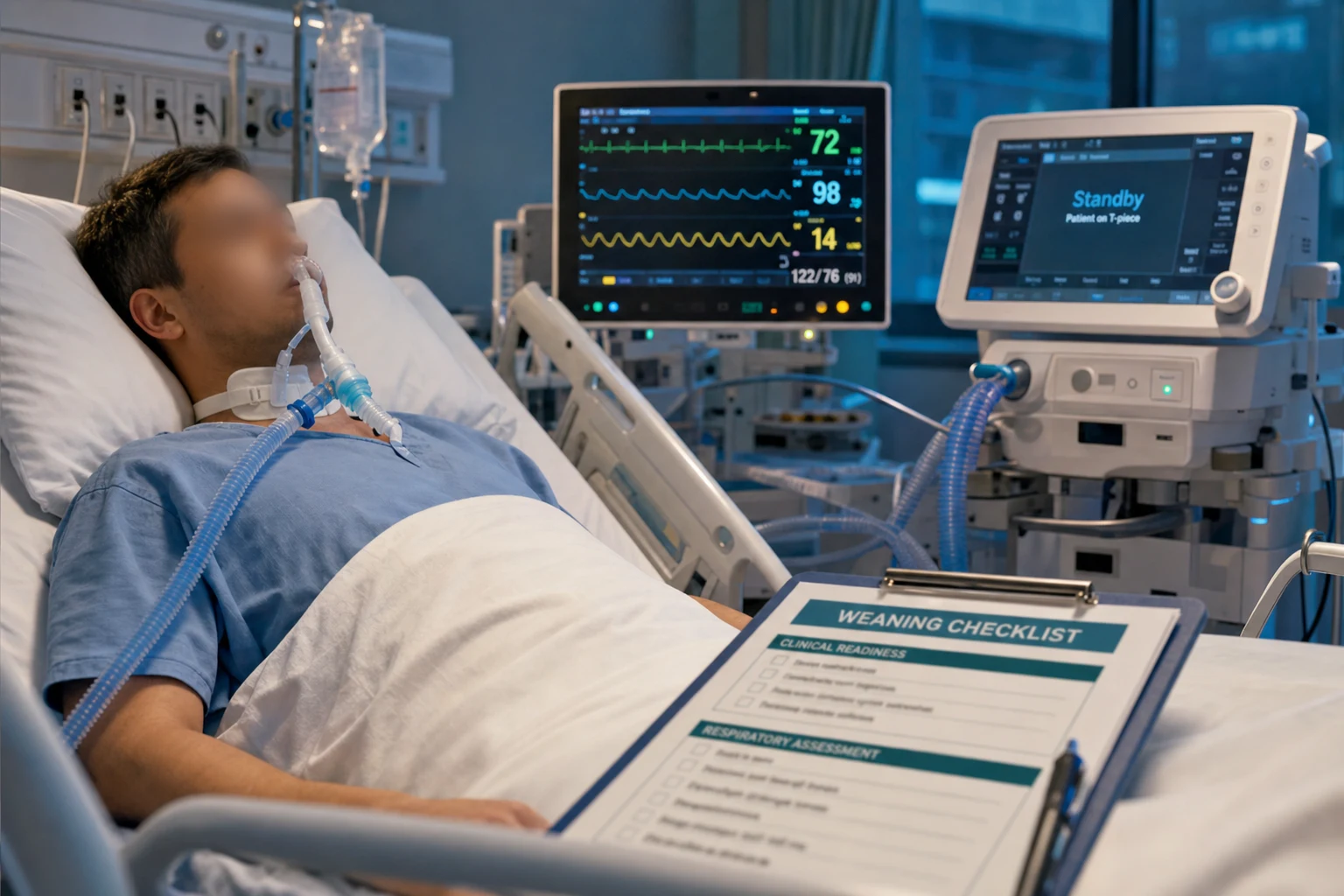
In the mechanically ventilated patient the bronchoscope sits inside the ETT and occupies a significant fraction of its cross-sectional area, which has two consequences the intensivist must actively manage.[1][2]
- Increase FiO2 to 1.0 (100%) before and during the procedure. Hypoxaemia is the commonest complication; the scope obstructs flow and the lavage fluid floods alveoli. Pre-oxygenase and keep FiO2 at 1.0 throughout, returning to baseline only after the scope is out and SpO2 has recovered.
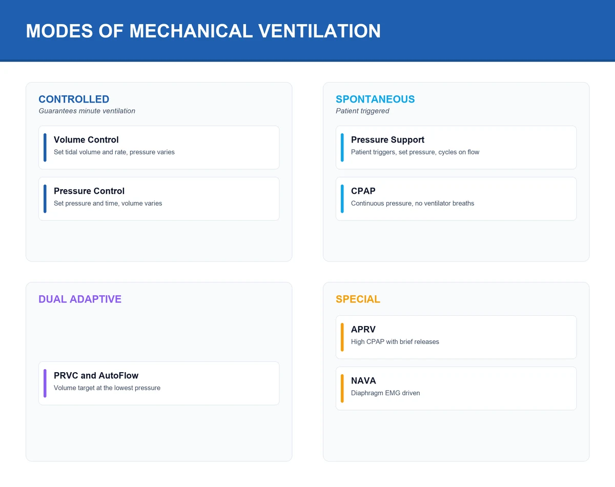
- Reduce tidal volume by ~50%. Because exhaled tidal volume falls (the scope takes up space and increases resistance), the ventilator will under-deliver effective ventilation and peak airway pressure rises. Set VT to roughly half the baseline, watch the peak pressure, and confirm that exhaled VT is being delivered. Some clinicians switch from volume-control to pressure-control for the procedure.
- Adjust PEEP. The BTS guideline and ICU practice support reducing PEEP during insertion (to limit air leak around the scope and reduce barotrauma), then titrating back up — a higher PEEP may be needed to maintain oxygenation during lavage. Watch for auto-PEEP (air-trapping) if the scope partially obstructs expiration.
- Use a swivel adapter with a self-sealing bronchoscopy port to maintain the circuit and minimise leak.[1]
Bronchoalveolar lavage (BAL) technique
BAL samples the cells and microbes of the distal airways and alveoli. Done well it has a high diagnostic yield for infection and cytology; done badly it returns only bronchial washings (lower yield). The technique standardised by the BTS guideline is summarised below.[1]
BAL technique — wedge, instil, recover
Step 1 — Choose the segment and wedge the scope
Advance the bronchoscope into the target subsegmental bronchus (in diffuse disease, the right middle lobe or lingula recover best; in localised disease, the radiologically abnormal area) until it wedges snugly, sealing the airway so that instilled fluid cannot reflux back into the proximal bronchus. A proper wedge is the single step that determines yield — an unwedged scope returns only bronchial washings.
Step 2 — Instil sterile saline in aliquots
Instil STERILE, NON-BACTERICIOSTATIC 0.9% saline at room temperature in aliquots — typically 3-5 aliquots of 20-50 mL each, for a total instilled volume of 100-200 mL. Inject through the working channel, then immediately apply gentle suction to recover each aliquot before instilling the next.
Step 3 — Recover and pool the return
Recover the lavage by gentle hand-suction into a sterile specimen trap (vigorous suction collapses the airway and reduces return). Typical return is 40-60% of instilled volume; a return of at least 5% (and ideally much more — 30-50 mL minimum) is required for valid culture and cytology. If return is poor, re-wedge or choose another segment.
Step 4 — Send the right tests
Send promptly to the lab (microbiology within 1-2 h; cytology can be refrigerated). Microbiology: Gram stain and culture, quantitative culture (greater than 10^4 CFU/mL significant for VAP), viral PCR (influenza, RSV, SARS-CoV-2, CMV), fungal culture and galactomannan (Aspergillus), AFB and TB-PCR, stains and PCR for Pneumocystis. Cytology: cell count and differential (eosinophils, neutrophils, lymphocytes), malignant cells, foamy macrophages (alveolar proteinosis), haemosiderin-laden macrophages (diffuse alveolar haemorrhage).
Step 5 — Return ventilator settings to baseline
Withdraw the scope, return FiO2 and tidal volume to baseline, and monitor SpO2 for 30-60 min (oxygenation often dips transiently after lavage as fluid is absorbed). Document the procedure, the segments lavaged, the return volume, and the samples sent.
BAL versus protected specimen brush (PSB)
| Feature | BAL | PSB (protected specimen brush) |
|---|---|---|
| Volume sampled | Large (millilitres of epithelial lining fluid) | Small (brush tip) |
| Sensitivity for VAP | High; quantitative threshold >10^4 CFU/mL | High; threshold >10^3 CFU/mL |
| Contamination risk | Moderate (passing upper airway) | Low (protected double-sheath catheter) |
| Use | Broader — microbiology + cytology | Narrower — quantitative bacteriology only |
Quantitative thresholds for VAP
Quantitative culture thresholds
Complications
Bronchoscopy is safe when performed with monitoring and oxygenation, but the complication list is fixed and examinable. The BTS guideline and the AQuIRE Registry provide the rates.[1][3]
Hypoxaemia (#1)
20-30% of procedures transiently
- Scope partially obstructs airway + BAL lavage fluid floods alveoli
- Prevent: FiO2 1.0, pause if SpO2 <88%, limit lavage volume
- More likely with baseline hypoxaemia, single-lung ventilation, PEEP dependence
Bleeding
After biopsy
- Transbronchial biopsy 5-10% clinically significant; endobronchial biopsy less
- Prevent: check platelets >50 and INR <1.5 before biopsy; withhold anticoagulants/antiplatelets
- Manage with ice-cold saline, topical adrenaline 1:20,000, balloon tamponade; position bleeding side down
Pneumothorax
Transbronchial biopsy
- 2-5% after transbronchial biopsy (forceps reaches the pleura)
- MANDATORY post-procedure chest X-ray after transbronchial biopsy
- Not needed after diagnostic BAL alone
Laryngospasm / bronchospasm
Scope irritation
- From scope contact with the larynx or bronchial tree
- Higher in asthmatics — pre-treat with a bronchodilator and ensure adequate topical anaesthesia
- Manage by withdrawing the scope, deepening anaesthesia, giving bronchodilators, and 100% oxygen
Arrhythmia
Hypoxia, vagal, sedation
- Atrial and ventricular ectopy, rarely sustained arrhythmia or arrest
- Driven by hypoxaemia, vagal stimulation (coughing/gagging), and sedation
- Monitor ECG continuously; correct hypoxia first
Bacteremia / fever
Cytokine release and transient bacteraemia
- Post-BAL fever is common and usually self-limiting (cytokine release from lavage)
- Transient bacteraemia occurs after bronchoscopy, especially with APC; routine prophylactic antibiotics are NOT needed for most patients (BTS)
- Rare cases of meningitis and endocarditis are described — antibiotic prophylaxis only for high-risk cardiac lesions if indicated
Coagulation requirements before biopsy
Transbronchial biopsy penetrates bronchial mucosa and may breach a small vessel (bleeding) or reach the visceral pleura (pneumothorax). Check and correct before biopsy:[1][6]
- Platelets greater than 50 x 10^9/L — give a platelet transfusion if lower.
- INR less than 1.5 — give fresh-frozen plasma or vitamin K.
- Uraemia (creatinine greater than 180 micromol/L) increases bleeding from platelet dysfunction — consider desmopressin (DDAVP) 0.3 mcg/kg.
- Withhold anticoagulants and reverse as needed (vitamin K for warfarin; idarucizumab for dabigatran; andexanet for apixaban/rivaroxaban; protamine for heparin). Hold antiplatelets where feasible. [1]
Specific scenarios
Mucus plug and atelectasis
A mucus plug occluding a lobar or main bronchus causes rapid collapse of the dependent lung and is one of the clearest urgent indications for ICU bronchoscopy. Typical settings: the post-operative patient (splinting, poor cough), the immobilised patient, the asthmatic with tenacious secretions, and the patient with haemoptysis and a blood clot.[1][2]
The procedure: identify the plug, fragment it with suction or forceps, lavage with saline to loosen it, and remove it piecemeal. Re-expansion is often immediate, with visible improvement in oxygenation and on chest X-ray. Bronchoscopy is indicated when lobar or whole-lung collapse has not resolved with physiotherapy, positioning, and suctioning.[2]
Haemoptysis
In haemoptysis the bronchoscope localises the bleeding and provides temporising control while definitive therapy (bronchial artery embolisation or surgery) is organised. Key manoeuvres (see the massive-haemoptysis topic for the full cascade): position the bleeding side down, use a large (8 mm+) ETT, apply ice-cold saline lavage and topical adrenaline 1:20,000, and place a balloon blocker for tamponade. Rigid bronchoscopy is preferred for massive bleeding — larger suction and access for tamponade. APC controls a visible bleeding point but is itself associated with a measurable rate of transient bacteraemia.[4][7]
Foreign body aspiration
Suspect aspiration in any patient with sudden choking, refractory wheeze or collapse, especially children, the elderly, alcoholics, and those with impaired swallow. The radiograph is often normal (organic foreign bodies are radiolucent). Bronchoscopy is both diagnostic and therapeutic: extract with forceps, a Dormia basket, or a snare. Rigid bronchoscopy is preferred in children (better airway control, larger instruments) and for large or friable objects; flexible bronchoscopy via ETT suffices for many adult cases.[5]
The immunocompromised host
In the immunocompromised ICU patient with pulmonary infiltrates, BAL has a high diagnostic yield and can direct specific therapy, avoiding empirical over-treatment. Send a broad panel: bacterial stains and culture, viral PCR (CMV, respiratory viruses), fungal culture and galactomannan (Aspergillus), AFB and TB-PCR, and stains/PCR for Pneumocystis jirovecii.[10]
Pneumocystis jirovecii
HIV / immunosuppression
- BAL sensitivity ~90-95% (higher than induced sputum)
- Send: methenamine silver stain, immunofluorescence, and PCR
- Cytology: foamy alveolar exudate
Invasive aspergillosis
Neutropenia / transplant
- BAL galactomannan (optical index >1.0) plus PCR; combine with serum galactomannan
- Cytology/histology: septate acute-angle hyphae
CMV pneumonitis
Transplant / HIV
- BAL CMV PCR; cytology for owl-eye inclusions
- Distinguish active disease from shedding using quantitation and clinical context
Disseminated TB / NTM
Any immunosuppressed host
- BAL AFB stain and culture, TB-PCR (Xpert MTB/RIF) with rapid rifampicin resistance
Transbronchial and cryobiopsy
Transbronchial lung biopsy (TBBx) samples parenchyma and is useful for interstitial disease and disseminated infection in the immunocompromised. A meta-analysis of TBBx in the ICU confirmed a useful diagnostic yield with a manageable complication profile (pneumothorax and bleeding as the main risks).[6] Transbronchial cryobiopsy uses a cryoprobe to freeze and avulse a larger sample of lung tissue — better for interstitial disease diagnosis than forceps TBBx — but carries a higher rate of bleeding and pneumothorax and is not routine; it is performed in selected centres.[9]
EBUS-TBNA and EUS
Endobronchial ultrasound with transbronchial needle aspiration (EBUS-TBNA) samples mediastinal and hilar lymph nodes through a specialised scope with a curvilinear ultrasound probe at the tip. Indications: lung-cancer staging, and diagnosing sarcoidosis, TB, and lymphoma. It is usually performed by a respiratory physician rather than the intensivist, but the ICU candidate should recognise when it adds value — particularly for unexplained mediastinal lymphadenopathy.[1]
Contraindications
There are few absolute contraindications; most are relative and centre on the ability to oxygenate and the bleeding risk.[1][2]
- Absolute — inability to maintain oxygenation adequately even with FiO2 at 1.0 (e.g. severe ARDS on high PEEP where removal of the scope is unsafe); lack of trained operator and resuscitation equipment.
- Relative (biopsy) — severe coagulopathy (INR greater than 1.5, platelets less than 50) that cannot be corrected; uraemia.
- Relative (procedure) — unstable haemodynamics, recent myocardial infarction (less than 48 h) or unstable arrhythmia, severe bronchospasm.
- Relative (BAL) — severe hypoxaemia on high FiO2 in which the lavage-induced desaturation would be dangerous — weigh the diagnostic gain against the risk. [1]
Key numbers to remember
Bronchoscopy in numbers
Risk stratification by indication
Therapeutic for obstruction
Therapeutic bronchoscopy for malignant central airway obstruction (stent/laser/dilation) carries airway-fire, bleeding, and post-obstruction-pulmonary-oedema risk; performed by interventional pulmonology with rigid-bronchoscopy backup. The AQuIRE Registry documents substantial palliative benefit despite the complexity.
Evidence and landmark references
Du Rand 2013 — BTS guideline for diagnostic flexible bronchoscopy
Thorax 2013
National evidence-based guideline (accredited by NICE) on diagnostic flexible bronchoscopy in adults
Key finding
Codified indications, consent, monitoring, sedation, and complication thresholds; confirmed the safety of bronchoscopy in ventilated patients with FiO2 1.0 and a swivel adapter, and set lidocaine ceiling at 8.2 mg/kg
Practice change
The operative standard for technique, sedation, and complications in adult bronchoscopy
Patolia 2021 — Bronchoscopy in ICU patients with respiratory failure
J Thorac Dis 2021
Comprehensive review of flexible bronchoscopy in intubated and non-intubated ICU patients
Key finding
Diagnostic yield and safety across indications (VAP, atelectasis, haemoptysis, foreign body, immunocompromised); ventilated patients require FiO2 1.0, reduced tidal volume, and a swivel adapter
Practice change
Defined the modern ICU bronchoscopy workflow and the expanding role of the intensivist
Ost/AQuIRE 2016 — Diagnostic yield and complications for peripheral lesions
Am J Respir Crit Care Med 2016
Multicentre registry (AQuIRE) of bronchoscopy for peripheral lung lesions
Key finding
Quantified diagnostic yield and the rates of pneumothorax (requiring chest tube ~4%) and significant bleeding; identified predictors of complications (degree of sedation, combined procedures, smaller airway)
Practice change
Risk-stratified peripheral-lesion bronchoscopy and informed consent
Ost/AQuIRE 2015 — Therapeutic bronchoscopy for malignant central airway obstruction
Chest 2015
Multicentre registry of therapeutic bronchoscopy (stent/laser/dilation) for malignant central airway obstruction
Key finding
High technical success in restoring airway patency with substantial improvement in dyspnoea and quality of life, even in advanced disease
Practice change
Established therapeutic bronchoscopy as an effective palliative intervention in malignant airway obstruction
Helgeson 2021 — Transbronchial biopsy in the ICU: systematic review
J Bronchol Interv Pulmonol 2021
Systematic review and meta-analysis of transbronchial forceps biopsy in the ICU
Key finding
Useful diagnostic yield for diffuse parenchymal disease with pneumothorax and bleeding as the main complications, manageable with standard precautions
Practice change
Quantified the yield and safety of TBBx in critically ill patients
Matveychuk 2014 — Bacteraemia after bronchoscopy with APC
Lung 2014
Prospective study of the incidence of bacteraemia following bronchoscopy with argon plasma coagulation
Key finding
Measurable rate of transient bacteraemia after APC, supporting the BTS position against routine prophylactic antibiotics but flagging the need for caution in high-risk cardiac patients
Practice change
Informed the antibiotic-prophylaxis policy for bronchoscopy
SAQ — Bronchoscopy in the ventilated ICU patient with suspected VAP
10 minutes · 10 marks
A 68-year-old man is intubated and ventilated (8.0 mm ETT, FiO2 0.5, PEEP 8) for severe CAP. On day 7 his FiO2 requirements rise from 0.4 to 0.6, purulent secretions increase, and a new right lower lobe infiltrate appears with a temperature of 38.9°C. The team asks you to perform a diagnostic bronchoscopy with bronchoalveolar lavage. Outline your preparation, technique, and interpretation.
SAQ — Massive haemoptysis: bronchoscopic control
10 minutes · 10 marks
A 55-year-old man with known bronchiectasis coughs up approximately 400 mL of bright red blood over 30 minutes and is dyspnoeic with SpO2 90% on 15 L/min via non-rebreather. He is awake, tachycardic (HR 118, BP 96/60). The examiners ask how you would use bronchoscopy as part of the immediate management of massive haemoptysis.
Clinical pearls
Red flags
References
- [1]Du Rand IA, Blaikley J, Booton R, Chaudhuri N, Gupta V, Khalid S, et al. British Thoracic Society guideline for diagnostic flexible bronchoscopy in adults: accredited by NICE Thorax, 2013.PMID 23860341
- [2]Patolia S, Farhat R, Subramaniyam R, et al. Bronchoscopy in intubated and non-intubated intensive care unit patients with respiratory failure J Thorac Dis, 2021.PMID 34527353
- [3]Ost DE, Ernst A, Lei X, Kovitz KL, Benzaquen S, Diaz-Mendoza J, et al. Diagnostic Yield and Complications of Bronchoscopy for Peripheral Lung Lesions. Results of the AQuIRE Registry Am J Respir Crit Care Med, 2016.PMID 26367186
- [4]Ost DE, Ernst A, Grosu HB, Lei X, Diaz-Mendoza J, Slade M, et al. Therapeutic bronchoscopy for malignant central airway obstruction: success rates and impact on dyspnea and quality of life Chest, 2015.PMID 25358019
- [5]Bajaj D, Sachdeva A, Deepak D. Foreign body aspiration J Thorac Dis, 2021.PMID 34527356
- [6]Helgeson SA, Imam JS, Burnside RC, Fernandez-Bussy S, Brigham TJ, Patel NM. Transbronchial Forceps Biopsy in the Intensive Care Unit: A Systematic Review and Meta-analysis J Bronchology Interv Pulmonol, 2021.PMID 33758151
- [7]Matveychuk A, Guber A, Talker O, Shitrit D. Incidence of bacteremia following bronchoscopy with argon plasma coagulation: a prospective study Lung, 2014.PMID 24770443
- [8]Alexander WJ, Baker GL, Hunker FD. Bacteremia and meningitis following fiberoptic bronchoscopy Arch Intern Med, 1979.PMID 443952
- [9]Rodrigues I, Estevao Gomes R, Coutinho LM, Rego MT, Machado F, Morais A. Diagnostic yield and safety of transbronchial lung cryobiopsy and surgical lung biopsy in interstitial lung diseases: a systematic review and meta-analysis Eur Respir Rev, 2022.PMID 36198419
- [10]Azoulay E, Russell L, Van de Louw A, Metaxa V, Bauer P, Povoa P, et al. Diagnosis of severe respiratory infections in immunocompromised patients Intensive Care Med, 2020.PMID 32034433
- [11]Solidoro P, Corbetta L, Patrucco F, Sorbello M, Piccioni F, D'amato L. Competences in bronchoscopy for Intensive Care Unit, anesthesiology, thoracic surgery and lung transplantation Panminerva Med, 2019.PMID 30394713
- [12]Weerawarna T, Mishra R, Tantray S, Bharti M, Mehta A, Bilaceroglu S. Flexible Bronchoscopy in the Intensive Care Unit: Controversies, Clinical Applications, and the Expanding Role of Intensivists J Clin Med, 2026.PMID 42355736