ICU · Respiratory
Acute severe community-acquired pneumonia: pulmonary complications and ARDS
Also known as CAP complicated by ARDS · Pneumonia-induced ARDS · Pulmonary ARDS vs non-pulmonary ARDS
Severe CAP can progress to ARDS (acute respiratory distress syndrome) — direct lung injury from pulmonary infection. Pulmonary ARDS (from pneumonia, aspiration) vs non-pulmonary ARDS (from sepsis, trauma, pancreatitis): pulmonary ARDS has more consolidation (less recruitable lung), worse response to PEEP, and may benefit less from some standard ARDS therapies. Diagnosis: Berlin definition (timing <1 week, bilateral infiltrates, not fully explained by cardiac failure, PaO2/FiO2 <300). Management: lung-protective ventilation (VT 6 mL/kg, plateau <30, driving pressure <15), higher PEEP if recruitable, prone positioning (PaO2/FiO2 <150), conservative fluid strategy, treat underlying pneumonia. Pulmonary ARDS may have higher mortality than non-pulmonary ARDS.
On this page & tools
Your progress
Saved locally on this device.
Target exams
Red flags
Severe community-acquired pneumonia (CAP) is the single commonest cause of ARDS worldwide. The same pneumonia that fills alveoli with inflammatory exudate can, in a subset of patients, trigger the runaway diffuse alveolar damage (DAD) cascade that defines ARDS — and beyond the lung, bacteraemic CAP seeds the pleura (effusion/empyema), destroys parenchyma (abscess), disseminates (endocarditis, meningitis, septic arthritis) and drives systemic organ failure (AKI, DIC). This topic follows that chain: how severe pneumonia becomes ARDS, how to ventilate it, and how to recognise and manage the complications that determine who survives. [1]
Pathophysiology: from severe pneumonia to ARDS
Severe CAP progresses to ARDS through a stereotyped cascade of direct alveolar injury. The sequence — alveolar macrophage activation → neutrophil influx → diffuse alveolar damage (DAD) → non-cardiogenic pulmonary oedema — is the histological and physiological signature of pulmonary ARDS, and explains both its (predominantly consolidative) imaging pattern and its (relatively poor) response to high PEEP. Pneumonia is the archetypal direct/pulmonary insult, in contrast to the indirect/non-pulmonary injury of extrathoracic sepsis, which produces oedema that is more recruitable. [1]
Pulmonary ARDS pathophysiology cascade
1. Inoculation and alveolar colonisation
Pathogen (S. pneumoniae, S. aureus incl. PVL-producing, Legionella, Gram-negatives, influenza/COVID/SARS viruses, atypicals) reaches the alveolus via micro-aspiration or inhalation. Pneumococcal cell-wall components, viral replication, and toxin production (PVL, pneumolysin) trigger the innate immune response.
2. Alveolar macrophage and epithelial activation
Alveolar macrophages release TNF-α, IL-1β, IL-6, IL-8 (neutrophil chemoattractant) and activate alveolar epithelial cells. IL-6 drives the systemic inflammatory response (CRP, fever) and predicts ARDS development. Type I pneumocyte injury exposes the basement membrane and disables the alveolar-capillary barrier.
3. Massive neutrophil influx and alveolitis
Neutrophils transmigrate into the alveolar space, releasing reactive oxygen species, proteases (elastase, MMPs), neutrophil extracellular traps (NETs) and leukotrienes. This unchecked inflammatory cascade injures both epithelium and endothelium — the histological hallmark of diffuse alveolar damage (DAD).
4. Diffuse alveolar damage (DAD) — three phases
EXUDATIVE (days 1-6): hyaline membranes (proteinaceous debris + necrotic type I pneumocytes), alveolar flooding. PROLIFERATIVE (days 7-14): type II pneumocyte proliferation, fibroblast migration, early organisation. FIBROTIC (>2 weeks): interstitial and intra-alveolar fibrosis — worse outcome, the substrate of "late/fibrotic" ARDS.
5. Non-cardiogenic pulmonary oedema
Endothelial injury increases alveolar-capillary permeability → protein-rich oedama floods the alveoli. Unlike cardiogenic oedema (low-protein, hydrostatic), this is a HIGH-protein exudate. PAOP is normal (<18). The oedema inactivates surfactant → atelectasis, reduced compliance, shunt.
6. Physiological consequences (the "baby lung")
Aerated, recruitable lung is drastically reduced ("baby lung", not stiff lung). Shunt and low V/Q → severe, often refractory hypoxaemia. Reduced compliance → high work of breathing. Pulmonary hypertension (hypoxia + vasoconstriction + microthrombi). Dead space rises in late disease. The lung is SMALL and HETEROGENEOUS — the rationale for low Vt and prone positioning.
Exudative phase
Days 1-6
- Hyaline membranes line alveoli (protein-rich exudate + necrotic type I pneumocytes)
- Interstitial and alveolar oedema, dense neutrophil infiltrate
- Surfactant inactivation, atelectasis, reduced compliance
- Clinical: acute hypoxaemia, bilateral infiltrates, shunt
Proliferative phase
Days 7-14
- Type II pneumocyte proliferation (attempts at re-epithelialisation)
- Fibroblast proliferation and collagen deposition begins
- Resolution of hyaline membranes, partial clearing of oedema
- Clinical: oxygenation may improve OR organisation/fibrosis may begin
Fibrotic phase
>2 weeks
- Interstitial and intra-alveolar fibrosis, microcystic change
- Pulmonary vascular remodelling, pulmonary hypertension
- Markedly reduced compliance, high dead space, hypercapnia
- Clinical: prolonged ventilation, worse mortality, "unresolving ARDS"
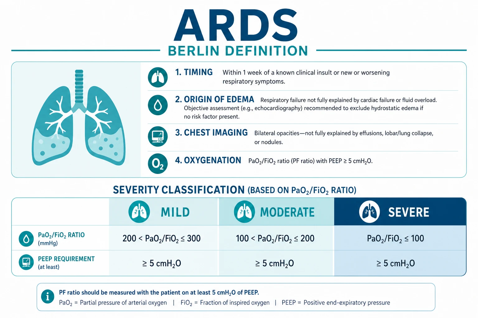
Diagnosis: the Berlin definition (2012)
Timing
Within 1 week
- Within 1 week of a known clinical insult OR new/worsening respiratory symptoms
- Chronic fibrotic lung disease or subacute processes (>1 week) are NOT ARDS
- CAP-ARDS usually declares within 24-72 h of pneumonia onset
Imaging
Bilateral
- Bilateral opacities on chest X-ray OR CT
- NOT fully explained by effusions, lobar/lung collapse, or nodules
- CT more sensitive than CXR; beware atelectasis mimicking oedema
- Unilateral dense pneumonia is NOT Berlin ARDS (but may still need protective ventilation)
Origin of oedema
Non-cardiogenic
- Respiratory failure NOT fully explained by cardiac failure or fluid overload
- Objective assessment (echocardiography) required if no obvious ARDS risk factor
- Cardiac failure and ARDS can COEXIST (e.g., sepsis with cardiomyopathy)
- PAOP >18 is no longer required (Berlin dropped the Swan requirement)
Oxygenation
P/F on PEEP >=5
- Mild: PaO2/FiO2 200-300
- Moderate: PaO2/FiO2 100-200
- Severe: PaO2/FiO2 <100
- P/F MUST be interpreted WITH the PEEP and FiO2 — always quote both
Pulmonary vs non-pulmonary ARDS
Pulmonary ARDS
Direct lung injury
- Causes: pneumonia (#1), aspiration, inhalation injury, near-drowning, lung contusion
- Morphology: more CONSOLIDATION (alveolar filling with inflammatory exudate) — less recruitable lung
- PEEP response: may be POOR (consolidated alveoli cannot be recruited — high PEEP overdistends remaining healthy lung)
- CT: focal pattern (dependent consolidation) — high PEEP worsens overdistension of non-dependent lung
- Mortality: may be higher than non-pulmonary ARDS (LUNG SAFE study)
Non-pulmonary ARDS
Indirect lung injury
- Causes: sepsis (#1 from non-pulmonary source), trauma, pancreatitis, transfusion (TRALI), burns, drug reaction
- Morphology: more INTERSTITIAL oedema (leaky capillaries) — more recruitable lung
- PEEP response: may be BETTER (oedematous alveoli can be recruited with PEEP)
- CT: non-focal pattern (diffuse bilateral infiltrates) — may tolerate higher PEEP
- Mortality: may be lower than pulmonary ARDS
Management
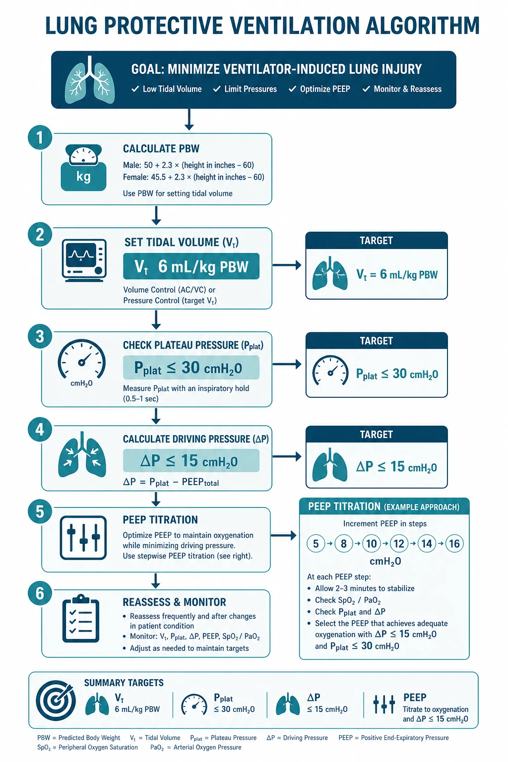
CAP with ARDS management
Confirm ARDS diagnosis
Berlin criteria: (1) Timing: within 1 week of known clinical insult or new/worsening respiratory symptoms. (2) Chest imaging: bilateral opacities not fully explained by effusions, lobar/lung collapse, or nodules. (3) Origin of oedema: respiratory failure not fully explained by cardiac failure or fluid overload (objective assessment — echo). (4) Oxygenation: mild (PaO2/FiO2 200-300 with PEEP >=5), moderate (100-200), severe (<100).
Lung-protective ventilation
VT 6 mL/kg predicted body weight (reduces VILI). Plateau pressure <30 cmH2O. Driving pressure <15 cmH2O (Amato: strongest predictor). PEEP: titrate based on recruitability — pulmonary ARDS may tolerate lower PEEP (consolidation not recruitable). Higher PEEP strategies (ALVEOLI, EXPRESS trials): no overall benefit but may help hyperinflammatory phenotype.
Prone positioning (PaO2/FiO2 <150)
PROSEVA trial (NEJM 2013): 16h/day prone for moderate-severe ARDS reduced 28-day mortality (16% vs 32.8%). Benefits: (1) Recruits dependent lung regions. (2) Reduces shunt. (3) More homogeneous ventilation. (4) Reduces VILI. For pulmonary ARDS (pneumonia): particularly beneficial (consolidated dependent lung is recruited by prone). Contraindications: spinal instability, recent abdominal surgery, pregnancy.
Conservative fluid strategy
FACTT trial: conservative fluid (target CVP <4) improved oxygenation and ventilator-free days. CAUTION: in septic shock (common with CAP-ARDS): balance fluid restriction against need for perfusion. Use dynamic monitoring (PLR, SVV) + vasopressors (noradrenaline) to minimise fluid while maintaining MAP >65. Lactate clearance guides adequacy.
Treat underlying pneumonia
Appropriate antibiotics (within 1h for septic shock). De-escalate at 48-72h based on cultures. Source control (drain pleural effusion/empyema). Antivirals if viral cause (oseltamivir for influenza, remdesivir for COVID-19). Corticosteroids: hydrocortisone 200 mg/day for severe CAP with ARDS (CAPE COD: reduced mortality).
Consider advanced therapies
VV-ECMO: for refractory ARDS (PaO2/FiO2 <80 despite optimised ventilation). CESAR trial: transfer to ECMO centre improved survival. Neuromuscular blockade: cisatracurium 48h for severe hypoxaemia (ACURASYS: improved outcomes; ROSE: no benefit — selective use). Inhaled pulmonary vasodilators (nitric oxide, epoprostenol): improve oxygenation temporarily — no mortality benefit. Bridge to recovery.
Ventilation targets and predicted body weight
The lung-protective strategy is titrated to predicted (ideal) body weight (PBW), not actual weight — ARDSNet PBW formulae: [1]
Lung-protective ventilation targets
- Male PBW = 50 + 0.91 × (height cm − 152.4)
- Female PBW = 45.5 + 0.91 × (height cm − 152.4) [1]
If plateau pressure >30 or driving pressure >15, reduce VT by 1 mL/kg (down to 4 mL/kg) before reducing PEEP. If raising PEEP increases the driving pressure, the lung is being overdistended — reduce PEEP. This is the bedside test of recruitability. [1]
PEEP optimisation
PEEP is the great double-edged sword of ARDS ventilation: it recruits oedematous alveoli (improving oxygenation and reducing shunt) but overdistends already-open lung (worsening VILI). In pulmonary ARDS the consolidated, dependent lung is poorly recruitable, so the same "high PEEP" strategy that helps non-pulmonary ARDS can cause harm. [1]
Lower PEEP strategy
Preferred in pulmonary/focal ARDS
- Rationale: consolidated alveoli are NOT recruitable — high PEEP overdistends the remaining healthy non-dependent lung
- ALVEOLI, EXPRESS, LOVS, EPVent trials: higher PEEP = NO overall mortality benefit
- Best for focal CT pattern (pulmonary ARDS, pneumonia): much consolidated lung is dead
- Set PEEP just above the lower inflection point of the pressure-volume curve, or use a low PEEP/FiO2 table
- If a PEEP rise increases ΔP, the lung is being overdistended — REDUCE PEEP
Higher PEEP strategy
May benefit diffuse/recruitable ARDS
- Rationale: oedematous alveoli ARE recruitable — PEEP keeps them open, reduces shunt, improves oxygenation
- Meta-analysis (Briel 2010): higher PEEP reduced mortality ONLY in moderate-severe ARDS (PaO2/FiO2 <200)
- Best for non-focal/diffuse CT pattern (non-pulmonary ARDS, sepsis, TRALI)
- Best PEEP identified by decremental PEEP trial; oesophageal-pressure-guided (PREVENT) — no benefit
- Caution: high PEEP in focal pulmonary ARDS worsens overdistension and reduces venous return (hypotension)
Landmark trials — what they showed
ARMA — ventilation with lower tidal volumes
Multicentre RCT (n=861)
Population: ALI/ARDS, PEEP >=5
Key finding
Lower VT reduced mortality (31% vs 39.8%, p=0.007) and increased ventilator-free days.
Practice change
Lung-protective ventilation (VT 6 mL/kg PBW) is the single best-supported mortality-reducing intervention in ARDS. Apply to EVERY ARDS patient from intubation.
PROSEVA — prone positioning in severe ARDS
Multicentre RCT (n=466)
Population: Moderate-severe ARDS, PaO2/FiO2 <150, FiO2 >=60%, PEEP >=5
Key finding
28-day mortality 16.0% vs 32.8% (HR 0.39, p<0.001); 90-day mortality 23.6% vs 41.0%. NNT ~6.
Practice change
Early, prolonged (>=16 h) prone positioning reduces mortality in moderate-severe ARDS. The ONLY ventilation adjunct besides low VT proven to reduce mortality.
FACTT — conservative vs liberal fluid management
Multicentre RCT (n=1000)
Population: ALI/ARDS
Key finding
No mortality difference, BUT conservative strategy improved oxygenation, increased ventilator-free days (14.6 vs 12.1) and ICU-free days, with NO increase in renal failure.
Practice change
A conservative fluid strategy improves lung outcomes without harm. Use in CAP-ARDS once shock has resolved; balance against ongoing septic shock resuscitation.
EOLIA — VV-ECMO in very severe ARDS
Multicentre RCT (n=249, stopped early)
Population: Very severe ARDS (PaO2/FiO2 <50 >3h, <80 >6h, or pH <7.25 with PaCO2 >=60)
Key finding
35% vs 46% (RR 0.76) — NOT statistically significant on primary ITT (p=0.09), but Bayesian reanalysis strongly favours ECMO; 28% crossover to rescue ECMO diluted the effect.
Practice change
VV-ECMO is a reasonable rescue for refractory severe ARDS (PaO2/FiO2 <80 despite optimised ventilation and proning). Refer EARLY to an ECMO centre — outcomes worsen with prolonged ventilation before cannulation.
ACURASYS vs ROSE — early neuromuscular blockade
Two multicentre RCTs (ACURASYS n=340; ROSE n=1006)
Population: Severe ARDS (PaO2/FiO2 <150)
Key finding
ACURASYS: 31.6% vs 40.7% (adjusted HR 0.68, p=0.049). ROSE: NO benefit (42.5% vs 42.8%), more akinesia/ICS compression.
Practice change
Routine early cisatracurium is NOT beneficial (ROSE). Reserve for severe refractory hypoxaemia with dangerous dyssynchrony/high drive — short course, deep sedation mandatory.
ART — alveolar recruitment manoeuvres (HARM)
Multicentre RCT (n=1010)
Population: Moderate-severe ARDS
Key finding
INCREASED 28-day mortality (27.8% vs 22.7%, p=0.041) and 6-month mortality.
Practice change
Do NOT routinely use aggressive recruitment manoeuvres or maximal PEEP titration in ARDS. If an RM is used for rescue, keep it brief and modest (e.g., 40 cmH2O x 40 s) with full haemodynamic monitoring.
VV-ECMO selection
When to refer for VV-ECMO in CAP-ARDS
Refer EARLY — phone the retrieval/ECMO centre before the patient is moribund. Contraindications: irreversible comorbidity, advanced age/frailty, ventilation >7-10 days (relative), anticoagulation contraindicated with active bleeding, limited vascular access. Mortality in EOLIA-style severe ARDS with ECMO ≈ 35-40%. CESAR (2009) showed transfer to an ECMO-capable centre improved survival versus continued conventional ventilation. [1]
Pulmonary and systemic complications of severe CAP
ARDS is just ONE of the complications of severe CAP. Pneumonia is a systemic illness: the same bacteraemia and inflammation that drive ARDS also seed the pleura, destroy lung parenchyma, disseminate to distant sites, and injure kidneys and coagulation. Active surveillance for each of these is mandatory in any ventilated CAP patient. [1]
Parapneumonic effusion and empyema
~40-60% of bacterial CAP has an associated pleural effusion; progression to empyema (~5%) dramatically worsens outcome. The key decision is SIMPLE vs COMPLICATED — made on pleural fluid pH, biochemistry and appearance. [1]
Uncomplicated (simple)
Transudative, sterile
- Pathophysiology: sterile sympathetic pleural effusion from adjacent inflammation
- Pleural fluid: clear, pH >7.20, LDH <1000, glucose >2.2 mmol/L, Gram stain negative
- Management: antibiotics for pneumonia; effusion usually resolves — NO chest tube
Complicated parapneumonic
Exudative, loculating
- Pathophysiology: bacterial invasion of pleural space, fibrin deposition, loculation begins
- Pleural fluid: cloudy/pus-like, pH <7.20 (or LDH >1000, glucose <2.2), Gram stain may be positive, culture may be negative after antibiotics
- Management: CHEST TUBE (tube thoracostomy) + antibiotics; ultrasound/CT to guide placement
Empyema (frank)
Pus in pleural space
- Pathophysiology: frank pus; bacterial multiplication, thick loculations, peeling on lung surface (rind)
- Pleural fluid: frank pus (pH not needed if pus visible); culture positive in ~60%
- Management: chest tube + intrapleural tPA/DNase OR VATS; prolonged antibiotics 2-6 weeks; trapped lung (rind) may need decortication
Management of parapneumonic effusion/empyema
Detect — ultrasound the chest
Bedside ultrasound (or CT) on every ventilated CAP patient. Look for: size, loculation (septations), echogenicity (hyperechoic = pus/cellular). Diagnostic thoracentesis if effusion >10 mm on lateral decubitus/ultrasound.
Sample pleural fluid
Send for: pH (in blood-gas syringe, on ice), Gram stain + culture, protein, LDH, glucose, cell count. Light’s criteria confirm exudate. pH <7.2 = complicated — chest tube. NEVER send pH if fluid is frank pus (treat as empyema regardless).
Insert chest tube (ultrasound-guided)
For complicated effusion/empyema: large-bore (24-32Fr) for pus, smaller pigtail acceptable for non-purulent loculated fluid. Ultrasound-guided to avoid organ injury. Aim for complete drainage.
If not draining/loculated: intrapleural tPA/DNase
MIST2 (NEJM 2011): combination alteplase (tPA) 10 mg BD + DNase 5 mg BD for 3 days reduced drainage, length of stay and surgical referral (tPA alone or DNase alone did NOT help — only the combination). MIST1 (streptokinase) was NEGATIVE.
If still failing: surgical referral (VATS)
Failure of tube + tPA/DNase → video-assisted thoracoscopic surgery (VATS) for debridement and breakdown of loculations. Open drainage/thoracotomy + decortication for chronic organised empyema with trapped lung (rind). Microbiology-guided antibiotics 2-6 weeks.
MIST2 — intrapleural tPA/DNase for pleural infection
Multicentre RCT, 2x2 factorial (n=210)
Population: Pleural infection (empyema + complicated parapneumonic effusion)
Key finding
Combination tPA + DNase increased drainage and reduced CXR opacity, surgical referral (4% vs 18%) and length of stay. EITHER ALONE was ineffective (DNase alone trended to harm).
Practice change
For loculated parapneumonic effusion/empyema failing tube drainage, use COMBINATION intrapleural tPA 10 mg BD + DNase 5 mg BD for 3 days. Do not use either agent alone; do not use streptokinase (MIST1 negative).
Lung abscess
A localised collection of pus within destroyed lung parenchyma — most often from aspiration of oropharyngeal contents (mixed anaerobes), necrotising pneumonia (S. aureus, Klebsiella, Pseudomonas, type-3 pneumococcus), or septic emboli. Different from empyema: an abscess is IN the lung, an empyema is IN the pleural space — distinguishing them (CT with contrast, enhancing rim) matters because drainage differs. [1]
Lung abscess
Pus within lung parenchyma
- Causes: aspiration (mixed anaerobes, #1), necrotising pneumonia (S. aureus incl. PVL, Klebsiella, Pseudomonas, type-3 pneumococcus), septic emboli
- Imaging: cavity WITH air-fluid level inside lung parenchyma; thick irregular wall, enhances with contrast
- Microbiology: anaerobes (Peptostreptococcus, Fusobacterium, Prevotella) + aerobes; send sputum, blood cultures
- Management: PROLONGED antibiotics (4-6 weeks) IV then PO; drainage only if large (>6 cm), failing therapy, or immunocompromise
Empyema
Pus in pleural space
- Causes: parapneumonic (#1), post-operative, haematogenous, oesophageal rupture
- Imaging: collection in pleural space, pleural split-sign, may have air-fluid level if bronchopleural fistula
- Microbiology: streptococci, staph, anaerobes, Gram-negatives
- Management: chest tube drainage is PRIMARY therapy (antibiotics alone fail); tPA/DNase or VATS for loculation
Management of lung abscess
Confirm diagnosis
CT chest with contrast: thick-walled cavity with air-fluid level, surrounded by consolidation, often dependent lobe (posterior upper / superior lower — aspiration territory). Send sputum Gram stain/culture, blood cultures, consider bronchoscopy (BAL) if no organism.
Antibiotics — prolonged course
Empiric: amoxicillin-clavulanate OR clindamycin (anaerobic cover) + ceftriaxone. Add MRSA cover (vancomycin/linezolid) if S. aureus risk. Tailor to culture. Duration 4-6 weeks (IV→PO when afebrile and improving) — much longer than uncomplicated pneumonia.
Drainage if indicated
Indications: abscess >6 cm, clinical deterioration on antibiotics, immunocompromise, rupture into pleura (empyema/bronchopleural fistula). Options: percutaneous catheter drain (image-guided), endoscopic drainage. Surgery (resection) rarely — for failure of medical + percutaneous therapy.
Exclude bronchial obstruction
A persistent/central abscess in a non-dependent lobe or a patient >40 yrs → bronchoscopy to exclude endobronchial obstruction (tumour, foreign body). An obstructing cancer behind a "pneumonia" will not resolve until the obstruction is relieved.
Watch for complications
Rupture → pyopneumothorax/empyema (needs chest tube ± surgery). Massive haemoptysis (erosion into vessel — bronchial artery embolisation). Metastatic spread (brain abscess — especially with virulent organisms).
Metastatic infection (haematogenous spread)
Bacteraemic pneumonia — particularly pneumococcal, staphylococcal (incl. S. aureus from skin/IVDU) and Gram-negative — seeds distant sites. A deteriorating CAP patient with new focal signs must be investigated for metastatic foci; an unrecognised endocarditis or cerebral abscess is a common and lethal miss. [1]
Infective endocarditis
New murmur + bacteraemia
- Risk: S. aureus bacteraemia (up to ~25% have endocarditis), prosthetic valve, IVDU, structural heart disease
- Features: new/changed murmur, embolic phenomena (splinter, Janeway, Osler), heart failure, conduction block
- Diagnosis: transthoracic echo (TTE) → if negative and high suspicion, transoesophageal echo (TOE); modified Duke criteria
- Management: organism-targeted IV antibiotics 4-6 weeks, surgical referral if heart failure/large vegetation/fungal
Meningitis
Bacterial CNS seeding
- Risk: pneumococcal bacteraemia (especially if asplenia), Listeria (elderly/immunocompromised)
- Features: headache, meningism, altered mental status, seizures; may be masked by ICU sedation
- Diagnosis: urgent CT (if focal signs/low GCS) then LP; CSF Gram stain, culture, cell count, protein/glucose, pneumococcal antigen/PCR
- Management: empirical ceftriaxone + vancomycin + dexamethasone (before/with first antibiotic); add ampicillin if Listeria risk
Septic arthritis / osteomyelitis
Joint/bone seeding
- Risk: S. aureus, pneumococcus, Gram-negatives; IVDU, prosthetic joint, diabetes
- Features: hot swollen joint, back pain (vertebral osteomyelitis/epidural abscess), refusing to bear weight
- Diagnosis: joint aspirate/blood cultures; MRI for osteomyelitis/epidural abscess
- Management: surgical washout for septic arthritis; prolonged organism-targeted antibiotics 4-6 weeks
Other foci
Don't forget
- Brain abscess / cerebral empyema: headache, focal deficit, seizures — CT/MRI
- Endophthalmitis: visual change, red eye — ophthalmology emergency
- Splenic / hepatic abscess, psoas abscess: CT abdomen/pelvis
- Prosthetic device infection (pacemaker, joint, graft): remove if possible
Acute kidney injury (sepsis-associated AKI)
Sepsis-associated AKI (SA-AKI) occurs in ~50% of septic shock and is an independent mortality multiplier. In CAP-ARDS it interacts viciously with the lungs: fluid accumulation worsens oxygenation (FACTT), and AKI limits the conservative fluid strategy itself. RRT may be needed; fluid overload on a stiff lung is catastrophic. [1]
Management of sepsis-associated AKI in CAP-ARDS
Prevent — resuscitate to perfusion, not to a number
Sepsis-induced hypoperfusion: restore intravascular volume with crystalloid (30 mL/kg bolus per SSC), then use dynamic measures (passive leg raise, SVV, fluid challenge) to avoid over-resuscitation. Noradrenaline to MAP >=65. Lactate clearance >=10%/h or capillary refill <=3 s as targets. Avoid nephrotoxins (NSAIDs, aminoglycosides, contrast where possible).
Detect — KDIGO staging
Stage 1: Cr >=1.5-1.9x baseline OR >=26.5 µmol/L in 48h OR UO <0.5 mL/kg/h for 6-12h. Stage 2: 2-2.9x OR UO <0.5 for >=12h. Stage 3: 3x OR Cr >=353.6 OR RRT OR UO <0.3 for >=24h or anuria >=12h.
Manage — fluids, vasopressors, source control
Once euvolaemic, AVOID further fluid — use vasopressors not fluid to maintain MAP. Stress-dose hydrocortisone if refractory shock (CAPE COD: hydrocortisone 200 mg/day in severe CAP). Treat the pneumonia (source control: drain empyema, debride abscess). Albumin 25% for refractory shock if fluid needed.
Renal replacement therapy
Indications (AEIOU): Acidosis (refractory), Electrolytes (K+ refractory), Intoxications, Overload (fluid — common in ARDS), Uraemia. In haemodynamically unstable ICU patients use CRRT (CVVHDF) — gentler, allows ultrafiltration to support the conservative fluid strategy. Dose 20-25 mL/kg/h.
Avoid harm
No low-dose dopamine (no benefit, harm). No furosemide to "convert" oliguric to non-oliguric AKI (no outcome benefit; may delay RRT). Glycaemic control 140-180 mg/dL. Avoid hyperchloraemic fluids (use balanced crystalloids — SMART trial: less AKI).
CRRT (CVVHDF)
Continuous
- Best for haemodynamically unstable ICU patient (septic shock, ARDS)
- Gentle solute removal, allows controlled ultrafiltration → supports conservative fluid strategy
- Less fluid shift, less cerebral oedema; anticoagulation with citrate (or heparin)
- Runs 24/7; dose 20-25 mL/kg/h effluent
Intermittent HD
Standard
- Best for haemodynamically stable patient or rapid clearance needs (toxin, severe hyperkalaemia)
- Faster solute/fluid removal → intradialytic hypotension (dangerous in ARDS/shock)
- Delivered 3-4x/week, 4h sessions
- Risk of dialysis disequilibrium with very high urea
Disseminated intravascular coagulation (DIC)
Sepsis is the commonest cause of DIC; pneumococcal and meningococcal bacteraemia are classic triggers. DIC is a consumptive coagulopathy: simultaneous microvascular thrombosis (organ failure) AND bleeding (platelet/factor depletion). It is NOT a primary bleeding disorder — it is dysregulated haemostasis driven by tissue-factor release and cytokine-mediated endothelial injury. [1]
Diagnosis and management of sepsis-associated DIC
Recognise the laboratory pattern
Sepsis + (a) thrombocytopaenia OR falling platelet trend, (b) prolonged PT/aPTT, (c) raised D-dimer, (d) low fibrinogen (late). Bleeding AND/OR thrombosis (purpura fulminans, digital ischaemia, line clotting).
Score with ISTH overt-DIC
Score (0-8): platelets (>100=0, 50-100=1, <50=2), elevated D-dimer (no increase=0, moderate=2, strong=3), prolonged PT (<3s=0, 3-6s=1, >6s=2), fibrinogen (>1 g/L=0, <1 g/L=1). Score >=5 = overt DIC (repeat daily).
TREAT THE CAUSE — sepsis source control
DIC is a manifestation of uncontrolled sepsis: appropriate antibiotics within 1h, drainage of empyema/abscess, surgical debridement of necrotic tissue. Without source control, no amount of blood products will correct DIC.
Component replacement for bleeding/high risk
Active bleeding (or pre-procedure / platelet <20 / fibrinogen <1.5): transfuse platelets (target >50), FFP (target PT/aPTT <1.5x normal), cryoprecipitate (target fibrinogen >1.5 g/L). Treat the patient, not the numbers — prophylactic transfusion in non-bleeding DIC is not indicated.
Consider anticoagulation
Thrombotic phenotype (purpura fulminans, line clotting, microvascular thrombosis): therapeutic heparin/LMWH. Trials (SCARLET — recombinant thrombomodulin) were negative overall but selected patients (coagulopathy + organ failure) may benefit. VTE prophylaxis should NOT be withheld unless actively bleeding.
Bleeding phenotype
Consumption
- Dominant problem: depletion of platelets and clotting factors
- Findings: oozing from lines/sites, GI/GU bleed, petechiae, epistaxis
- Platelets low, fibrinogen low, PT/aPTT prolonged
- Management: platelets, FFP, cryoprecipitate; treat sepsis
Thrombotic phenotype
Microvascular thrombosis
- Dominant problem: widespread intravascular activation → organ ischaemia
- Findings: purpura fulminans, digital ischaemia/necrosis, AKI, ARDS, line clotting
- Platelets low (consumption), D-dimer markedly raised, fibrinogen may be normal
- Management: therapeutic heparin + treat sepsis; thrombomodulin in selected patients
Clinical pearls
Exam-exhaustive pearls: ARDS pathophysiology and complications
Red flags
Prognosis
CAP-ARDS outcomes by the numbers
Exam practice
SAQ — Severe CAP with ARDS and complications
10 minutes · 10 marks
A 58-year-old man with a 5-day history of productive cough, fever and dyspnoea is intubated for type 1 respiratory failure. On PEEP 10, FiO2 0.9, SpO2 90%. CXR shows dense bilateral consolidation with a moderate left pleural effusion. Ventilator: plateau 32 cmH2O, driving pressure 18. BP 95/60 on noradrenaline 0.3 µg/kg/min, lactate 3.1. Platelets 88, INR 1.8, fibrinogen 1.3 g/L, creatinine 220 (baseline 80). Urine pneumococcal antigen positive.
References
- [1]Martin-Loeches I, Torres A. Severe community-acquired pneumonia Eur Respir Rev, 2022.PMID 36517046
- [2]Bellani G, et al. Notum palmitoleoyl-protein carboxylesterase regulates Fas cell surface death receptor-mediated apoptosis via the Wnt signaling pathway in colon adenocarcinoma Bioengineered, 2021.PMID 34402722
- [3]ARDS Definition Task Force, Ranieri VM, Rubenfeld GD, et al. Acute respiratory distress syndrome: the Berlin Definition JAMA, 2012.PMID 22797452
- [4]The Acute Respiratory Distress Syndrome Network. Ventilation with lower tidal volumes as compared with traditional tidal volumes for acute lung injury and the acute respiratory distress syndrome N Engl J Med, 2000.PMID 10793162
- [5]Guérin C, Reignier J, Richard JC, et al. Prone positioning in severe acute respiratory distress syndrome N Engl J Med, 2013.PMID 23688302
- [6]Wiedemann HP, Wheeler AP, Bernard GR, et al. Azathioprine in dermatology: the past, the present, and the future J Am Acad Dermatol, 2006.PMID 16908341
- [7]Combes A, Hajage D, Capellier G, et al. Extracorporeal Membrane Oxygenation for Severe Acute Respiratory Distress Syndrome N Engl J Med, 2018.PMID 29791822
- [8]Peek GJ, Mugford M, Tiruvoipati R, et al. Efficacy and economic assessment of conventional ventilatory support versus extracorporeal membrane oxygenation for severe adult respiratory failure (CESAR): a multicentre randomised controlled trial Lancet, 2009.PMID 19762075
- [9]Papazian L, Forel JM, Gacouin A, et al. Neuromuscular blockers in early acute respiratory distress syndrome N Engl J Med, 2010.PMID 20843245
- [10]Moss M, Huang DT, Brower RG, et al. Early Neuromuscular Blockade in the Acute Respiratory Distress Syndrome N Engl J Med, 2019.PMID 31112383
- [11]Amato MBP, Meade MO, Slutsky AS, et al. Driving pressure and survival in the acute respiratory distress syndrome N Engl J Med, 2015.PMID 25693014
- [12]Cavalcanti AB, Suzumura ÉA, Laranjeira LN, et al. Circulation of DENV2 and DENV4 in Aedes aegypti (Diptera: Culicidae) mosquitoes from Praia, Santiago Island, Cabo Verde J Insect Sci, 2017.PMID 28973490
- [13]Rahman NM, Maskell NA, West A, et al. Intrapleural use of tissue plasminogen activator and DNase in pleural infection N Engl J Med, 2011.PMID 21830966
- [14]Dequin PF, Meziani F, Quenot JP, et al. The effect of Nateglinide and Octreotide on follicular morphology and free radical scavenging system in letrazole-induced rat model of PCOS Eur Rev Med Pharmacol Sci, 2022.PMID 36524509
- [15]Matthay MA, Zemans RL, Zimmerman GA, et al. Acute respiratory distress syndrome in adults: diagnosis, outcomes, long-term sequelae, and management Lancet, 2022.PMID 36070788
- [16]Fan E, Brodie D, Slutsky AS. Acute Respiratory Distress Syndrome: Advances in Diagnosis and Treatment JAMA, 2018.PMID 29466596