ICU · Respiratory
Acute severe community-acquired pneumonia: overview and integration
Also known as CAP integrated management · CAP ICU curriculum summary · Pneumonia fellowship overview
This topic integrates the entire CAP ICU curriculum — from community presentation through ICU admission to long-term recovery. Provides a high-level framework for understanding how all the CAP subtopics fit together as a coherent management pathway. Key integration points: (1) Recognition and triage (severity scoring → admission level). (2) Resuscitation (antibiotics + fluids + oxygen + vasopressors). (3) Ventilation strategy (lung-protective, prone, ECMO). (4) Antimicrobial management (empiric → targeted → de-escalation). (5) Complication management (ARDS, sepsis, AKI, empyema). (6) Rehabilitation (early mobilisation → pulmonary rehab). (7) Discharge and follow-up (GP, CXR, vaccination, smoking cessation). (8) Quality improvement (audit metrics, readmission rate).
On this page & tools
Your progress
Saved locally on this device.
Target exams
Red flags
Integrated framework
CAP ICU management — 8 interconnected domains
1. Recognition and triage
PRESENTATION: fever, cough, dyspnoea, consolidation on CXR. SEVERITY SCORING: CURB-65 (bedside), PSI (comprehensive), IDSA/ATS (ICU triage). ADMISSION LEVEL: outpatient (score 0-1), ward (score 2-3), HDU (score 3 with risk factors), ICU (1 major OR 3+ minor criteria). KEY: lower threshold for ICU in elderly, pregnant, immunocompromised.
2. Resuscitation (Hour-1 bundle)
ANTIBIOTICS within 1 hour (severe CAP) or 4 hours (ward CAP). CULTURES before antibiotics if possible (blood + sputum + urinary antigens + respiratory virus PCR). FLUIDS: 30 mL/kg crystalloid if hypotensive/shocked. Balanced crystalloid preferred. OXYGEN: target SpO2 92-96% (88-92% COPD). VASOPRESSORS: noradrenaline for septic shock (MAP >65). CORTICOSTEROIDS: hydrocortisone 200 mg/day for severe CAP (CAPE COD).
3. Ventilation strategy
IF requiring mechanical ventilation: LUNG-PROTECTIVE: VT 6 mL/kg PBW, plateau <30, driving pressure <15. PEEP: titrated to oxygenation and compliance. PRONE POSITIONING: 16h/day if PaO2/FiO2 <150 (PROSEVA). NMBA: cisatracurium 48h if severe hypoxaemia (selective). iNO/epoprostenol: temporary oxygenation improvement (bridge). VV-ECMO: for refractory ARDS (PaO2/FiO2 <80 despite optimisation). CONSERVATIVE FLUIDS: FACTT strategy.
4. Antimicrobial management
EMPIRIC: ceftriaxone 2g IV + azithromycin 500mg IV. ADD: vancomycin/linezolid if MRSA risk. CHANGE: piperacillin-tazobactam if Pseudomonas risk. ADD: oseltamivir if influenza suspected/confirmed. DE-ESCALATE at 48-72h: based on culture results + clinical response. DURATION: 5-7 days (7-14 for Legionella, Pseudomonas, S. aureus). PROCALTONIN: <0.25 or >80% drop = stop antibiotics. KEY: stewardship reduces resistance + C. diff + cost.
5. Complication management
ARDS: lung-protective ventilation, prone, steroids, ECMO. SEPTIC SHOCK: noradrenaline, add vasopressin, hydrocortisone if refractory. AKI: avoid nephrotoxins, optimise haemodynamics, CRRT if indicated. EMPYEMA: chest tube + antibiotics ± tPA/DNase ± VATS. BACTERAEMIA: check echocardiogram (S. aureus → 25% endocarditis). DIC: treat underlying cause, blood products. ELECTROLYTES: correct K, Mg, phosphate.
6. Rehabilitation (from day 1)
ICU PHASE: passive ROM day 1 → sit → stand → walk when stable. ABCDEF bundle (minimise sedation, early mobility, prevent delirium). NUTRITION: early enteral within 48h. WARD PHASE: progressive ambulation, incentive spirometry, chest physio. DISCHARGE PHASE: pulmonary rehabilitation referral, exercise prescription. PICS PREVENTION: minimise delirium, minimise sedation, glycaemic control, early mobilisation.
7. Discharge and follow-up
DISCHARGE CRITERIA: afebrile 24-48h, improving symptoms, tolerating oral, SpO2 stable, able to mobilise, home support available, oral antibiotics available. EDUCATION: teach-back, written materials (year 6 reading level), warning signs, recovery timeline, medication management, smoking cessation, vaccination. FOLLOW-UP: GP at 1 week, CXR at 6 weeks (resolution — non-resolving = investigate), ICU clinic at 2-3 months (if ICU admitted — PICS assessment). VACCINATION: pneumococcal + influenza before discharge.
8. Quality improvement
METRICS: time to first antibiotic (<1h severe), blood culture rate, CURB-65 documentation rate, de-escalation rate, 30-day readmission rate (<15%), SMR (<1), VAP rate (<5/1000 vent days), hand hygiene compliance (>90%), corticosteroid use rate (severe CAP). AUDIT: monthly review of metrics → identify areas for improvement → PDSA cycle → re-measure. LEARNING: root cause analysis for unexpected outcomes. EDUCATION: ongoing staff education on evidence-based CAP management.
SAQ — Severe CAP: the first 6 hours
10 minutes · 10 marks
A 59-year-old man presents with 3 days of fever, productive cough and dyspnoea. He is confused, RR 32, SpO2 88% on room air, BP 86/52, HR 124, temperature 38.8°C. CXR shows right upper and middle lobe consolidation. CURB-65 is 4. The examiners ask you to outline the integrated management of severe community-acquired pneumonia in the first 6 hours and justify each step.
SAQ — CAP complications and the deteriorating patient
10 minutes · 10 marks
A 72-year-old woman with severe pneumococcal CAP is on day 4 of appropriate antibiotics. She develops a swinging fever, pleuritic chest pain and a rising CRP. Bedside ultrasound shows a large right pleural effusion with septations. The examiners ask you to discuss the parapneumonic effusion, then a separate complication — her septic shock that is now refractory to noradrenaline.
Clinical pearls
Red flags
The integrated CAP management pathway — presentation to recovery
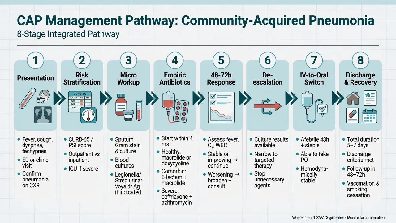
The fellowship exam expects CAP to be discussed as a single coherent narrative — the patient who walks (or is wheeled) into the emergency department with a cough, traverses the ICU, and is discharged months later still recovering. The pathway below expands each of the eight integrated domains into its practical, examinable detail. Each stage feeds the next: a missed microbiological diagnosis makes de-escalation impossible; a delayed first antibiotic converts curable pneumonia into septic shock; an unrecognised non-resolving infiltrate hides a malignancy. Integration is the point. [1]
flowchart TD
    A[Presentation<br/>cough + fever + dyspnoea + CXR infiltrate] --> B[Risk stratify<br/>CURB-65 / PSI / ATS-IDSA]
    B --> C{Admission level}
    C -->|low risk| D1[Outpatient<br/>oral amoxycillin ± macrolide]
    C -->|moderate| D2[Ward<br/>IV beta-lactam ± macrolide]
    C -->|severe / ICU criteria| D3[ICU<br/>Hour-1 bundle + ventilation]
    D2 --> E[Microbiology<br/>cultures BEFORE antibiotics]
    D3 --> E
    E --> F[Empiric antibiotics<br/>within 1 h if severe]
    F --> G[Monitor response 48-72 h<br/>clinical + PCT]
    G --> H[De-escalate / stop<br/>culture-directed]
    H --> I[IV to oral switch<br/>afebrile + improving + tolerating PO]
    I --> J[Discharge<br/>stable + home support]
    J --> K[Follow-up<br/>GP 1 wk, CXR 6 wk, ICU clinic 2-3 mo]
    K --> L[Recovery<br/>3-6 mo fatigue, PICS screen]
Stage 1 — Presentation: recognising CAP
CAP is defined as an acute infection of the pulmonary parenchyma acquired in the community (or diagnosed within 48 hours of hospital admission — after 48 h it is hospital-acquired, HAP). The diagnosis is syndromic, not microbiological: most patients are treated empirically because the causative organism is never identified in 40-60% of cases even with maximal sampling.[2]
The four diagnostic pillars of CAP at presentation
| Pillar | Requirement | Notes / pitfalls |
|---|---|---|
| Acute onset | New or worsening respiratory symptoms (cough, sputum, dyspnoea, pleuritic chest pain) usually <7-14 days | Subacute onset (weeks) suggests TB, fungal infection, malignancy, or organising pneumonia — NOT typical bacterial CAP |
| Systemic features | Fever (often >38°C), rigors, myalgia, malaise; confusion in the elderly may be the ONLY sign | Absence of fever does NOT exclude CAP — up to 30% of elderly/immunocompromised patients are afebrile. Hypothermia is a poor prognostic sign |
| Chest imaging | New pulmonary infiltrate (consolidation, ground-glass, interstitial) on CXR or CT not fully explained by another process | CXR is the standard; CT is more sensitive (picks up ~10% of CAP with normal CXR). A normal CXR with strong clinical suspicion = repeat in 24-48 h or CT. Beware: early dehydration, profound neutropenia, Pneumocystis can give a normal initial CXR |
| Inflammatory markers | Raised CRP, procalcitonin, white cell count (or a left shift / band form even if total WCC normal) | CRP >100 mg/L supports bacterial aetiology; PCT >0.25 ng/mL supports bacterial infection and may guide antibiotics; a normal CRP and PCT makes typical bacterial CAP less likely (but not impossible) |
Recognising CAP at the bedside — the five-step check
- CONFIRM ACUTE RESPIRATORY ILLNESS — new cough, purulent sputum, dyspnoea, pleuritic pain within the last 2 weeks. Ask about preceding viral illness, influenza season, sick contacts, travel, animal exposure, aspiration risk
- EXAMINE FOR FOCAL SIGNS — tachypnoea (the most sensitive sign), tachycardia, hypoxia, focal crackles/bronchial breathing, dullness to percussion (effusion/consolidation), pleural rub. Confusion in the elderly = CAP until proven otherwise
- CHEST X-RAY — look for lobar consolidation (typical: pneumococcus), multilobar (severe / staphylococcal / Klebsiella), interstitial (atypical/viral/Pneumocystis), cavitation (S. aureus, Klebsiella, anaerobes, TB), pleural effusion (parapneumonic). A PA + lateral film increases sensitivity
- OXYGENATION AND BLOODS — arterial or venous blood gas, FBC, U&E, LFTs, CRP, procalcitonin, lactate. Hypoxia (SpO2 <92%), raised lactate (>2 mmol/L), uraemia, hyponatraemia (Legionella), thrombocytopenia/DIC, and deranged LFTs all worsen prognosis
- DOCUMENT AND GRADE — apply CURB-65 at the bedside; if admitted, calculate the PSI. Document imaging, severity, and an explicit site-of-care decision. A severity score that is calculated but does not change WHERE the patient goes is useless
Typical vs atypical pneumonia pathogens — the classic exam distinction
| Feature | Typical (pyogenic) | Atypical |
|---|---|---|
| Pathogens | Streptococcus pneumoniae (commonest cause overall), Haemophilus influenzae, Moraxella catarrhalis, Klebsiella pneumoniae, Staphylococcus aureus, Group A streptococcus | Mycoplasma pneumoniae, Legionella pneumophila, Chlamydia pneumoniae, Chlamydia psittaci (birds), Coxiella burnetii (Q fever, livestock), influenza and other respiratory viruses (now grouped functionally with atypicals) |
| Onset | Abrupt, rigors, high fever, productive purulent sputum | Often subacute, 'walking pneumonia', dry cough, prominent systemic/headache/GI symptoms |
| CXR | Lobar consolidation | Patchy/interstitial, often looks worse than the patient |
| Sputum Gram stain | Plentiful organisms and neutrophils | Often negative (do not grow on standard media — need special culture, PCR, or serology) |
| Distinguishing clues | — | Legionella: hyponatraemia, diarrhoea, confusion, deranged LFTs, CK elevation, β-lactam failure. Mycoplasma: young adults, erythema multiforme, cold agglutinins (haemolytic anaemia), bullous myringitis. Psittacosis: bird exposure, splenomegaly. Q fever: phase I/II antibodies, hepatitis, endocarditis |
| Empiric cover | β-lactam (ceftriaxone, amoxycillin, benzylpenicillin) | Add a macrolide (azithromycin, clarithromycin) or doxycycline — atypicals have no cell wall so β-lactams are useless |
Stage 2 — Risk stratification: choosing the site of care
The single most important early decision in CAP is where to treat the patient — home, ward, HDU, or ICU. Three validated scores are used in concert, each answering a different question. The fellowship candidate must know all three and — crucially — their limitations (no score replaces clinical judgement, especially in the elderly, pregnant, immunocompromised, or socially isolated).[2][4][5]
The three CAP severity scores — CURB-65 vs PSI vs IDSA-ATS
| Feature | CURB-65 | PSI (PORT score / Pneumonia Severity Index) | IDSA/ATS minor-major criteria |
|---|---|---|---|
| Purpose | Bedside triage: who needs hospital? | Comprehensive mortality prediction; identifies low-risk outpatients | ICU triage: who needs intensive care? |
| Components | Confusion, Urea >7 mmol/L, RR >30, BP (SBP <90 or DBP <60), age >65 (1 point each) | Demographics (age, sex), nursing home, 5 comorbidities, 5 physical exam findings, 7 lab/imaging findings — summed into 5 risk classes (I-V) | 2 MAJOR: invasive ventilation, septic shock requiring vasopressors. 9 MINOR: RR >30, PaO2/FiO2 <250, multilobar infiltrates, confusion, uraemia, WCC <4, platelets <100, hypothermia <36, hypotension needing aggressive fluids |
| Scoring | 0-5, simple arithmetic | 20-step calculation (often done by app) | Count major and minor criteria separately |
| Interpretation | 0-1: outpatient; 2: inpatient; 3-5: severe, consider ICU | Classes I-II: outpatient; III: brief inpatient/observation; IV-V: inpatient (V often ICU) | 1 major OR >=3 minor criteria = severe CAP = ICU admission |
| Strengths | Fast, memorable, reproducible, validates well for mortality | Most accurate mortality prediction; the reference standard for outpatient safety | Specifically designed for ICU triage; predicts need for ICU, not just mortality |
| Weaknesses | Under-scores young patients with severe physiological derangement (e.g., a 25-year-old septic patient scores 0-1 but needs ICU); age dominates; omits oxygenation | Cumbersome; age dominates; under-scores young patients with severe physiology; requires labs | Minor criteria thresholds debated; scoring all 9 minors can delay ICU; some advocate the simpler 2-major-only rule |
| When to use | ED triage, in the ambulance, at first contact | Formal admission decision, discharge planning | Once admitted: who needs ICU/HDU vs ward |
Risk stratification and site-of-care decision — practical sequence
- CALCULATE CURB-65 AT THE BEDSIDE — takes 30 seconds. Score 0-1: consider outpatient with safety-net advice and 48 h review. Score 2: admit to ward. Score >=3: severe — admit, calculate PSI, assess for ICU
- IF ADMITTED, CALCULATE PSI AND CHECK IDSA/ATS CRITERIA — PSI refines the mortality prediction; IDSA/ATS criteria answer the ICU question. 1 major criterion (need for invasive ventilation OR septic shock on vasopressors) = ICU immediately — no further scoring needed
- ASSESS THE 9 IDSA-ATS MINOR CRITERIA — if >=3 minor criteria present, manage in ICU/HDU. Some experts use a lower threshold (>=2) in the elderly or immunocompromised. Do NOT delay antibiotics while counting
- APPLY CLINICAL OVER-RIDE — admit/upgrade if any of: poor social circumstances, unable to tolerate oral, immunocompromise, pregnancy, suspected sepsis, hypoxia not corrected by oxygen, recent influenza, end-organ dysfunction. The score is a guide, not a verdict
- REASSESS FREQUENTLY — a patient can deteriorate rapidly (especially in the first 24 h). Repeat CURB-65 and clinical assessment at every nursing handover and on any change. Have an early warning score (e.g., MEWS/NEWS2) running continuously
Fine 1997 — The Pneumonia Severity Index / PORT score (PMID 9031800)
Design
Prospective observational cohort study, 2,287 inpatients with CAP, derivation + validation, then validated in >38,000 patients
Aim
Derive a prediction rule to identify patients with CAP at LOW risk of 30-day mortality who could be safely treated as outpatients
The rule
A two-step algorithm: step 1 screens for low-risk Class I (age <50, no comorbidity or derangement); step 2 assigns points for age, nursing home, 5 comorbidities (neoplastic, liver, CHF, cerebrovascular, renal), 5 exam findings (altered mental status, RR >30, SBP <90, temp <35 or >40, pulse >125), 7 labs/imaging (pH <7.35, BUN >30, Na <130, glucose >250, haematocrit <30, PaO2 <60 or SpO2 <90, pleural effusion)
Five risk classes
Class I-V. Mortality: I 0.1%, II 0.6%, III 0.9%, IV 9.3%, V 27.0%
Bottom line
PSI is the reference standard for CAP mortality prediction. Classes I-II can be safely managed as outpatients; IV-V require inpatient care. Cumbersome to calculate at the bedside (use an app), and age dominates — it can under-score severe physiology in young patients. Used alongside CURB-65 for site-of-care decisions
Lim 2003 — CURB-65 derivation (PMID 12746012)
Design
Prospective study of 1,068 patients with suspected CAP in three UK hospitals; derivation and validation of a simple severity score
The score
Five equally weighted points: **C**onfusion, **U**rea >7 mmol/L, **R**espiratory rate >=30, **B**lood pressure (SBP <90 or DBP <=60), age >=65
Mortality by score
Score 0: 0-1.5%; score 1: 2.7%; score 2: 6.8%; score 3: 14%; score 4: 27.8%; score 5: 27-40%
Recommended use
0-1: low risk — consider outpatient. 2: moderate — inpatient (ward). >=3: severe — consider ICU
Bottom line
CURB-65 is the bedside workhorse — fast, memorable, reproducible, and validated internationally. Its weakness is that age is heavily weighted (an unwell 25-year-old scores 0) and it omits oxygenation. Use with clinical judgement and the PSI/IDSA-ATS criteria
Stage 3 — Microbiological workup: culture before antibiotics
The microbiological workup serves two purposes: (1) to refine therapy (de-escalate empiric cover to a targeted agent); and (2) for public health (notifiable pathogens, outbreak detection, resistance surveillance). The cardinal rule — draw cultures BEFORE the first antibiotic dose — is frequently violated in the rush to give the sepsis bundle, halving the yield of blood cultures within minutes.[1][2]
[1]Diagnostic tests in CAP — yield, timing, and what they tell you
| Test | Yield / timing | What it changes |
|---|---|---|
| Blood cultures (x2 sets) | Positive in 10-25% admitted CAP, 25-50% severe CAP; before antibiotics | The reference standard for bacteraemia; identifies the organism AND its sensitivities. Mandatory in severe CAP, ICU admission, and before de-escalation |
| Sputum Gram stain and culture | Useful only if a good-quality sample (low squamous epithelial cells, <10/low-power field; >25 neutrophils); ~50-60% yield when adequate | Confirms typical pathogens (pneumococcus, H. influenzae) and detects resistant organisms. S. aureus or Gram-negative bacilli in sputum should prompt consideration of MRSA/Pseudomonas cover |
| Urinary pneumococcal antigen | Sensitivity 70-80% in bacteraemic pneumococcal CAP; specificity >90% | Rapid (minutes). Particularly useful if antibiotics already given (sterilises cultures). Recommended in severe CAP |
| Urinary Legionella antigen | Detects only Legionella serogroup 1 (80-90% of cases); sensitivity 70-90% for serogroup 1, near-100% specificity | Rapid. Mandatory in severe CAP and in any CAP with suggestive features (hyponatraemia, diarrhoea, confusion, β-lactam failure, travel). Negative antigen does NOT exclude non-serogroup-1 Legionella |
| Respiratory virus PCR panel | Influenza A/B, RSV, SARS-CoV-2, parainfluenza, metapneumovirus, adenovirus — nasopharyngeal swab or BAL | Viral CAP (especially influenza and COVID-19) is common and changes therapy (oseltamivir for influenza within 48 h of onset; antivirals/immunomodulation for COVID). Co-infection with bacteria is common in viral CAP — still give antibacterials |
| Atypical serology (paired) | Acute and convalescent (4-6 weeks apart) — >4-fold rise is diagnostic; single acute titres unreliable | Retrospective diagnosis of Mycoplasma, Chlamydia, Coxiella, Legionella non-serogroup-1. Useless for acute management but valuable for epidemiology and confirming the cause. PCR (where available) is faster |
| Mycoplasma/Chlamydia PCR | Throat swab or sputum PCR — sensitive, rapid | Useful during outbreaks; confirms atypical cover is needed |
| Sputum AFB / TB PCR (Xpert MTB/RIF) | If cough >2 weeks, weight loss, night sweats, risk factors (born/travelled in endemic area, immunocompromise, incarceration, alcohol misuse) | Excludes/diagnoses pulmonary TB; Xpert also detects rifampicin resistance. Send in any non-resolving or atypical CAP |
| Arterial blood gas | Hypoxia, hypercapnia, acidosis | Quantifies respiratory failure and guides oxygen targets and ventilation |
| HIV test (with consent) | Routine in severe CAP, recurrent CAP, or unusual pathogens | Pneumococcal bacteraemia and severe CAP are AIDS-defining; HIV is a common unrecognised comorbidity |
Pre-antibiotic microbiological bundle in severe CAP — the one-hour window
- TWO SETS OF BLOOD CULTURES — separate venepuncture sites, aerobic + anaerobic bottles, ~20 mL per set, before any antibiotic. Label the exact time. In septic shock, do not delay antibiotics >45 min waiting — take one set then treat
- EXPECTORATED SPUTUM — instruct the patient to deep-cough a fresh sample (not saliva); if unobtainable, consider induced sputum (nebulised hypertonic saline) or endotracheal aspirate if intubated. Send for Gram stain, culture, and sensitivity. In immunocompromised, add AFB and fungal stains
- URINARY ANTIGENS — pneumococcal AND Legionella. These are rapid, high-specificity, and unaffected by prior antibiotics. Mandatory in severe CAP
- NASOPHARYNGEAL SWAB FOR RESPIRATORY VIRUS PCR — influenza A/B, RSV, SARS-CoV-2 (and broader panel if available). Critical during viral seasons — empiric oseltamivir within 48 h of symptom onset reduces influenza mortality
- CHEST IMAGING AND PARAPNEUMONIC EFFUSION SAMPLING — if effusion >10 mm on lateral decubitus film or ultrasound, diagnostic thoracentesis (send for pH, protein, LDH, glucose, Gram stain, culture, cell count). pH <7.2 = complicated parapneumonic effusion / empyema — chest tube
- CONSIDER BRONCHOSCOPY / BAL — if intubated, immunocompromised, failing therapy, or suspected Pneumocystis/opportunistic infection. BAL increases yield substantially and is safe in expert hands
- BASELINE BLOODS — FBC, U&E, LFTs, CRP, procalcitonin, lactate, coagulation, troponin (myocardial infarction and atrial fibrillation are common CAP complications), ABG. Repeat CRP/PCT at 48-72 h to track response
Stage 4 — Empiric antibiotics: the first hour
The first antibiotic dose is the single most important intervention in severe CAP. Empiric therapy is started before the organism is known, chosen to cover the likely pathogens based on severity, comorbidities, and local resistance patterns. The cardinal principle: cover broadly and early, then narrow once the pathogen is identified.[1][2][3]
Empiric antibiotic regimens for CAP — by severity and risk
| Scenario | Empiric regimen | Rationale |
|---|---|---|
| Outpatient (CURB-65 0-1), no comorbidity | Amoxycillin 1 g PO tds (or doxycycline 100 mg bd if penicillin-allergic). Add a macrolide (clarithromycin 500 mg bd) if atypical suspected | S. pneumoniae is the commonest pathogen and remains sensitive to penicillin in most of ANZ/UK. Macrolide adds atypical cover if Mycoplasma, Legionella, or Chlamydia suspected |
| Outpatient with comorbidity (COPD, CHF, diabetes, CKD, malignancy, immunosuppression) | Amoxycillin-clavulanate 875/125 mg PO tds + macrolide OR doxycycline; alternative: respiratory fluoroquinolone (moxifloxacin) monotherapy | Broader cover for H. influenzae, Moraxella, enteric Gram-negatives, and atypicals. Comorbid patients have higher resistance risk and worse outcomes |
| Inpatient ward (CURB-65 2-3), non-severe | Benzylpenicillin 1.2 g IV q6h (or ceftriaxone 1-2 g IV od) + azithromycin 500 mg IV/PO od; alternative: respiratory fluoroquinolone monotherapy | IV β-lactam + macrolide covers typicals + atypicals. Ceftriaxone preferred if aspiration, meningitis concern, or penicillin-resistant pneumococcus suspected |
| Severe CAP / ICU (IDSA-ATS criteria) | Ceftriaxone 2 g IV od + azithromycin 500 mg IV od, OR piperacillin-tazobactam 4.5 g IV q6h + azithromycin (if Pseudomonas risk) | Broad β-lactam + macrolide is the standard severe CAP regimen; reduces mortality vs β-lactam alone (macrolide has immunomodulatory and anti-Legionella benefit) |
| Severe CAP with MRSA risk (post-influenza, end-stage renal disease, IVDU, recent healthcare, known colonisation) | ADD vancomycin 15-25 mg/kg q8-12h (target AUC 400-600) or linezolid 600 mg q12h | S. aureus CAP is post-viral, necrotising, and rapidly progressive. Linezolid preferred by some (better lung penetration, toxin suppression). Add to standard regimen |
| Severe CAP with Pseudomonas risk (bronchiectasis, severe COPD, repeated antibiotics, recent hospitalisation) | Piperacillin-tazobactam (or ceftazidime, cefepime, meropenem) + macrolide (covers Pseudomonas AND atypicals). ADD MRSA cover if also at risk | Standard ceftriaxone does NOT cover Pseudomonas. Use an anti-pseudomonal β-lactam and ensure atypical cover with the macrolide |
| Suspected/confirmed influenza | ADD oseltamivir 75 mg PO bd (or zanamivir if unable to take PO), ideally within 48 h of symptom onset; continue 5 days, longer if immunocompromised | Oseltamivir reduces influenza mortality even when given late in critically ill patients. Empiric oseltamivir in severe CAP during influenza season until PCR returns |
The severe CAP hour-1 bundle — the resuscitative antibiotic
- RECOGNISE SEVERE CAP — IDSA-ATS criteria met (1 major OR >=3 minor) OR clinical judgement. Start the clock — antibiotics target is <1 h from recognition
- TAKE CULTURES RAPIDLY (within 45 min if possible) — two sets blood cultures, sputum, urinary antigens, respiratory virus swab. Do NOT delay antibiotics beyond 1 h for cultures if shocked
- GIVE THE EMPIRIC ANTIBIOTIC IMMEDIATELY — ceftriaxone 2 g IV + azithromycin 500 mg IV over the first hour. Add vancomycin/linezolid if MRSA risk; switch to piperacillin-tazobactam if Pseudomonas risk; add oseltamivir in flu season
- PARALLEL RESUSCITATION — oxygen to target SpO2 92-96% (88-92% if COPD), 30 mL/kg crystalloid bolus if hypotensive/lactate >2, noradrenaline if shock persists, transfuse if Hb <70, control source (drain empyema)
- REASSESS AT 1 HOUR, 3 HOURS, 6 HOURS — lactate clearance (>10%), MAP >65, urine output >0.5 mL/kg/h, mental status. Persisting shock despite adequate fluids = add vasopressin, consider hydrocortisone (CAPE COD)
- DOCUMENT THE TIME OF FIRST ANTIBIOTIC — auditable. Time-to-antibiotic <1 h in severe CAP is a key quality metric and correlates with survival
Kumar 2006 — Antibiotic timing in septic shock (PMID 16625125)
Design
Retrospective cohort, 2,154 adults with septic shock (CAP a major source) across 14 ICUs and 28 EDs in Canada and the USA
Exposure
Time from first documented hypotension to initiation of EFFECTIVE antimicrobial therapy
Primary outcome
Hospital mortality
Key result
Mortality was 25% when effective antibiotics were given in the FIRST hour, rising 7.6 percentage points (OR ~1.12) per hour of delay thereafter; by 6 h mortality exceeded 60%. Effect was independent of severity, source, and organ failure
Bottom line
In septic shock — including severe CAP — the first antibiotic is a resuscitation drug. Give effective empiric antibiotics WITHIN ONE HOUR of recognising shock. This is the single strongest temporal-outcome relationship in sepsis literature and underpins the Surviving Sepsis hour-1 bundle
Stage 5 — Monitoring the response: 48-72 hours and procalcitonin
Once empiric therapy is started, the question becomes: is the patient getting better? The classical teaching is that clinical response is assessed at 48-72 hours. Failure to improve by day 3-5 triggers a structured re-evaluation (differential diagnosis of non-resolving CAP). Modern practice increasingly uses procalcitonin (PCT) to support both the decision to continue and to stop antibiotics.[7][8]
Markers of clinical response in CAP — day-by-day assessment
| Marker | Improving | Worrying / non-response |
|---|---|---|
| Fever | Defervescence over 48-72 h (typical) | Persistent or recurrent fever at day 3-5 — reassess |
| Respiratory rate / oxygenation | RR falling, SpO2 rising, FiO2 weaning, P/F improving | Persisting tachypnoea, escalating FiO2/PEEP — consider ARDS, effusion, PE, wrong organism |
| Haemodynamics | Off vasopressors, MAP stable, lactate clearing | Persisting/rebounding shock — wrong drug, source not controlled (empyema, abscess), endocarditis |
| White cell count / CRP | CRP falls by >50% in 4-5 days in responding CAP | CRP static or rising — resistant organism, complication, wrong diagnosis |
| Procalcitonin (PCT) | Falls by >80% from baseline, or <0.25 ng/mL = stop antibiotics | Persistently elevated — ongoing bacterial infection; reassess |
| Mental state | Confusion resolving | Worsening confusion — hypoxia, sepsis, hyponatraemia, alcohol withdrawal, PICS |
| Radiology | CXR improvement lags clinical (often worse at 48 h — do NOT treat the X-ray) | Progressive multilobar infiltrates — ARDS, Staph/Klebsiella necrotising, wrong organism, alternative diagnosis |
| Functional status | Mobilising, eating | Bed-bound, not eating — frailty, complications, prolonged recovery |
Schuetz 2018 — Procalcitonin-guided antibiotic stewardship IPD meta-analysis (PMID 29790814)
Design
Individual patient data meta-analysis of 26 randomised controlled trials, 6,708 patients with acute respiratory tract infections (CAP a major subgroup)
Intervention
Procalcitonin algorithm: start antibiotics if PCT >0.25-0.5 ng/mL; encourage STOPPING antibiotics when PCT falls by >80% from baseline OR reaches <0.25 ng/mL
Primary outcome
30-day mortality: 8.6% (PCT-guided) vs 9.6% (control) — PCT was NON-INFERIOR (and possibly slightly superior)
Antibiotic exposure
PCT-guided patients had 2.4 fewer days of antibiotic exposure (95% CI 2.0-2.7) and lower antibiotic-related side effects
Bottom line
Procalcitonin-guided antibiotic stewardship is SAFE (no excess mortality) and reduces antibiotic duration and adverse effects across respiratory infections including CAP. The algorithm: stop when PCT falls by >80% from peak OR is <0.25 ng/mL. ProACT (below) showed it does NOT help in ED CAP specifically — PCT is a stewardship tool, not a diagnostic one
ProACT 2018 — Procalcitonin-guided antibiotics for LRTI (PMID 29791812)
Design
Multicentre randomised controlled trial (PETAL Network), 1,656 ED patients with suspected lower respiratory tract infection including CAP
Intervention
Procalcitonin assay disclosed to clinicians with a stop-antibiotic recommendation (PCT <0.1, or >80% drop) vs usual care (PCT blinded)
Primary outcome
Adverse outcomes (death, ICU admission, complications, repeat antibiotics) at 30 days: NO difference. Antibiotic exposure reduced modestly but not as much as expected
Bottom line
In unselected ED lower-respiratory-tract infection, **disclosing a procalcitonin result did NOT improve outcomes** and only modestly reduced antibiotic use — partly because clinicians over-rode the algorithm and partly because viral LRTI is common. PCT is best used as a STEWARDSHIP adjunct in HOSPITALISED CAP to support stopping antibiotics, NOT as a stand-alone diagnostic test in the ED
Monitoring response to CAP therapy — the 72-hour reassessment
- AT 48-72 HOURS, ASSESS CLINICAL RESPONSE — has fever resolved? Is the patient off supplemental oxygen or weaning? Has RR normalised? Is the patient eating and mobilising? Are CRP and PCT falling?
- IF IMPROVING — continue current antibiotics, plan de-escalation based on cultures, plan IV-to-oral switch. Reassess duration using PCT (stop when >80% drop or <0.25)
- IF NOT IMPROVING BY DAY 3-5 — apply the differential of non-resolving CAP (see below). Do NOT simply 'escalate empirically' without a diagnosis
- REVIEW MICROBIOLOGY — has an organism been identified? Resistant organism? Unsuspected pathogen (Legionella, TB, PJP, fungal)? Co-infection?
- REVIEW COMPLICATIONS — empyema (drain), lung abscess, ARDS, metastatic infection (endocarditis, meningitis, septic arthritis), venous thromboembolism, superimposed HAP/VAP
- REVIEW THE DIAGNOSIS — is it really CAP? Consider PE (mimics CAP, common in immobilised/hypercoagulable), pulmonary oedema, vasculitis (GPA), malignancy with post-obstructive pneumonia, organising pneumonia, drug-induced pneumonitis, alveolar haemorrhage
- REVIEW HOST FACTORS — immunocompromise (HIV, neutropenia, immunosuppression), aspiration risk, untreated comorbidity (uncontrolled diabetes, malnutrition)
- CONSIDER BRONCHOSCOPY — for microbiology in non-resolving, immunocompromised, intubated, or strong suspicion of opportunistic pathogen
Differential diagnosis of non-resolving CAP at 72 hours
| Category | Causes | Action |
|---|---|---|
| Wrong organism / resistant | MRSA, Pseudomonas, ESBL, atypical covered inadequately (Legionella missed), TB, fungal (Aspergillus, Pneumocystis, Cryptococcus, endemic fungi) | Review cultures, extend empiric cover, send induced sputum/BAL for AFB and fungal stains; consider bronchoscopy |
| Wrong diagnosis | Pulmonary embolism, pulmonary oedema, vasculitis (GPA, EGPA), organising pneumonia, malignancy with post-obstructive infection, alveolar haemorrhage, drug-induced pneumonitis, eosinophilic pneumonia | CTPA, echo, autoimmune screen (ANCA, ANA, anti-GBM), bronchoscopy ± biopsy |
| Complication | Empyema, lung abscess, ARDS, metastatic infection (endocarditis — repeat blood cultures, echo), venous thromboembolism, superimposed HAP/VAP, parapneumonic effusion | Ultrasound/CT chest, thoracentesis, chest tube, echocardiogram, repeat imaging |
| Host factors | Immunocompromise (undisclosed HIV, neutropenia, immunosuppression), untreated comorbidity (uncontrolled diabetes, malnutrition, alcohol misuse, chronic liver disease), aspiration (recurrent) | HIV test, full immune workup, optimise comorbidity, swallow assessment, MDT discussion |
| Pharmacological | Inadequate dose, poor lung penetration, drug fever, antibiotic-related (e.g., linezolid, β-lactam fever), wrong duration | Review dosing, therapeutic drug monitoring (vancomycin AUC), consider line-related bacteraemia |
Stage 6 — De-escalation: narrowing to the pathogen
De-escalation is the antimicrobial stewardship backbone of CAP management: once a pathogen and its sensitivities are known, narrow empiric broad cover to the most targeted effective agent. This reduces resistance selection, Clostridioides difficile infection, antibiotic toxicity, and cost — without compromising outcomes. The procalcitonin algorithm and the 5-day duration evidence support this.[2][7]
Antibiotic duration in CAP — pathogen-specific
| Scenario | Recommended duration | Evidence / comment |
|---|---|---|
| Uncomplicated CAP, good response | 5 days (stop if afebrile 48-72 h AND clinically stable) | Short courses (<=5-7 days) are non-inferior to longer in most CAP; many patients are over-treated. Stop when clinical stability criteria met |
| S. pneumoniae, H. influenzae (typical, sensitive) | 5-7 days | Afebrile 48-72 h + clinically stable = stop |
| Legionella pneumophila | 7-14 days (some use 14-21 in immunocompromised) | Intracellular pathogen; macrolide or fluoroquinolone. Severe disease warrants longer course |
| Mycoplasma / Chlamydia pneumoniae | 7-14 days | Macrolide, doxycycline, or fluoroquinolone |
| Pseudomonas aeruginosa | 7-14 days (some 14-21 if bacteraemic/necrotising) | Re-assess daily; longer if slow response or complicated (abscess, empyema) |
| S. aureus CAP (MSSA / MRSA) | 7-14 days minimum; longer if bacteraemia, endocarditis, metastatic infection, cavitation | Always look for endocarditis (echo), exclude metastatic abscesses; bacteraemia often warrants >=14 days even after first negative blood culture if persistent |
| Bacteraemic CAP with metastatic infection (endocarditis, meningitis, osteomyelitis, septic arthritis) | Extended — 4-6 weeks for endocarditis, 2 weeks for meningitis, etc. | The pneumonia is a manifestation of a deeper infection; treat the metastatic focus by its own standard |
| Empyema / lung abscess | 4-6 weeks (often longer, until radiological resolution) | Source control (drain) is more important than antibiotic duration |
| PCT-guided stopping | Stop when PCT falls by >80% from peak OR reaches <0.25 ng/mL | Non-inferior to fixed-duration; reduces exposure. Use as a stewardship adjunct in hospitalised CAP |
De-escalation of CAP antibiotics — step by step
- DAILY MICROBIOLOGY REVIEW — as cultures and PCR results return (typically 48-72 h), identify the causative organism and its sensitivities
- NARROW THE SPECTRUM — if pneumococcus sensitive to penicillin, switch ceftriaxone to benzylpenicillin or amoxycillin. If Legionella, stop the β-lactam, continue the macrolide/fluoroquinolone. If influenza only, stop antibacterials once bacterial co-infection excluded (rare in adults — usually continue a short course)
- STOP REDUNDANT COVER — discontinue MRSA cover (vancomycin/linezolid) if no MRSA; discontinue Pseudomonas cover (piperacillin-tazobactam) if no Pseudomonas; discontinue oseltamivir once influenza PCR negative (outside season)
- USE PROCALCITONIN — when PCT falls >80% from peak OR is <0.25 ng/mL and the patient is clinically stable, stop antibiotics. Recheck PCT 6-12 h later; restart ONLY if clearly rising and clinically deteriorating
- SET A STOP DATE / REVIEW DATE AT THE START — every antibiotic prescription has a documented indication, review date, and stop date. Stewardship is a system, not an afterthought
- DAILY 'ANTIBIOTIC TIME-OUT' — on ICU rounds: 'Does this patient still need this antibiotic, at this dose, for this duration?' If no — stop, narrow, or switch
Stage 7 — IV-to-oral switch and discharge readiness
The intravenous-to-oral switch is a key milestone in CAP recovery: it marks clinical stability, enables ward/step-down care, and is a prerequisite for discharge. The principle is simple — once the patient can absorb oral medication and is clinically improving, oral antibiotics achieve equivalent serum concentrations to IV for the common CAP pathogens (good oral bioavailability: fluoroquinolones, linezolid, clindamycin, metronidazole, doxycycline, fluconazole).[2]
Criteria for IV-to-oral switch in CAP
| Criterion | Threshold | Comment |
|---|---|---|
| Clinical improvement | Cough, dyspnoea, RR improving | Subjective + objective improvement over 24 h |
| Afebrile | Temperature <37.8°C for 24-48 hours | Two consecutive readings >8 h apart. Persistent fever suggests complication — investigate before switching |
| Haemodynamic stability | Off vasopressors, SBP >90, MAP >65, lactate normal | No active shock |
| Tolerating oral intake | Eating and drinking, no vomiting, no ileus, no malabsorption | Essential — oral antibiotics are useless if not absorbed |
| Mental status | Improving, able to take oral meds reliably | Confusion precludes reliable oral dosing |
| Respiratory support weaning | Stable on low-flow nasal prongs or room air; not escalating FiO2 | A patient needing HFNC/NIV/IMV is NOT ready for switch |
| Inflammatory markers | Falling CRP and PCT | Falling CRP (e.g., >50% drop from peak) supports the switch |
| No uncontrolled complication | No new empyema, abscess, endocarditis, metastatic infection | Source control complete |
IV-to-oral switch and discharge readiness — the practical sequence
- CHECK ALL SWITCH CRITERIA DAILY from day 2 — when criteria met, switch from IV to oral. Use a drug with high oral bioavailability: amoxycillin, amoxycillin-clavulanate, clarithromycin/azithromycin, doxycycline, moxifloxacin/levofloxacin, linezolid. Match the IV organism and sensitivities
- MONITOR FOR 24 HOURS AFTER SWITCH — confirm clinical stability on oral therapy before discharge. Most failures occur in this window
- CONFIRM DISCHARGE READINESS CRITERIA — afebrile 24-48 h, clinical stability for 24 h on oral antibiotics, tolerating oral, SpO2 stable on room air (or back to baseline), able to mobilise safely, no new complications, home support available, oral antibiotic available, follow-up arranged
- PATIENT EDUCATION — teach-back on warning signs (worsening dyspnoea, recurrent fever, haemoptysis, pleuritic chest pain), recovery timeline (fatigue common for weeks), completion of the antibiotic course, smoking cessation referral, vaccination plan
- PRESCRIPTIONS — oral antibiotics to complete the planned course (e.g., total 5-7 days), analgesia, VTE prophylaxis if immobile, inhalers if newly-diagnosed COPD/asthma, salbutamol for bronchospasm
- VACCINATE BEFORE DISCHARGE — pneumococcal (PCV13/PPSV23 per local schedule), influenza (in season), COVID-19 boosters, and ensure Tdap/herpes zoster/RSV are up to date per age. Vaccination before discharge dramatically improves uptake
- ARRANGE FOLLOW-UP — GP review at 1 week; CXR at 6 weeks (resolution — non-resolving infiltrate mandates investigation); ICU follow-up clinic at 2-3 months for those ICU-admitted (PICS screening — cognitive, psychological, physical); pulmonary rehabilitation referral for those with persisting breathlessness
Stage 8 — Discharge, follow-up, and recovery
CAP is not 'cured' at discharge. Recovery is prolonged — fatigue for 3-6 months, cognitive impairment for 6-12 months, and some patients never fully recover (post-intensive-care syndrome, PICS). The discharge and follow-up phase is where long-term outcomes are won or lost: vaccination, smoking cessation, pulmonary rehabilitation, and active PICS screening reduce readmission and restore function.[1][2]
The recovery timeline after severe CAP / ICU admission
| Timeframe | Typical recovery | What to do |
|---|---|---|
| 0-2 weeks | Acute symptoms (cough, fever, dyspnoea) resolving; profound fatigue, weakness, sleep disturbance | Early mobilisation in hospital; pulmonary rehabilitation referral; smoking cessation; vaccination before discharge |
| 2-6 weeks | Improving but persistent fatigue, reduced exercise tolerance, possible depressive symptoms, new-onset anxiety | GP review; progressive ambulation; reassurance; screen for depression/anxiety; check CXR at 6 weeks for resolution |
| 6 weeks — CXR follow-up | CXR should be resolving or resolved | Non-resolving infiltrate at 6 weeks = investigate (malignancy, TB, bronchiectasis, immunodeficiency, organising pneumonia). Smokers and over-50s particularly warrant CT |
| 1-3 months | Most physical recovery; cognitive symptoms (memory, concentration) may persist; ICU follow-up clinic for those ICU-admitted | ICU follow-up clinic — PICS screening (cognitive, psychological, physical), PTSD screen, medication review, sexual function discussion |
| 3-6 months | Fatigue usually resolving; cognitive often improving; some have persisting anxiety/depression | Pulmonary rehabilitation completion; ongoing psychosocial support |
| 6-12 months | Most patients functionally recovered; cognitive recovery continues; some have permanent impairment | Vaccination update; address residual rehabilitation needs; if still symptomatic, investigate for chronic lung damage (fibrosis, bronchiectasis) |
Discharge bundle for severe CAP — the complete checklist
- CLINICAL STABILITY — afebrile 24-48 h, stable SpO2 on room air (or baseline), tolerating oral antibiotics for 24 h, mobilising safely, no active complications
- ANTIBIOTIC PLAN — oral course to complete planned duration; written plan; clear advice on adherence and what to do if symptoms recur
- VACCINATION — pneumococcal, influenza (seasonal), COVID-19 booster, and any overdue vaccines (Tdap, herpes zoster, RSV) given BEFORE discharge
- SMOKING CESSATION — brief intervention, nicotine replacement therapy, referral to quitline — the single most effective prevention of recurrent CAP and the underlying COPD/lung cancer risk
- FOLLOW-UP ARRANGED — GP at 1 week, CXR at 6 weeks, ICU follow-up clinic at 2-3 months (if ICU-admitted), pulmonary rehabilitation referral
- PATIENT EDUCATION — teach-back on warning signs, recovery timeline, medication management, written materials at year-6 reading level, contact details for worsening symptoms
- SOCIAL/COMMUNITY SUPPORT — home help, meals, transport, carer involvement as needed; early discharge coordinator involvement for complex patients
- DOCUMENT QUALITY METRICS — time-to-antibiotic, blood culture rate, de-escalation rate, length of stay, 30-day readmission — feed into the audit cycle
CAPE COD 2023 — Hydrocortisone in severe CAP (PMID 36652352)
Design
Multicentre randomised, double-blind, placebo-controlled trial, 800 patients with severe CAP (PSI class IV-V) in French ICUs
Intervention
Hydrocortisone 200 mg/day for 4 days (or 8 days if shock), then tapered, vs placebo — started within 24 h of admission
Primary outcome
Day-28 treatment failure (death, mechanical ventilation, refractory shock, or prolonged vasopressor use): significantly lower with hydrocortisone
Key result
Treatment failure reduced from 20% (placebo) to 12% (hydrocortisone), driven largely by fewer patients needing mechanical ventilation. More hyperglycaemia in the steroid group (expected)
Bottom line
Low-dose hydrocortisone (200 mg/day) in severe CAP reduces treatment failure, particularly the need for mechanical ventilation. Joins dexamethasone (DEXA-ARDS) as steroid evidence in severe lower respiratory infection. Adjunct — never a substitute for antibiotics and source control
Cross-cutting integration — how each stage affects every other
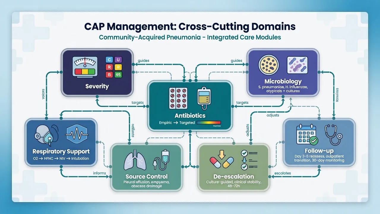
The fellowship examiner will test integration. The matrix below is the high-yield mental model: every domain of CAP management interacts with every other. A decision in one stage constrains or enables decisions in others. [1]
The CAP integration matrix — how the eight stages interact
| Domain | Affects | Is affected by |
|---|---|---|
| Recognition | Determines site of care and speed of bundle | Under-recognition in elderly (confusion, falls) delays everything downstream |
| Risk stratification | Threshold for ICU, intensity of monitoring, breadth of empiric cover | Over- or under-scoring leads to wrong admission level; young sick patients under-scored by CURB-65 |
| Microbiology | Enables de-escalation, public health notification, contact tracing | Sterilised by early antibiotics; inadequate sampling leads to prolonged empiric therapy, no de-escalation |
| Empiric antibiotics | Speed and breadth affect mortality (Kumar), resistance, C. diff | Driven by severity, comorbidity, local resistance; constrained by allergy, organ failure |
| Monitoring response | Triggers de-escalation, switch, discharge, or escalation (ECMO, complication) | Confounded by steroids (mask fever), radiographic lag, PCT kinetics |
| De-escalation | Stewardship, resistance, C. diff, length of stay, cost | Depends entirely on quality microbiology upstream; PCT supports it |
| IV-to-oral switch | Enables ward/step-down and discharge | Requires clinical stability, GI function, no active complication |
| Discharge and follow-up | Readmission, PICS, vaccination, smoking cessation, long-term mortality | Earlier safe discharge depends on all upstream stages executed well |
The integrated CAP viva — how to answer the examiner's 'discuss your management' question
- FRAME THE PATIENT — 'A 68-year-old smoker presents with 3 days of fever, purulent cough, dyspnoea, RR 32, SpO2 88% room air, BP 96/60, CURB-65 score 4, CXR shows right middle and lower lobe consolidation. This is severe CAP with sepsis.'
- STRUCTURE THE ANSWER IN THE EIGHT DOMAINS — 'I will discuss recognition, risk stratification, microbiology, empiric antibiotics, monitoring, de-escalation, switch, and discharge as a continuous pathway.'
- EMPHASISE TIME-CRITICAL INTERVENTIONS FIRST — 'Within the first hour I will take cultures and give ceftriaxone 2 g + azithromycin 500 mg IV, oxygen to SpO2 92-96%, 30 mL/kg crystalloid, and noradrenaline if shocked. Each hour of antibiotic delay increases mortality.'
- LINK DOMAINS EXPLICITLY — 'Cultures before antibiotics enable de-escalation at 48-72 h; procalcitonin guides duration; clinical stability at 48-72 h enables the oral switch and discharge planning; vaccination and smoking cessation before discharge reduce recurrence.'
- ANTICIPATE COMPLICATIONS — 'If he develops ARDS I will institute lung-protective ventilation (Vt 6 mL/kg PBW), prone 16 h/day if P/F <150, and consider ECMO if refractory. If AKI, I will optimise haemodynamics and consider CRRT. If empyema, drain. If endocarditis, extend antibiotics.'
- DESCRIBE THE RECOVERY PHASE — 'Recovery takes months; I will screen for PICS at the ICU follow-up clinic, repeat CXR at 6 weeks to exclude malignancy, and refer to pulmonary rehabilitation.'
- CLOSE WITH QUALITY — 'I will audit time-to-antibiotic, blood culture rate, de-escalation rate, and 30-day readmission in my unit as ongoing quality improvement.'
Additional clinical pearls
Additional red flags
Mnemonic and exam summary
CURB-65 — the bedside CAP severity score
IDSA-ATS severe CAP criteria — invasive or septic, or 3 minor criteria
The one-minute exam answer — severe CAP from presentation to discharge
| Stage | Key action | Time / target |
|---|---|---|
| 1. Recognise | New respiratory symptoms + CXR infiltrate + fever/inflammatory markers; CURB-65 | Minutes |
| 2. Risk stratify | CURB-65 (bedside) + PSI (admission) + IDSA-ATS (ICU); 1 major OR >=3 minor = ICU | <30 min |
| 3. Microbiology | 2 sets blood cultures, sputum, urinary pneumococcal + Legionella antigens, respiratory virus PCR — BEFORE antibiotics | Within 45 min |
| 4. Empiric antibiotics | Ceftriaxone 2 g IV + azithromycin 500 mg IV (+ vancomycin/linezolid if MRSA risk; + piperacillin-tazobactam if Pseudomonas risk; + oseltamivir in flu season) | Within 1 hour of recognition |
| 5. Monitor response | Clinical (fever, RR, SpO2, CRP) at 48-72 h; procalcitonin to guide duration | 48-72 h |
| 6. De-escalate | Narrow to pathogen once cultures return; stop redundant cover; set stop date | Daily from day 2 |
| 7. IV-to-oral switch | When clinically improved + afebrile 48 h + tolerating oral + stable SpO2 | Typically day 3-5 |
| 8. Discharge | Stable + completed/will complete oral course + home support + vaccinated + follow-up arranged | When criteria met |
References summary
The integrated CAP pathway rests on landmark trials and guidelines across severity scoring (Fine PSI, Lim CURB-65), antibiotic timing (Kumar), empiric therapy (Metlay/ATS-IDSA, Martin-Loeches severe CAP), corticosteroids (CAPE COD), procalcitonin-guided stewardship (Schuetz, ProACT), and the evolving recognition that recovery from severe CAP extends for months (PICS, vaccination, smoking cessation). Mastery for the fellowship exam means connecting these into one narrative — a patient traversing from ED presentation through ICU to long-term recovery — and articulating how each evidence-based intervention fits the integrated whole. [1]
References
- [1]Martin-Loeches I, Torres A. Severe community-acquired pneumonia Eur Respir Rev, 2022.PMID 36517046
- [2]Metlay JP, et al. Notum palmitoleoyl-protein carboxylesterase regulates Fas cell surface death receptor-mediated apoptosis via the Wnt signaling pathway in colon adenocarcinoma Bioengineered, 2021.PMID 34402722
- [3]Kumar A, Roberts D, Wood KE, et al. Duration of hypotension before initiation of effective antimicrobial therapy is the critical determinant of survival in human septic shock Crit Care Med, 2006.PMID 16625125
- [4]Fine MJ, Auble TE, Yealy DM, et al. Phacomatosis pigmentokeratotica: a patient with the rare melanocytic-epidermal twin nevus syndrome Dermatology, 1997.PMID 9031800
- [5]Lim WS, van der Eerden MM, Laing R, et al. The impact of ectopic pregnancy: a 16-year follow-up study Health Care Women Int, 2003.PMID 12746012
- [6]Dequin PF, Meziani F, Quenot JP, et al. (CAPE COD Network) Aspirin or Low-Molecular-Weight Heparin for Thromboprophylaxis after a Fracture N Engl J Med, 2023.PMID 36652352
- [7]Schuetz P, Wirz Y, Sager R, et al. A de novo mutation in PRICKLE1 associated with myoclonic epilepsy and autism spectrum disorder J Neurogenet, 2018.PMID 29790814
- [8]Huang DT, Yealy DM, Filbin MR, et al. (ProACT Trial) Automated oestrus detection using multimetric behaviour recognition in seasonal-calving dairy cattle on pasture N Z Vet J, 2018.PMID 29791812