ICU · Respiratory
Acute severe community-acquired pneumonia: respiratory failure and ventilator management
Also known as CAP with respiratory failure · Pneumonia requiring mechanical ventilation · Lung-protective ventilation in pneumonia · Type 1 respiratory failure from CAP · Hypoxaemic respiratory failure in pneumonia
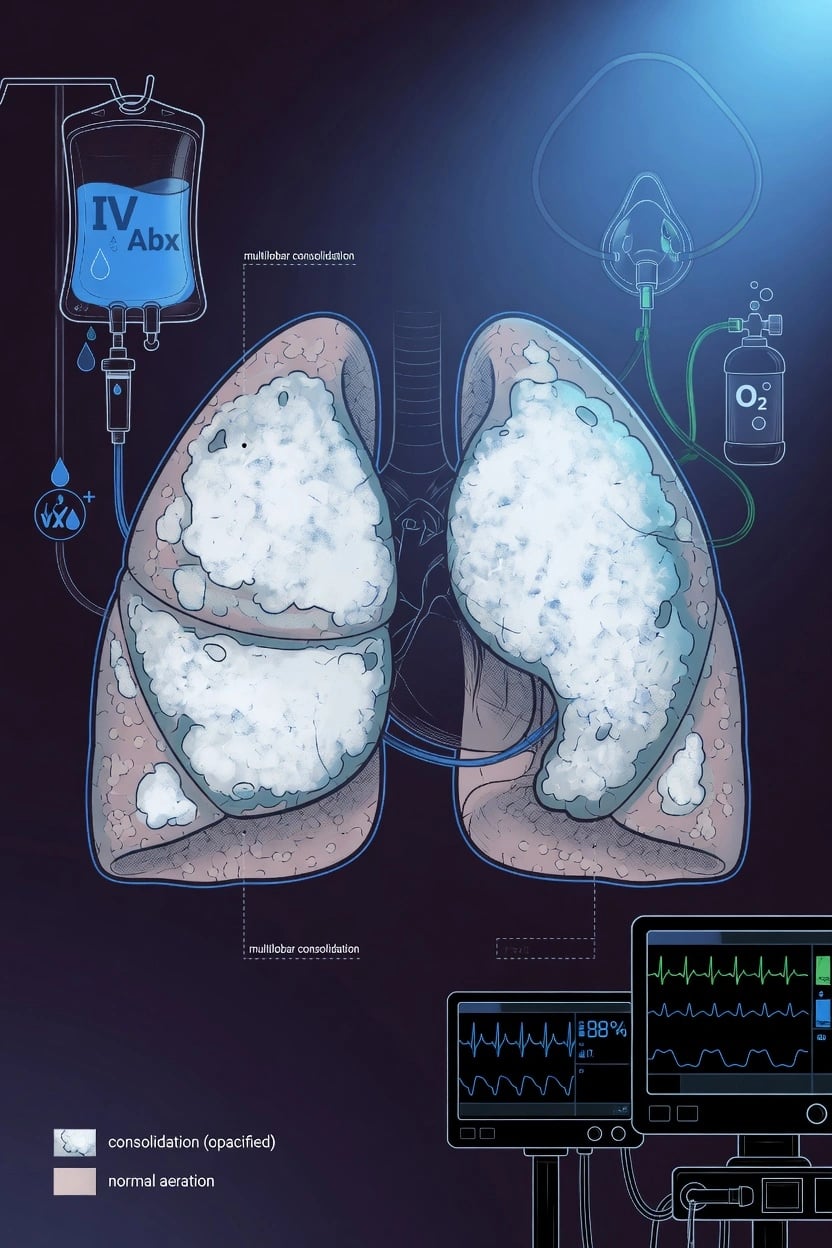
Severe CAP with respiratory failure requires mechanical ventilation in 30-60% of ICU admissions. Ventilation strategy: lung-protective ventilation (VT 6 mL/kg PBW, plateau <30, PEEP titrated). Consider prone positioning if PaO2/FiO2 <150. Open-lung vs permissive atelectasis: no clear benefit of recruitment manoeuvres in pneumonia (unlike ARDS). Antibiotic therapy: ceftriaxone + azithromoline (or moxifloxacin), broadened for MDR risk. Corticosteroids: adjunct for severe CAP (hydrocortisone 200 mg/day). De-escalate antibiotics at 48-72h based on cultures. Wean as pneumonia resolves. Target SpO2 92-96%.
On this page & tools
Your progress
Saved locally on this device.
Target exams
Red flags
Type 1 respiratory failure from CAP — the pathophysiology
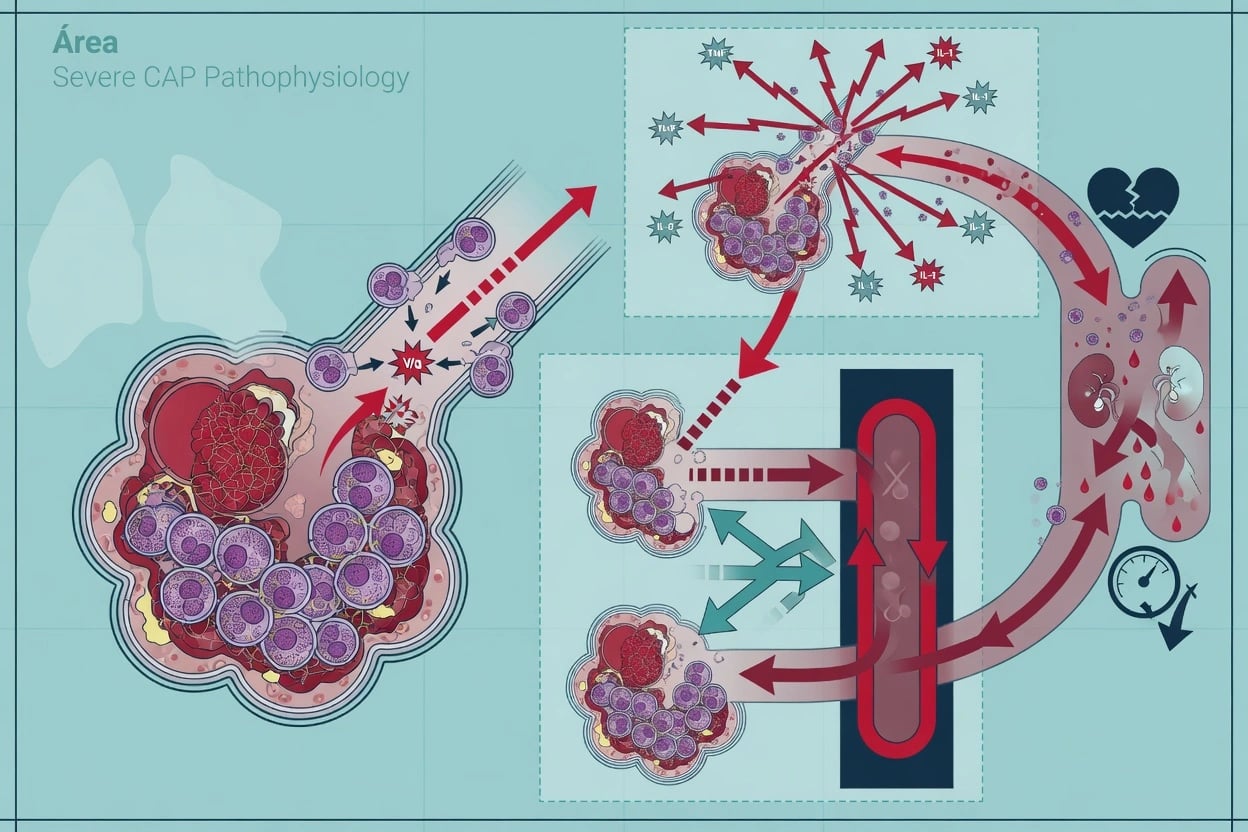
Severe CAP produces type 1 (hypoxaemic) respiratory failure — a PaO2 below 60 mmHg (SpO2 below 90 per cent) with a normal or low PaCO2. It is the dominant respiratory-failure pattern in pneumonia and arises from the consolidated, exudate-filled alveoli that the pathogen has flooded with neutrophils, protein-rich oedema and organisms. [1]
Two mechanisms dominate, and distinguishing them is the key to understanding why oxygen alone may fail:[1]
- V/Q mismatch (low V/Q — perfusion exceeds ventilation) — the commonest mechanism. Blood flows past alveoli choked with consolidation but the units are still partially ventilated, so the hypoxaemia is partially correctable with supplemental oxygen. This is the pneumonia that responds to a face mask or HFNC.
- Intrapulmonary shunt (true shunt, V/Q = 0) — perfused alveoli that are completely unventilated (consolidated or collapsed). Shunted blood bypasses the gas-exchange surface entirely, so increasing FiO2 does not raise the PaO2. This is the refractory hypoxaemia of extensive or multilobar CAP, the physiology that pushes the patient towards PEEP, recruitment and intubation. [1]
The A-a gradient is markedly elevated in CAP (a normal A-a gradient with hypoxaemia would point to hypoventilation, e.g. opiate toxicity — not pneumonia). The transition from V/Q-mismatch hypoxaemia (oxygen-responsive) to shunt physiology (oxygen-refractory) is the clinical signal that the patient is crossing from ward-level to ICU-level respiratory failure and that non-invasive strategies are about to fail.[2]
Type 2 (hypercapnic) failure is UNCOMMON in pure CAP — a rising PaCO2 in pneumonia signals respiratory-muscle fatigue and exhaustion (impending arrest), a coexisting COPD/asthma overlay, or a complication (pneumothorax, large pleural effusion, air trapping). It is a red flag, not an expected feature. [1]
Type 1 vs type 2 respiratory failure in the pneumonia patient
| Feature | Type 1 (hypoxaemic) | Type 2 (hypercapnic) |
|---|---|---|
| Definition | PaO2 <60 mmHg (SpO2 <90%), PaCO2 normal or low | PaCO2 >45 mmHg (with pH <7.35 if acute) |
| A-a gradient | Elevated (the hallmark of pneumonia) | Normal (pure) — elevated if COPD + CAP overlap |
| Mechanism in CAP | V/Q mismatch + shunt across consolidated alveoli | Muscle fatigue/exhaustion, COPD overlay, complication |
| Typical PaCO2 | Low (hyperventilation from hypoxaemic drive) | High (failing ventilation) |
| Oxygen response | Partial (V/Q mismatch) to refractory (shunt) | Improves PaO2 but may worsen CO2 in COPD retainers |
| First-line support | HFNC (FLORALI), escalate to intubation | NIV/BiPAP (if COPD overlay), controlled O2 |
| Target SpO2 | 92-96% | 88-92% (controlled oxygen in CO2 retainers) |
| Significance in CAP | The expected, common pattern | A RED FLAG — exhaustion, COPD, or complication |
The oxygen escalation ladder
Oxygen therapy in severe CAP is a stepwise escalation, matched to the severity of hypoxaemia and the work of breathing. The ladder runs from low-flow variable-performance devices through fixed-performance Venturi masks, to high-flow nasal cannula, non-invasive ventilation, and finally invasive mechanical ventilation. The decision to step up is driven by the SpO2 target (92-96% for most, 88-92% for COPD retainers) AND by the work of breathing / trend — a patient holding an acceptable SpO2 but with a rising respiratory rate and accessory-muscle use is failing and must be escalated.[2]
The oxygen escalation ladder in severe CAP
| Device | FiO2 range | Flow | Approximate PEEP | Best use in CAP |
|---|---|---|---|---|
| Nasal cannula | 24-44% | 1-6 L/min (≈+4% per L/min) | None | Mild hypoxaemia (SpO2 90-92%); comfortable, patient eats/talks |
| Simple face mask | 35-60% | 5-10 L/min (needs ≥5 to flush CO2) | None | Moderate hypoxaemia; imprecise FiO2 |
| Venturi mask | 24-60% (precise, fixed) | Set by entrainment valve | None | Precise FiO2 — the choice for COPD/CO2 retainers with CAP overlap |
| Non-rebreather mask | 60-90% (highest non-invasive) | 10-15 L/min + reservoir | None | Severe hypoxaemia as a bridge; one-way valves prevent rebreathing |
| HFNC | Up to 100% (precise) | 30-60 L/min | 3-5 cmH2O | Moderate-severe hypoxaemia (FLORALI) — first-line ICU device |
| NIV (CPAP/BiPAP) | Up to 100% | Machine-driven | 5-15 cmH2O | COPD + CAP overlap; cardiogenic oedema; select immunocompromised |
| Invasive ventilation | 21-100% | Variable | Variable | Refractory hypoxaemia, exhaustion, GCS <8, failed HFNC/NIV |
When to step up the oxygen ladder in severe CAP
Step 1 — Nasal cannula (2-6 L/min)
For MILD hypoxaemia (SpO2 90-92%). Comfortable, patient eats and talks. Each L/min adds ≈4% FiO2. FiO2 varies with the patient's breathing pattern (faster breathing entrains more room air → lower FiO2). Inadequate for true severe CAP.
Step 2 — Simple face mask (5-10 L/min, FiO2 35-60%)
For moderate hypoxaemia. MUST run at ≥5 L/min to flush exhaled CO2 from the mask (prevent rebreathing). Still an imprecise, variable-performance device. A temporary measure while preparing higher support.
Step 3 — Venturi mask (precise 24-60%)
A FIXED-performance device delivering a precise FiO2 by the oxygen-entrainment principle (the set FiO2 is independent of patient flow). The device of choice for the COPD / CO2-retaining patient with CAP, where a target SpO2 of 88-92% and a controlled FiO2 prevent oxygen-induced hypercapnia.
Step 4 — Non-rebreather mask (10-15 L/min, FiO2 60-90%)
The HIGHEST FiO2 achievable without intubation. A reservoir bag with one-way valves supplies oxygen-rich gas during inspiration and prevents rebreathing. A BRIDGE while preparing HFNC, NIV or intubation — not a destination. Use 100% FiO2 for the rapidly desaturating patient, then wean.
Step 5 — HFNC (30-60 L/min, FiO2 up to 100%)
The first-line ICU device for moderate-severe hypoxaemic CAP. Five mechanisms: precise FiO2, dead-space washout, low-level PEEP (3-5 cmH2O), heated humidification, reduced inspiratory resistance. FLORALI: reduced intubation in pneumonia. Monitor with the ROX index (intubate if <3.85). Better tolerated than NIV — patient can talk and eat.
Step 6 — NIV (CPAP or BiPAP)
Selectively. CPAP for alveolar recruitment in pure hypoxaemia (limited evidence, helmet preferred). BiPAP for the COPD/CO2-retaining patient with CAP and a rising PaCO2. CAUTION in de-novo hypoxaemic CAP without hypercapnia: face-mask NIV fails in 30-50% and may DELAY intubation (Confalonieri 1999 — only the COPD subgroup benefited).
Step 7 — Intubation and mechanical ventilation
Definitive airway and ventilatory support for: GCS <8 (cannot protect airway), refractory hypoxaemia (PaO2/FiO2 <150 despite HFNC/NIV), respiratory acidosis (pH <7.25), or exhaustion (paradoxical breathing, falling Vt, somnolence). Perform RSI with pre-oxygenation; then lung-protective ventilation (Vt 6 mL/kg PBW, PEEP 5-10, plateau <30).
HFNC in pneumonia — mechanisms, FLORALI, and the ROX index
High-flow nasal cannula (HFNC) is the first-line non-invasive device for the patient with severe CAP and moderate hypoxaemic respiratory failure. It delivers up to 60 L/min of heated, humidified, oxygen-blended gas through large-bore nasal prongs, sitting between standard oxygen and NIV. Its value in pneumonia rests on five physiological mechanisms rather than a single effect:[3]
- Low-level PEEP (3-5 cmH2O) — the high flow through the nasal passages generates positive pressure that splints open alveoli, recruits consolidated units, and reduces work of breathing. Modest, but real.
- Dead-space washout — the high flow flushes CO2-rich gas from the anatomical dead space (nasopharynx, trachea, bronchi), so each breath draws more fresh gas. This reduces the work of breathing and is especially valuable when pneumonia increases physiological dead space.
- Heated humidification (37 °C, 100% humidity) — pneumonia produces copious secretions; dry cold oxygen impairs ciliary function and thickens mucus. HFNC optimises mucociliary clearance and secretion management, a direct benefit in purulent pneumonia.
- Reduced inspiratory resistance — the high flow meets (and slightly exceeds) the patient's inspiratory demand, so the patient does not have to pull gas through high-resistance nasal passages. This offloads the fatiguing respiratory muscles.
- Precise, titrated FiO2 (21-100%) — unlike a simple mask, HFNC delivers a known oxygen concentration regardless of the patient's breathing pattern. [1]
The evidence — FLORALI
The FLORALI trial (Frat, NEJM 2015) randomised 310 patients with acute hypoxaemic respiratory failure (PaO2/FiO2 <300, non-hypercapnic) to HFNC, standard oxygen, or NIV (BiPAP).[3]
- Overall 28-day intubation rates were not significantly different (HFNC 38% vs standard 47% vs NIV 50%), though HFNC was lowest.
- In the predefined subgroups — pneumonia and PaO2/FiO2 <200 — HFNC significantly reduced intubation (35% vs 53% with standard oxygen) and showed a mortality benefit at 90 days.
- HFNC was better tolerated, with fewer ventilator-free days lost. [1]
The lesson for CAP: HFNC is a legitimate first-line device that may avoid intubation, especially in the pneumonia subgroup and the moderate-severe range — but it must be monitored, and the failing patient must be intubated promptly. [1]
The ROX index — predicting HFNC success or failure
The ROX index (Roca, J Crit Care 2016) = SpO2 / FiO2 ÷ respiratory rate, developed specifically in pneumonia patients on HFNC.[4]
- ROX ≥ 4.88 at 2 hours predicts HFNC success (continue).
- ROX < 3.85 at 2, 6, or 12 hours predicts the need for intubation — escalate.
- Values between 3.85 and 4.88 require close re-assessment and trend-watching. [1]
The ROX index is the bedside tool that prevents the cardinal error of HFNC: delayed intubation. A falling ROX index (rising respiratory rate, falling SpO2/FiO2) over the first hours signals failure and should trigger RSI, not another device. [1]
[1]NIV in pneumonia — the controversy
Non-invasive ventilation (NIV — CPAP or BiPAP) has a narrower, more contested role in CAP than HFNC. The physiological appeal is real — positive pressure recruits collapsed alveoli (CPAP), augments ventilation (BiPAP), and offloads the respiratory muscles — but the evidence in de-novo hypoxaemic CAP is mixed, and the chief harm is delayed intubation. [1]
Where NIV helps in CAP
- COPD with CAP overlay — the strongest indication. When a COPD patient develops pneumonia and tips into hypercapnic (type 2) respiratory failure, BiPAP (IPAP 10-15, EPAP 4-6) augments ventilation, clears CO2, and reduces intubation. PLANT-trial physiology: BiPAP reduces intubation and mortality in COPD with respiratory acidosis, and the CAP subgroup benefits.
- Immunocompromised CAP — selected patients (neutropenic, transplant) may benefit from early NIV to avoid the infectious complications of intubation, but modern practice increasingly favours early HFNC or controlled intubation rather than prolonged NIV trials.
- Cardiogenic pulmonary oedema masquerading as CAP — if the bilateral infiltrates are oedema, CPAP is first-line (3CPO). [1]
Where NIV fails (and harms) in CAP
- De-novo hypoxaemic CAP without hypercapnia — the face-mask NIV failure rate is 30-50%, and the harm of NIV in this group is the delayed intubation it causes. A patient who fails a prolonged NIV trial and is then intubated has worse outcomes than one intubated promptly.
- The Confalonieri 1999 RCT (CPAP vs standard oxygen in severe CAP) showed benefit only in the COPD subgroup; in pure hypoxaemic CAP there was no mortality benefit and a trend to harm.[9]
- Helmet CPAP (Patel 2016, JAMA) markedly improves NIV success in hypoxaemic failure (intubation 18% vs 62% with face mask) — if NIV is attempted for de-novo hypoxaemic CAP, the helmet interface is preferred over the face mask.
The practical rule
In severe CAP, prefer HFNC first. Reach for NIV (BiPAP) when there is a COPD/CO2-retention overlay with a rising PaCO2, or use helmet CPAP if HFNC is failing and the patient is not yet an intubation candidate. Set a time limit (1-2 hours) and intubate at the first sign of NIV failure — do not let a non-invasive trial drift into a delayed, high-risk intubation. [1]
HFNC vs NIV vs intubation in severe CAP
| HFNC | NIV (CPAP/BiPAP) | Invasive ventilation | |
|---|---|---|---|
| FiO2 | Up to 100%, precise | Up to 100% | Up to 100% |
| PEEP | 3-5 cmH2O (low-level) | 5-15 cmH2O (adjustable) | Fully adjustable |
| Best CAP role | Moderate hypoxaemia (P/F 200-300) | COPD + CAP with hypercapnia; helmet CPAP | Refractory, exhausted, GCS <8 |
| Tolerance | Excellent (talks, eats) | Moderate (claustrophobia, leaks) | Requires sedation/paralysis |
| Key trial | FLORALI (reduced intubation in pneumonia) | Confalonieri 1999 (COPD subgroup only) | ARDSNet, PROSEVA |
| Failure risk | 38% (FLORALI); monitor ROX | 30-50% in de-novo hypoxaemia | — |
| Main harm | Delayed intubation if failing | Delayed intubation, mask complications, aspiration | VAP, sedation, VILI |
| Monitor | ROX index (intubate if <3.85) | Trend, ABG at 1 h, intubate if failing | Lung-protective settings |
When to intubate in CAP respiratory failure
The decision to intubate in severe CAP rests on four pillars: airway, oxygenation, ventilation, and exhaustion. Waiting for a single hard threshold invites a crash intubation; the skilled clinician recognises the trajectory — a patient who is deteriorating despite escalating non-invasive support needs intubating now, not after the next blood gas. [1]
Indications for intubation in severe CAP
Airway compromise
GCS <8 — the patient cannot protect the airway (aspiration risk), from hypoxia, hypercapnia, or septic encephalopathy. Also: copious secretions the patient cannot clear, or loss of gag/cough reflex.
Refractory hypoxaemia
PaO2/FiO2 <150 despite optimised HFNC or NIV. OR SpO2 <90% despite FiO2 ≥60%. This is shunt physiology — the consolidated lung cannot oxygenate blood, and only PEEP from mechanical ventilation will recruit units. Increasing FiO2 further will not help.
Respiratory acidosis
pH <7.25 from CO2 retention, persisting despite NIV/BiPAP. A rising PaCO2 in CAP is muscle fatigue or a complication (pneumothorax, air trapping) — the failing ventilatory pump needs mechanical replacement.
Exhaustion (pre-terminal)
Paradoxical breathing (abdomen moves INWARD on inspiration — diaphragm failure), falling tidal volume, decreasing respiratory rate (bradypnoea — the fatigued patient slows down), somnolence, and silent chest. This is imminent respiratory arrest — intubate IMMEDIATELY, do not wait for the blood gas.
Haemodynamic instability
Septic shock with rising lactate, worsening acidosis, and increasing vasopressor requirement — the oxygen debt demands intubation to reduce the work of breathing (which consumes a large fraction of cardiac output in the distressed patient) and to deliver lung-protective ventilation.
Failed non-invasive trial
HFNC with ROX index <3.85 at 2-12 h, or NIV failure (rising RR, falling SpO2, rising PaCO2 after 1-2 h). The cardinal error is a delayed intubation after a prolonged, failing non-invasive trial — outcomes are worse than prompt intubation.
Rapid sequence intubation (RSI) in severe CAP
The pneumonia patient is haemodynamically fragile, hypoxaemic, and difficult to pre-oxygenate — the classic "can't intubate, can't oxygenate" risk is high. RSI (rapid sequence intubation — no mask ventilation, cricoid pressure, a fast-acting induction agent plus a fast-acting paralytic, with a prepared back-up airway plan) is the standard. The CAP-specific modifications matter: [1]
RSI for severe CAP — the CAP-specific checklist
1. Prepare and pre-oxygenate
Pre-oxygenate with 100% FiO2 (HFNC kept ON during the attempt — "apnoeic oxygenation" via HFNC maintains oxygenation during the apnoeic period). A pneumonia lung desaturates FAST — the FRC is reduced by consolidation and the shunt fraction is high, so the safe apnoea time may be under a minute. Have a video laryngoscope and a supraglottic-airway/basket rescue ready. Position head-up.
2. Induction — KETAMINE preferred
Ketamine (1-2 mg/kg IV) is the preferred induction agent in severe CAP: it PRESERVES bronchial tone and sympathetic drive (bronchodilator effect — useful when pneumonia coexists with bronchospasm/COPD/asthma), supports blood pressure in septic shock (sympathetic stimulation — unlike propofol which causes hypotension), and maintains respiratory drive. AVOID propofol in septic/hypotensive CAP (severe hypotension). Etomidate is acceptable but suppresses cortisol (relevant if considering hydrocortisone for refractory shock).
3. Paralysis — AVOID succinylcholine if hyperkalaemic
Succinylcholine (1-1.5 mg/kg) is fast-onset/fast-offset but RAISES serum potassium by ≈0.5 mmol/L. In severe CAP with rhabdomyolysis, sepsis-induced hyperkalaemia, crush injury, or burns >24 h, this can provoke malignant arrhythmia — use ROCURONIUM (1.2 mg/kg) instead. Rocuronium is also preferred when sugammadex reversal is available. Rocuronium does not raise potassium and is the standard paralytic in septic CAP.
4. Intubate and confirm
Direct or video laryngoscopy; pass the tube; CONFIRM with waveform capnography (CO2 trace — the gold standard), bilateral chest rise, and chest auscultation. Secure the tube. Obtain a post-intubation CXR to confirm depth and rule out right-main-stem intubation, aspiration, or pneumothorax.
5. Post-intubation
Sedate and analgese (propofol + fentanyl, or midazolam + fentanyl; add dexmedetomidine early for delirium-sparing sedation). Set lung-protective ventilation (Vt 6 mL/kg PBW, PEEP 5-10, plateau <30, FiO2 titrated to SpO2 92-96%). Send a tracheal aspirate for culture (diagnostic — ventilator-associated sampling). Start/review antibiotics. Add hydrocortisone 200 mg/day for severe CAP.
Induction and paralytic agents for RSI in severe CAP
| Agent | Dose | Advantages in CAP | Cautions in CAP |
|---|---|---|---|
| Ketamine (preferred) | 1-2 mg/kg IV | Bronchodilator (preserves bronchial tone), supports BP in shock, maintains respiratory drive | Transient emergence phenomena (less relevant if sedated post-RSI); hypersalivation (mitigated by glycopyrrolate) |
| Propofol | 1.5-3 mg/kg IV | Smooth, antiemetic, reduces ICP | Severe hypotension — AVOID in septic/shocked CAP |
| Etomidate | 0.2-0.3 mg/kg IV | Haemodynamically neutral | Adrenal suppression (single-dose — give hydrocortisone if shock) |
| Succinylcholine | 1-1.5 mg/kg IV | Fast onset/offset (40-60 s) | RAISES K+ ≈0.5 mmol/L — AVOID if hyperkalaemia (rhabdo, sepsis, burns >24h, crush) |
| Rocuronium (preferred paralytic) | 1.2 mg/kg IV | No potassium rise, reversible with sugammadex | Longer offset than sux (relevant if difficult airway); irreversible if no sugammadex |
Ventilator settings for pneumonia
Once intubated, the pneumonia lung needs lung-protective ventilation. The consolidated, heterogeneous lung is small ("baby lung") and prone to ventilator-induced lung injury (VILI) from overdistension of the healthy regions and cyclic collapse/reopening (atelectrauma) of the diseased regions — the same biotrauma pathway as ARDS, whether or not the patient formally meets ARDS criteria. [1]
Initial ventilator settings for severe CAP
| Setting | Recommendation | Rationale |
|---|---|---|
| Mode | Volume-controlled (VC) or pressure-controlled (PC) | VC guarantees tidal volume (lung protection); PC may improve gas distribution in heterogeneous lung |
| Tidal volume | 6 mL/kg PBW (4-8 range) | Lung-protective — reduces VILI even WITHOUT ARDS. PBW = 50 + 2.3 × (height in inches − 60) for men; adjust for women |
| Plateau pressure | <30 cmH2O | Limits overdistension (barotrauma/volutrauma) of the healthy, compliant lung regions |
| Driving pressure (ΔP) | <15 cmH2O (plateau − PEEP) | The best predictor of survival (Amato 2015) — reflects the stress on the baby lung |
| PEEP | 5-10 cmH2O, titrate to oxygenation | Splints open alveoli, improves oxygenation, reduces atelectrauma. CAUTION: high PEEP may overdistend healthy regions in heterogeneous pneumonia (unlike uniform ARDS) |
| Respiratory rate | 12-20/min (adjust for PaCO2) | Target PaCO2 35-45 (permissive hypercapnia acceptable if pH ≥7.25 — avoids injurious Vt/pressure) |
| FiO2 | Minimum for SpO2 92-96% | Avoid hyperoxia (HOT-ICU, ICU-ROX — no benefit, possible harm). Liberal oxygen increases mortality |
| I:E ratio | 1:2 (or inverse 1:1 if severe) | Inverse ratio improves oxygenation (longer inspiratory time) but risks air trapping and haemodynamic compromise |
| Target SpO2 | 92-96% (88-92% if COPD retainer) | Conservative oxygenation — no benefit to hyperoxia |
PBW calculation matters: tidal volume is set on predicted (ideal) body weight, NOT actual body weight — using actual weight over-ventilates (and injures) the lung. Measure height and calculate PBW. [1]
Ventilation strategy for pneumonia
Initial settings
Lung-protective
- Mode: volume-controlled (VC) or pressure-controlled (PC)
- Tidal volume: 6 mL/kg predicted body weight (PBW) — even without ARDS, reduces lung injury
- PEEP: start 5-8, titrate to oxygenation (avoid overdistension of healthy lung regions)
- FiO2: minimum to maintain SpO2 92-96% (or PaO2 >60)
- Plateau pressure: <30 cmH2O
- Rate: 12-20 (adjust for PaCO2 target 35-45)
If worsening (PaO2/FiO2 <150)
Escalation
- Increase PEEP cautiously (may worsen overdistension in heterogeneous lung)
- Prone positioning (16h/day — improves oxygenation, reduces mortality in ARDS-like physiology)
- Consider neuromuscular blockade (cisatracurium infusion — reduces ventilator dyssynchrony)
- ECCO2R (extracorporeal CO2 removal) or VV-ECMO if refractory
- Recruitment manoeuvres: controversial in pneumonia (may worsen overdistension)
Management of treatment failure
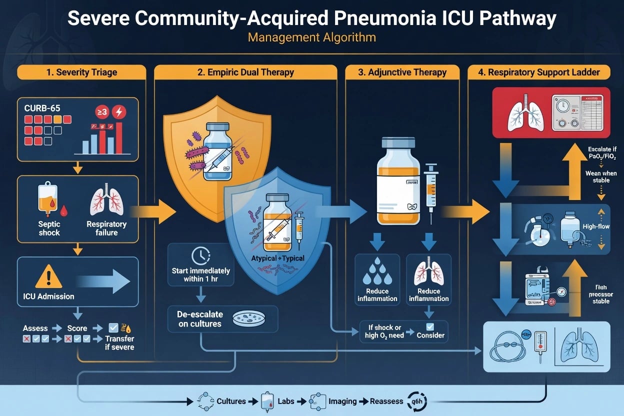
What to do when CAP doesn't improve at 72 hours
Reassess diagnosis
Is it really pneumonia? Consider: pulmonary embolism, pulmonary oedema, ARDS (non-pulmonary sepsis), vasculitis, alveolar haemorrhage, drug reaction, eosinophilic pneumonia. Check: CT chest (pattern, distribution, complications).
Check for complications
Empyema/parapneumonic effusion (thoracentesis — pH, culture). Lung abscess (CT — cavity with air-fluid level). ARDS (PaO2/FiO2 <300 with bilateral infiltrates — different ventilation strategy). Septic emboli (endocarditis — echo, blood cultures). Metastatic infection (meningitis, endocarditis).
Review antibiotic coverage
Are the right organisms covered? Consider: atypicals (Legionella, Mycoplasma — urinary antigen, serology), MRSA (add vancomycin/linezomid), Pseudomonas (add anti-pseudomonal), fungal (Aspergilla in immunocompromised), viral (influenza, COVID-19 — antivirals), TB. Check culture results and sensitivities. Consider bronchoalveolar lavage (BAL) if sputum inadequate.
Optimise source control
Drain pleural effusion if large or empyema (chest tube). Drain abscess (percutaneous or surgical). Remove/replace infected central line. Treat concurrent infection (UTI, cholangitis, endocarditis).
Consider corticosteroids
Hydrocortisone 200 mg/day (continuous infusion) for severe CAP with high inflammatory burden. CAPE COD trial (2023): hydrocortisone reduced 28-day mortality in severe CAP. Also beneficial for PJP, influenza pneumonia. Continue for 7 days or until improvement.
The landmark trials — oxygen, ventilation, and adjuncts in CAP
FLORALI
NEJM 2015
310 pts with acute hypoxaemic respiratory failure (PaO2/FiO2 <300, non-hypercapnic) — HFNC vs standard O2 vs NIV (BiPAP)
Key finding
Overall intubation: HFNC 38% vs standard 47% vs NIV 50% (NS overall). In the pneumonia and PaO2/FiO2 <200 subgroups, HFNC significantly reduced intubation (35% vs 53%) and 90-day mortality. HFNC better tolerated.
Practice change
HFNC is first-line for moderate-severe hypoxaemic respiratory failure, especially pneumonia and PaO2/FiO2 <200
ROX index (Roca 2016)
J Crit Care 2016
Prospective cohort — pneumonia patients on HFNC; derived SpO2/FiO2 ÷ RR
Key finding
ROX ≥4.88 at 2 h predicts HFNC success; ROX <3.85 predicts the need for intubation. Provides a bedside tool to time intubation and avoid the harm of a delayed, failing HFNC trial
Practice change
Use the ROX index to monitor HFNC and trigger intubation when <3.85
ARDSNet
NEJM 2000
861 pts with ALI/ARDS — Vt 6 mL/kg PBW + plateau <30 vs Vt 12 mL/kg + plateau <50
Key finding
Low tidal volume REDUCED mortality (31% vs 39.8%, 22% relative reduction) and increased ventilator-free days
Practice change
Lung-protective ventilation (Vt 6 mL/kg PBW, plateau <30) is the standard for ALL severe lung injury, including pneumonia with or without ARDS
PROSEVA
NEJM 2013
466 pts with severe ARDS (PaO2/FiO2 <150) — prone ≥16 h/day vs supine, early
Key finding
Prone positioning REDUCED 28-day mortality (16.0% vs 32.8%, NNT 6) with no increase in complications
Practice change
Prone ≥16 h/day for moderate-severe ARDS (PaO2/FiO2 <150) — including CAP-ARDS
CAPE COD
NEJM 2023
RCT — hydrocortisone 200 mg/day (continuous infusion) for 7 days vs placebo in severe CAP requiring ICU
Key finding
REDUCED 28-day treatment failure (need for ventilation, vasopressors, or death). Stopped early for efficacy. Strongest evidence for corticosteroids in bacterial CAP
Practice change
Hydrocortisone 200 mg/day for 7 days is adjunctive therapy in severe CAP (ICU, high inflammatory burden)
HOT-ICU
NEJM 2021
2928 pts with acute hypoxaemic respiratory failure — lower (PaO2 60 mmHg) vs higher (PaO2 90 mmHg) oxygenation target
Key finding
No difference in 90-day mortality (lower 42.4% vs higher 42.8%). No subgroup benefit. Conservative targets are safe
Practice change
Target SpO2 92-96% (PaO2 ~60-80) — avoid liberal oxygen; no benefit and possible harm from hyperoxia
Confalonieri 1999
Am J Respir Crit Care Med 1999
RCT — CPAP vs standard oxygen in severe CAP
Key finding
CPAP improved oxygenation and dyspnoea but NO mortality benefit overall; benefit confined to the COPD subgroup. Frames NIV as a COPD-overlay therapy, not a de-novo hypoxaemic CAP therapy
Practice change
NIV in CAP is selective — benefit mainly when COPD/CO2-retention overlays the pneumonia
Amato 2015 (driving pressure)
NEJM 2015
Individual-patient-data meta-analysis of ARDS ventilation trials
Key finding
Driving pressure (ΔP = plateau − PEEP) was the ventilatory variable most strongly associated with survival — independent of Vt and PEEP. ΔP <15 cmH2O safest
Practice change
Monitor and minimise driving pressure (target <15 cmH2O) alongside Vt 6 mL/kg and plateau <30
Advanced ventilation — when P/F <150
When the pneumonia patient develops moderate-severe ARDS (PaO2/FiO2 <150) despite lung-protective ventilation, a stepped escalation is applied. The aim is to improve oxygenation and reduce VILI without harming the failing lung further.[2]
Escalation ladder when pneumonia progresses to severe ARDS
Optimise PEEP
Titrate PEEP using a PEEP/FiO2 table or, better, by compliance (driving pressure). Aim for the PEEP that minimises ΔP (best compliance). CAUTION in pneumonia: the heterogeneous lung means high PEEP may overdistend healthy regions and worsen oxygenation — judge by response, not by a number.
Prone positioning (≥16 h/day)
For PaO2/FiO2 <150. PROSEVA: 16% absolute mortality reduction. Mechanism: recruits dorsal (dependent, collapsed) lung, redistributes perfusion, reduces shunt, homogenises ventilation. Continue daily until PaO2/FiO2 >150 with reasonable FiO2/PEEP.
Neuromuscular blockade
Cisatracurium infusion for 48 h in severe hypoxaemia (PaO2/FiO2 <150). ACURASYS: reduced mortality; ROSE: no routine benefit. Use selectively for severe dyssynchrony, high driving pressure, or profound hypoxaemia. Ensure deep sedation first (NMB without sedation is unacceptable).
Conservative fluid strategy
FACTT: a fluid-restrictive strategy (target even-to-negative fluid balance once shock resolves) improved oxygenation and ventilator-free days. Balance against perfusion — use dynamic monitoring (PLR, SVV) and vasopressors to minimise fluid while maintaining MAP ≥65.
VV-ECMO
Rescue for refractory hypoxaemia (PaO2/FiO2 <80) or severe hypercapnic acidosis despite optimised ventilation + proning. EOLIA (2018): non-significant, but Bayesian re-analysis suggests possible benefit. Transfer to an ECMO centre early (CESAR).
SAQ — Choosing the oxygen/ventilation interface in CAP respiratory failure
10 minutes · 10 marks
A 66-year-old woman (BMI 31) presents with 2 days of fever, rigors and progressive dyspnoea. RR 34, SpO2 86% on a non-rebreather at 15 L/min, BP 102/64, HR 118, GCS 15. CXR shows multilobar consolidation. ABG: pH 7.32, PaCO2 34, PaO2 56, lactate 2.4. The examiners ask you to choose the respiratory support strategy and outline thresholds for escalation.
SAQ — Non-resolving pneumonia and ventilator-associated problems
10 minutes · 10 marks
A 71-year-old man intubated for severe CAP (ceftriaxone + azithromycin) has had no improvement after 72 hours. Temperature 38.5°C, purulent secretions, FiO2 has risen from 0.5 to 0.7, and a new left lower lobe infiltrate is seen on CXR. Plateau pressure has risen from 24 to 32 cmH2O. The examiners ask you to systematically evaluate failure to improve and address the rising plateau pressure.
Clinical pearls
Red flags
References
- [1]Martin-Loeches I, Torres A. Severe community-acquired pneumonia Eur Respir Rev, 2022.PMID 36517046
- [2]Ferrer M, et al. Notum palmitoleoyl-protein carboxylesterase regulates Fas cell surface death receptor-mediated apoptosis via the Wnt signaling pathway in colon adenocarcinoma Bioengineered, 2021.PMID 34402722
- [3]Frat JP, Thille AW, Mercat A, et al.; FLORALI Study Group; REVA Network. High-flow oxygen through nasal cannula in acute hypoxemic respiratory failure N Engl J Med, 2015.PMID 25981908
- [4]Roca O, Messika J, Caralt B, et al. Predicting success of high-flow nasal cannula in pneumonia patients with hypoxemic respiratory failure: The utility of the ROX index J Crit Care, 2016.PMID 27481760
- [5]Acute Respiratory Distress Syndrome Network (ARDSNet). Handling of hazardous materials Ann Emerg Med, 2000.PMID 10613956
- [6]Guérin C, Reignier J, Richard JC, et al.; PROSEVA Study Group. Prone positioning in severe acute respiratory distress syndrome N Engl J Med, 2013.PMID 23688302
- [7]Dequin PF, Meziani F, Quenot JP, et al.; CAPE COD Network. Aspirin or Low-Molecular-Weight Heparin for Thromboprophylaxis after a Fracture N Engl J Med, 2023.PMID 36652352
- [8]Schjørring OL, Klitgaard TL, Perner A, et al.; HOT-ICU Investigators. Discovery and Characterisation of Highly Cooperative FAK-Degrading PROTACs Angew Chem Int Ed Engl, 2021.PMID 34416073
- [9]Confalonieri M, Potena A, Carbone G, et al. Irritated lesion. What does it mean when an inflamed scaly plaque becomes red-brown in color? Geriatrics, 1999.PMID 10451644