ICU · Respiratory / ventilation
Acute Severe COPD Exacerbation: NIV and Invasive Ventilation — Comprehensive ICU Management
Also known as AECOPD · Acute exacerbation of COPD · COPD type 2 respiratory failure · Bilevel NIV in COPD · Plant trial NIV · Permissive hypercapnia · Auto-PEEP ventilation · Anthonisen criteria · Controlled oxygen COPD
An acute severe exacerbation of COPD (AECOPD) causing hypercapnic (type 2) respiratory failure is an ICU emergency built on three pillars: controlled oxygen (SpO2 88-92 per cent to avoid oxygen-induced hypercapnia), a medical bundle (nebulised salbutamol 5 mg with ipratropium 500 mcg, prednisolone 40 mg for 5 days, and antibiotics when Anthonisen criteria are met), and escalating respiratory support driven by the arterial pH. Bilevel non-invasive ventilation (BiPAP, IPAP 10-15, EPAP 4-6, titrate up) is first-line for the acidotic patient (pH <7.35) — the Plant trial (Lancet 2000) and the Lightowler Cochrane meta-analysis (BMJ 2003) prove it cuts intubation, mortality, and length of stay. Intubate when NIV fails (pH <7.25 after one hour of NIV, worsening GCS, respiratory arrest); on the ventilator use a low rate (10-12), long expiration (I:E 1:3-1:4), external PEEP just below intrinsic PEEP, and permissive hypercapnia (pH >7.20). Early mobilisation and pulmonary rehabilitation referral complete the pathway.
On this page & tools
Your progress
Saved locally on this device.
1 MCQ with explanations
Target exams
Red flags
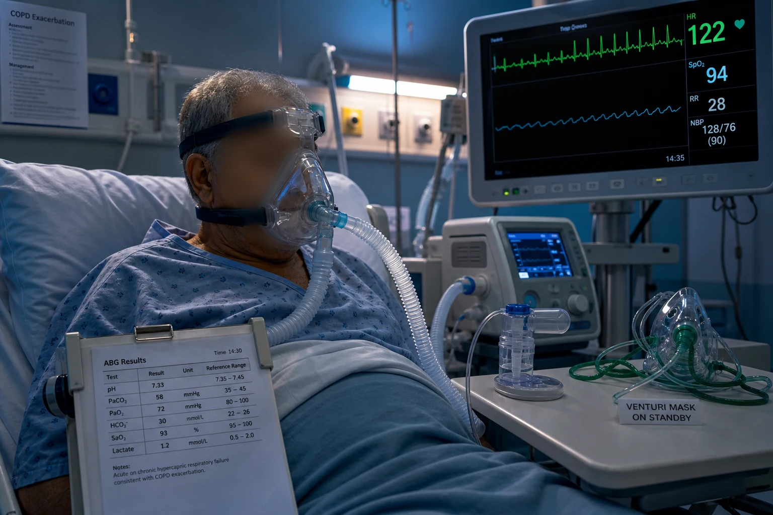
Definition and GOLD context
An acute exacerbation of COPD (AECOPD) is an acute, sustained worsening of dyspnoea, cough, and/or sputum beyond day-to-day variation that prompts a change in regular medication. The GOLD 2024 report defines it as an event characterised by increased respiratory symptoms, and emphasises that exacerbations drive disease progression, hospitalisation, mortality, and healthcare cost.[1]
GOLD stratifies exacerbation severity by the clinical impact:
- Mild — treated with short-acting bronchodilators only (SABA/SAMA).
- Moderate — requires antibiotics and/or systemic corticosteroids in addition to bronchodilators.
- Severe — the patient has acute respiratory failure needing ventilatory support (NIV or invasive mechanical ventilation) or develops haemodynamic instability; this is the domain of the ICU.[1]
The NICE NG115 guideline aligns with GOLD: it recommends controlled oxygen, systemic corticosteroids, antibiotics guided by sputum purulence, and NIV for hypercapnic respiratory failure, and stresses early pulmonary rehabilitation after discharge.[1]
In the ICU the entity of interest is the severe AECOPD with hypercapnic (type 2) respiratory failure and respiratory acidosis — the low-pH, high-CO2 patient for whom bilevel NIV was designed and proven.[1][2]
Causes and triggers — infection is number one
Around 70 per cent of exacerbations are infective, with the remainder due to environmental exposure or a non-infective complication. Identifying and treating the trigger is as important as supporting ventilation.[1]
Infection (~70%)
The dominant trigger
- Viral (~50%): rhinovirus, influenza, parainfluenza, RSV, coronavirus, adenovirus
- Bacterial (~30-50%): Haemophilus influenzae, Streptococcus pneumoniae, Moraxella catarrhalis; Pseudomonas aeruginosa in severe disease/bronchiectasis
- Co-infection (viral + bacterial) is common and drives a larger FEV1 fall
- Defines the rationale for antibiotics when sputum is purulent (Anthonisen)
Environmental (~10%)
Non-infective irritant
- Air pollution (PM2.5, ozone, NO2) and temperature drops
- Occupational dust/fume exposure
- Cigarette smoking continuation
- Often coexists with infection; treat supportively
Unknown (~30%)
Cryptogenic
- No pathogen or irritant identified
- Eosinophilic phenotype may dominate (steroid-responsive)
- Blood eosinophil count can guide ICS use in stable disease
The mimics
Do not miss
- Pneumothorax (sudden unilateral deterioration)
- Pulmonary embolism (disproportionate hypoxia, tachycardia, immobility)
- Cardiac failure / arrhythmia (atrial fibrillation, cor pulmonale, ACS)
- Pleural effusion, lobar collapse from mucus plugging, aspiration
Pathophysiology — why the COPD patient retains CO2
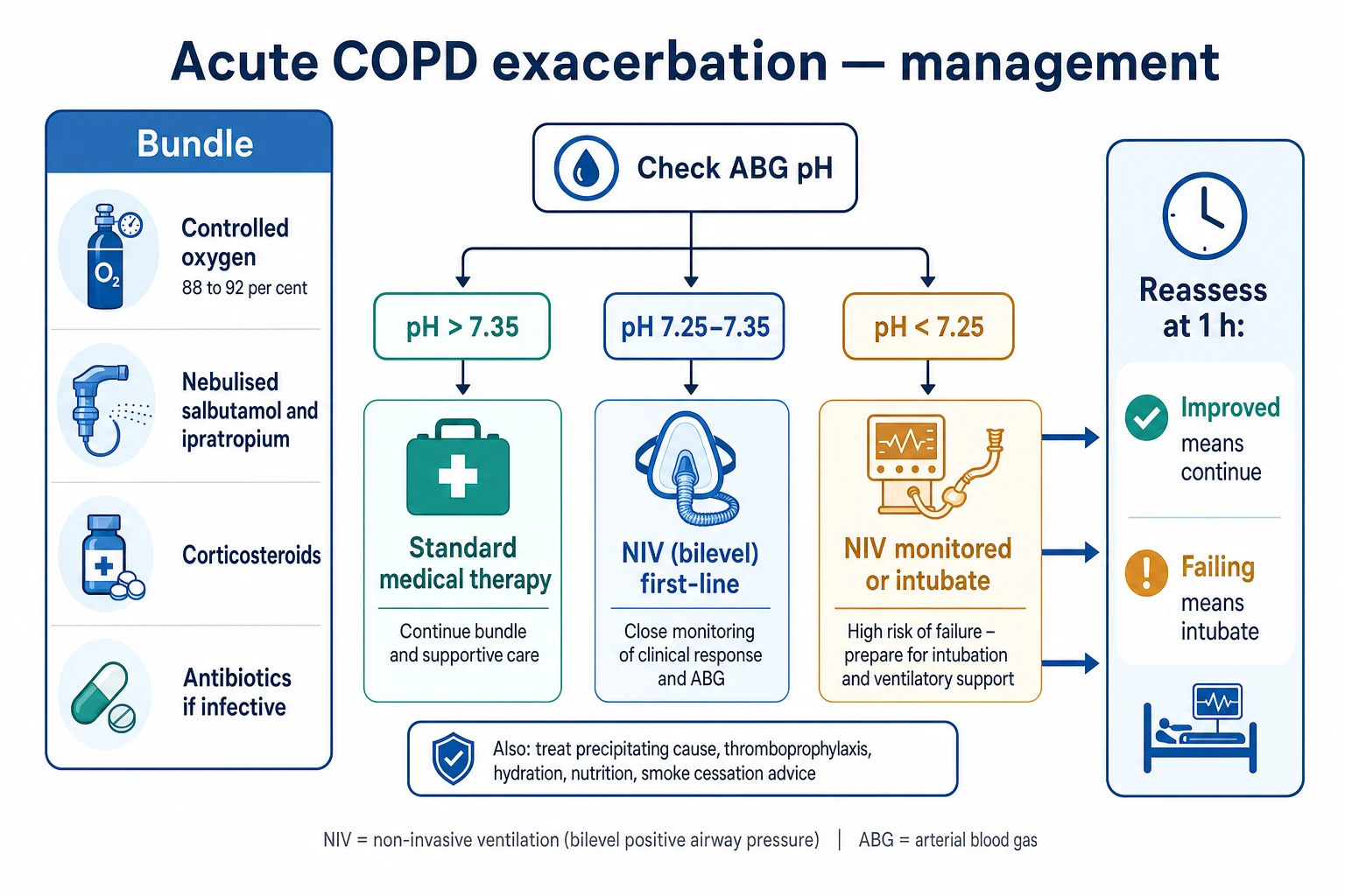
The respiratory failure of an AECOPD is type 2 (hypercapnic) with a respiratory acidosis. Four mechanisms converge, and oxygen therapy can worsen all of them:[1]
1. Increased load, reduced capacity. Bronchospasm, mucosal oedema, and secretions raise airway resistance; dynamic hyperinflation (gas trapping) adds an elastic threshold load — the patient must generate a pressure equal to the intrinsic PEEP (auto-PEEP) before any airflow begins. The respiratory muscles fatigue, alveolar ventilation falls, and PaCO2 climbs. [1]
2. V/Q mismatch. Areas of low ventilation relative to perfusion increase physiological dead space and venous admixture, raising PaCO2 and lowering PaO2. [1]
3. The Haldane effect. Deoxygenated haemoglobin carries CO2 readily (as carbamino compounds and bicarbonate). Oxygenating haemoglobin releases this CO2. In the ventilation-limited patient the released CO2 cannot be cleared, so PaCO2 rises acutely — the molecular basis of oxygen-induced hypercapnia. [1]
4. Loss of hypoxic pulmonary vasoconstriction. A high FiO2 abolishes hypoxic vasoconstriction, redistributing blood to poorly ventilated units and increasing dead space. [1]
Clinical presentation
The patient is usually a known COPD sufferer (though AECOPD may be the first presentation). Typical features: [1]
- Breathlessness worsening over hours to days, with accessory muscle use, pursed-lip breathing, and inability to speak in full sentences
- Tachypnoea initially, then bradypnoea as the patient tires — a pre-arrest sign; a silent chest signals exhaustion and impending arrest
- Wheeze and a prolonged expiratory phase
- Productive cough with increased sputum volume or purulence if infective
- Confusion or drowsiness from CO2 narcosis (a falling GCS is a relative contraindication to NIV and a trigger to intubate)
- Signs of haemodynamic compromise (tachycardia, hypotension, atrial fibrillation) in the advanced case
- Warm peripheries, a bounding pulse, and asterixis (CO2 flap) in severe hypercapnia [1]
A sudden unilateral deterioration with tracheal deviation suggests tension pneumothorax — treat it before treating the exacerbation. [1]
Severity — the arterial blood gas drives everything
The arterial blood gas is the single most important investigation. The pH defines severity and dictates the level of respiratory support.[1][2]
AECOPD severity by arterial pH (click each)
pH <7.25
Severe acidosis. NIV may still work but must be in a monitored setting (HDU/ICU); have a low threshold to intubate. Co-existing obtundation, agitation, or haemodynamic instability favours intubation.
Outcomes by pH band and NIV use
Investigations
- Arterial blood gas — the pivotal test. Record pH, PaCO2, PaO2, bicarbonate, base excess. A raised bicarbonate confirms chronic CO2 retention now acutely decompensated. Recheck after one hour of NIV.
- Chest X-ray — find the trigger and the mimics: pneumothorax, consolidation, pulmonary oedema, pleural effusion.
- ECG — atrial fibrillation and right heart strain (cor pulmonale) are common; exclude myocardial ischaemia.
- Full blood count, electrolytes, CRP — polycythaemia from chronic hypoxia, hypokalaemia from beta-agonist therapy, an infective marker.
- Sputum and blood cultures — if infective, taken before antibiotics.
- Troponin and BNP — if a cardiac contribution is suspected.
- Venous blood gas — a useful screen; a normal venous pH makes a significant acidosis unlikely, but an arterial gas is definitive.
- Theophylline level — only if the patient takes a methylxanthine. [1]
Management — the bundle and the escalation
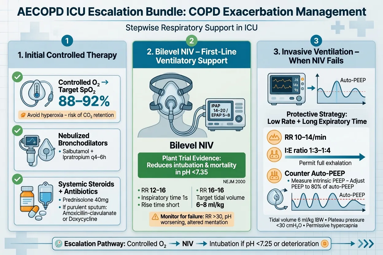
Management runs in parallel: the medical bundle (oxygen, bronchodilators, steroids, antibiotics) is applied to every patient, while respiratory support is escalated according to the ABG pH.[1][1]
The medical bundle
Controlled oxygen
First step
- Venturi mask 24-28 per cent, titrate to SpO2 88-92 per cent
- Avoid high-flow oxygen — worsens hypercapnia (Haldane effect, V/Q mismatch)
- Recheck ABG 30-60 min after any change in FiO2
- During NIV, set FiO2 to the same SpO2 target
Bronchodilators
Salbutamol + ipratropium
- Nebulised salbutamol 5 mg + ipratropium 500 mcg
- Frequency q1-4h in the severe case, settling to q6h; continuous if refractory
- In the pre-NIV patient drive the nebuliser with AIR, not oxygen (avoids hypercapnia)
- In the ventilated patient give via the in-line nebuliser circuit
- IV salbutamol has no advantage and more toxicity — avoid
Corticosteroids
Prednisolone 40 mg
- Prednisolone 40 mg PO daily for 5 days; no taper needed
- IV hydrocortisone 100 mg if oral absorption is uncertain
- Speeds recovery, reduces early relapse, improves FEV1 and dyspnoea
- Prolonged (>14 day) courses add hyperglycaemia, myopathy, infection — avoid
Antibiotics
If Anthonisen criteria
- Give for Anthonisen type 1 or 2 (increased volume + purulence +/- dyspnoea); purulent sputum alone is sufficient (NICE)
- First-line: amoxicillin-clavulanate 500/125 mg PO TDS, or doxycycline 100 mg BD
- Severe/ICU: broaden cover; add Pseudomonas cover if risk factors (recent hospital/antibiotics, bronchiectasis, severe airflow limitation)
- 5-7 days; tailor to cultures; a procalcitonin-guided strategy reduces antibiotic exposure
The escalation pathway — controlled oxygen to NIV to intubation
Respiratory support is ABG-driven. The FlowSteps below trace the complete pathway from the acidotic ward patient to the intubated ICU patient.[1][2]
The escalation pathway — controlled oxygen to NIV to intubation
Step 1 — Controlled oxygen + medical bundle
Venturi 24-28 per cent to SpO2 88-92 per cent. Salbutamol 5 mg + ipratropium 500 mcg nebulised in AIR. Prednisolone 40 mg. Antibiotics if Anthonisen criteria met. Take an ABG. This is the floor of every pathway.
Step 2 — Start NIV if pH 7.25-7.35
Bilevel (BiPAP): IPAP 10-15, EPAP 4-6 cmH2O, FiO2 to SpO2 88-92 per cent, well-fitting full-face (oronasal) mask, backup rate ~12-14. The Plant trial (2000) showed early NIV here reduces intubation (15% vs 27%) and mortality; the Lightowler meta-analysis (2003) confirms reduced mortality (RR 0.41), intubation (RR 0.42), and stay. Do not wait for the patient to tire.
Step 3 — Titrate NIV up
Increase IPAP by 2 cmH2O every 10-15 min (toward 20 cmH2O) until the tidal volume rises, the respiratory rate falls, and the PaCO2 drops. Keep EPAP at 4-6 (raise only if oxygenation is the problem). Recheck ABG at 1 hour.
Step 4 — Reassess at 1 hour
Improved (rising pH, falling PaCO2 and RR, less distress) = continue and wean. Failing (pH <7.25 and not improving, worsening GCS, rising RR, agitation) = NIV failure — do NOT prolong a failing trial; move to intubation. A delayed intubation after failed NIV is a higher-risk intubation.
Step 5 — Intubate for NIV failure or contraindication
Intubate for: pH <7.25 failing NIV after 1 hour; respiratory arrest or peri-arrest; obtundation (GCS <8, cannot protect airway); copious secretions NIV cannot clear; haemodynamic instability; agitation precluding NIV. Use RSI; anticipate hypotension on induction (preload-dependent, high intrathoracic pressure).
Step 6 — Ventilate for permissive hypercapnia
Volume-controlled, Vt 6-8 mL/kg IBW, rate 10-12, I:E 1:3 or 1:4 (long expiration), external PEEP ~75-80 per cent of intrinsic PEEP (typically 5 cmH2O). Accept a PaCO2 rise provided pH >7.20. Watch for dynamic hyperinflation — disconnect to vent auto-PEEP if hypotensive.
Bilevel NIV — settings and technique
NIV in AECOPD is bilevel (BiPAP), not CPAP. CPAP alone does not unload the work of breathing against auto-PEEP and is not first-line for COPD. The IPAP (inspiratory positive airway pressure) unloads the fatigued muscles and boosts tidal volume; the EPAP (expiratory positive airway pressure) splints the airways open and counteracts intrinsic PEEP, lowering the pressure the patient must generate to trigger a breath.[2][4]
BiPAP settings for AECOPD — starting values and targets
Technique points:
- Use a well-fitting oronasal (full-face) mask first; switch to a nasal or total-face mask if leak or claustrophobia is a problem.
- Let the patient hold the mask to the face to build tolerance before strapping it on.
- Set a backup respiratory rate (~12-14) so the ventilator supports an apnoeic or bradypnoeic patient, but allow spontaneous triggering.
- Humidify, suction regularly, and allow brief "sip and peel-off" breaks for the cooperative patient.
- Recheck the ABG at one hour — the pH trend at one hour is the best predictor of NIV success.[1]
Contraindications to NIV
Absolute (or near-absolute) — favour intubation: respiratory arrest, inability to protect the airway (low GCS, copious secretions), severe agitation or confusion precluding cooperation, haemodynamic instability, facial trauma/surgery, upper airway obstruction. [1]
Relative — proceed with caution in a monitored area: pH <7.25, pneumonia on CXR, and the patient who fails to improve at one hour.[2]
NIV versus invasive ventilation — when to choose which
Bilevel NIV
First-line for pH 7.25-7.35
- Reduces intubation, mortality, and length of stay (Plant 2000; Lightowler 2003)
- Lower nosocomial pneumonia; preserves cough, speech, swallowing
- Requires a cooperative, airway-protective patient
- Fails ~15-25 per cent; recognise failure early and intubate
Invasive ventilation
For NIV failure / contraindication
- Indicated for obtundation, arrest, refractory acidosis, haemodynamic instability
- Full control of ventilation and secretions; sedation/analgesia
- Higher risk: ventilator-associated pneumonia, dynamic hyperinflation, auto-PEEP arrest, weaning difficulty
- Mortality still significant (~20-25%) but better than an unrecognised failing NIV trial
When to intubate — recognising NIV failure
NIV fails in roughly 15-25 per cent of AECOPD patients. The art is recognising failure early, because a delayed intubation after a failed NIV trial carries a higher risk than a timely one.[1][2]
Intubate when any of the following are present: [1]
- pH <7.25 that is not improving after one hour of optimised NIV — the single most important objective criterion.
- Worsening GCS / decreasing consciousness — CO2 narcosis; the patient cannot protect the airway.
- Respiratory arrest or peri-arrest — bradypnoea, silent chest, loss of respiratory effort.
- Haemodynamic instability — hypotension, sustained tachycardia, or new arrhythmia.
- Copious secretions that NIV cannot clear, or agitation/confusion precluding mask tolerance.
- Failure to improve at the one-hour reassessment (worsening pH, rising PaCO2, rising respiratory rate).[1]
Invasive ventilation — the COPD rules
The goal in the intubated COPD patient is to deliver an adequate tidal volume while leaving enough time for full exhalation, so gas is not trapped and auto-PEEP does not accumulate. Failure to do this produces dynamic hyperinflation, hypotension, barotrauma, and the auto-PEEP arrest.[1]
Invasive ventilation settings for the intubated COPD patient
Set a low tidal volume
Vt 6-8 mL/kg ideal body weight. A high Vt overdistends diseased lungs and lengthens expiration, worsening gas trapping.
Set a low respiratory rate
Rate 10-12 per minute. A fast rate shortens expiration and is the commonest cause of dangerous auto-PEEP. Accept permissive hypercapnia (pH >7.20) rather than increasing the rate.
Set a long expiratory time
I:E ratio 1:3 to 1:4. Increase inspiratory flow to shorten inspiration and lengthen expiration — the single most useful adjustment when auto-PEEP is rising.
Set external PEEP ~75-80% of intrinsic PEEP
Typically 5 cmH2O. External PEEP below the intrinsic level reduces the threshold load of auto-PEEP without adding to it. Too much external PEEP worsens hyperinflation. Measure intrinsic PEEP with an expiratory hold.
Avoid dynamic hyperinflation
Watch the expiratory flow trace: if flow has not returned to zero before the next breath, gas is being trapped. Danger signs: hypotension (especially post-intubation), rising plateau pressure, slow capillary refill. Disconnecting the circuit transiently vents trapped gas and unmasks the cause of hypotension.
Accept permissive hypercapnia
PaCO2 may rise to 70-90 mmHg provided pH >7.20. Do NOT increase Vt or rate to normalise CO2 — that causes volutrauma and gas trapping. Permissive hypercapnia is CONTRAINDICATED in raised intracranial pressure.
Weaning and early mobilisation
Once the patient is improving — pH normalising, PaCO2 falling, secretions controlled, trigger treated — begin weaning NIV: reduce IPAP in 2 cmH2O steps, increase off-periods, and use a Hudson mask for meals. Do not stop NIV abruptly. [1]
For the intubated patient, use daily sedation breaks and a spontaneous breathing trial (SBT) once FiO2 is <40 per cent, PEEP <8, and the patient is triggering. COPD patients are at high risk of prolonged ventilation; a protocolised weaning approach and early mobilisation reduce ventilator-days. Early mobilisation — sitting out of bed, marching on the spot, active limb exercises within 24-48 hours of stability — prevents deconditioning and steroid myopathy and is part of standard ICU rehabilitation.[1][1]
Evidence and landmark trials
PLANT (Plant)
Lancet 2000
236 pts, pH 7.25-7.35 AECOPD on general respiratory wards — early NIV vs standard therapy
Key finding
Intubation 15% NIV vs 27% control (p=0.05); in-hospital mortality 4.7% lower; fewer complications; shorter stay
Practice change
Early bilevel NIV became first-line for the acidotic AECOPD on the ward
Lightowler (Cochrane)
BMJ 2003
8 RCTs meta-analysis — NPPV + usual care vs usual care for hypercapnic AECOPD
Key finding
Reduced mortality (RR 0.41), intubation (RR 0.42), treatment failure (RR 0.51), and hospital stay (-3.24 days); rapid improvement in pH, PaCO2, RR at 1 hour
Practice change
NIV confirmed as first-line intervention coupled with usual medical care in all suitable AECOPD patients
Brochard 1995
NEJM 1995
85 pts AECOPD — NIV vs standard therapy
Key finding
Fewer complications, shorter stay, trend to lower mortality; intubation reduced
Practice change
Confirmed NIV as superior to standard therapy in moderate-severe AECOPD
Anthonisen
Ann Intern Med 1987
173 pts, 362 exacerbations — antibiotic vs placebo (RCT, double-blind, crossover)
Key finding
Antibiotic raised success rate (68% vs 55%) and reduced failure-with-deterioration (10% vs 19%); benefit greatest with all three criteria (type 1)
Practice change
Established the Anthonisen criteria that still guide antibiotic use in AECOPD
Prognosis
AECOPD outcomes
Hospital mortality for an acidotic AECOPD treated with NIV is around 8-10 per cent, rising to 20-25 per cent when invasive ventilation is required.[1][2] Readmission within 90 days is common (~20 per cent) and predicts poorer long-term survival. Poor prognostic features include advanced age, low BMI, comorbid cardiac disease, ongoing smoking, and severe baseline airflow obstruction. Long-term non-invasive domiciliary ventilation, smoking cessation, optimised inhaler therapy, and pulmonary rehabilitation reduce future exacerbations and are part of the discharge bundle.[1][1]
Clinical pearls
Red flags
Exam SAQ
SAQ — Acidotic AECOPD on the ICU threshold
10 minutes · 10 marks
A 74-year-old with severe COPD has three days of purulent sputum and dyspnoea. RR 34, SpO2 85% on air, GCS 14. ABG on 28% Venturi: pH 7.27, PaCO2 86 mmHg, PaO2 62 mmHg, HCO3 36. CXR hyperinflated, no pneumothorax.
References
- [1]Plant PK, Owen JL, Elliott MW. Early use of non-invasive ventilation for acute exacerbations of chronic obstructive pulmonary disease on general respiratory wards: a multicentre randomised controlled trial Lancet, 2000.PMID 10859037
- [2]Lightowler JV, Wedzicha JA, Elliott MW, Ram FS. Non-invasive positive pressure ventilation to treat respiratory failure resulting from exacerbations of chronic obstructive pulmonary disease: Cochrane systematic review and meta-analysis BMJ, 2003.PMID 12543832
- [3]Anthonisen NR, Manfreda J, Warren CP, Hershfield ES, Harding GK, Nelson NA. Antibiotic therapy in exacerbations of chronic obstructive pulmonary disease Ann Intern Med, 1987.PMID 3492164
- [4]Brochard L, Mancebo J, Wysocki M, Lofaso F, Conti G, Rauss A, Simonneau G, Benito S, Gasparetto A, Lemaire F. Noninvasive ventilation for acute exacerbations of chronic obstructive pulmonary disease N Engl J Med, 1995.PMID 7651472