ICU · Respiratory / ventilation
ECMO Complications — Clotting, Haemolysis, Infection, Neurological
Also known as ECMO complications · ECMO bleeding · Circuit thrombosis · ECMO haemolysis · ECMO infection · ECMO neurological complications · ECMO stroke · Heparin-induced thrombocytopenia · HIT · Plasma-free haemoglobin · Harlequin syndrome · North-south syndrome · Distal perfusion cannula · Pump thrombosis
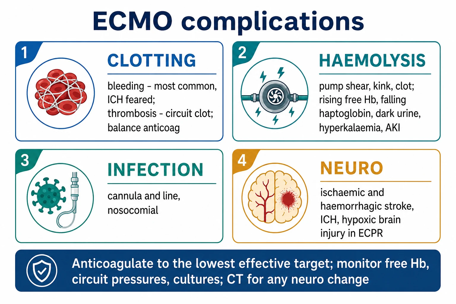
ECMO is a high-risk therapy whose complications fall into four categories. Clotting: bleeding is the commonest serious complication (anticoagulation plus large cannulae; intracranial haemorrhage the most feared), balanced against circuit thrombosis from under-anticoagulation. Haemolysis: from pump shear, kinked cannulae, or an oxygenator clot — a rising plasma-free haemoglobin, falling haptoglobin, dark urine, hyperkalaemia, and haemoglobinuric AKI. Infection: cannula-site, bloodstream, and nosocomial. Neurological: ischaemic and haemorrhagic stroke and intracranial haemorrhage, plus the hypoxic brain injury of ECPR. Anticoagulate to the lowest effective target; monitor the circuit pressures, the free haemoglobin, the cultures, and have a low threshold for a CT for any neurological change.
On this page & tools
Your progress
Saved locally on this device.
Target exams
Overview & definition
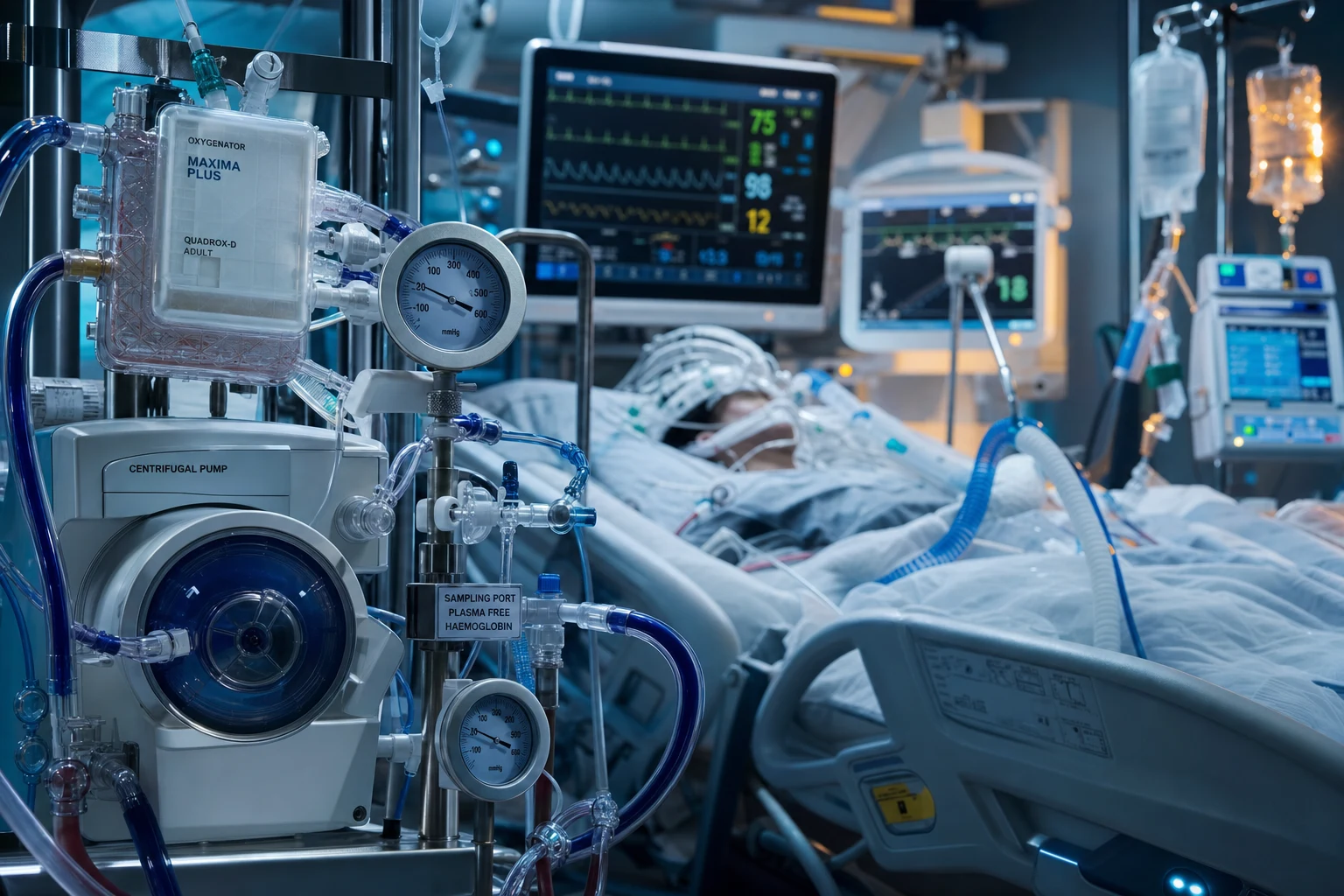
[1]ECMO is a high-risk therapy: the patient is anticoagulated, carries large-bore cannulae, and has blood exposed to an artificial surface and a pump. Its complications fall into four categories — clotting (bleeding and thrombosis), haemolysis, infection, and neurological injury — alongside the mechanical and vascular complications of the circuit and the cannulae (limb ischaemia, LV distension, the Harlequin syndrome, air embolism).[1][1][1]
The unifying mechanism is contact activation: the moment native blood meets the polymethylpentene oxygenator fibres, the pump rotor, and the PVC tubing, the extrinsic and intrinsic coagulation cascades, complement, and the inflammatory pathways are all triggered, platelets are consumed, and a high-shear environment fragments red cells and cleaves von Willebrand factor. Every complication of ECMO can be traced back to this interface between blood and the artificial surface — which is why anticoagulation, circuit design, and meticulous surveillance are the three pillars of safe ECMO practice.[1][1]
Clotting — bleeding and thrombosis
The central tension of ECMO is the anticoagulation balance: anticoagulate enough to prevent clot in the circuit, little enough to avoid catastrophic bleeding.[1][1]
- Bleeding is the commonest serious complication — from the systemic anticoagulation, the large-bore cannulae, and the uraemic platelet dysfunction of critical illness. Sites include the cannulation sites, surgical wounds, the gastrointestinal tract, and — the most feared — intracranial haemorrhage. Management: anticoagulate to the lowest effective target, transfuse and correct coagulopathy and thrombocytopenia, and for major bleeding stop the anticoagulation and give factor replacement (or protamine for heparin).[1]
- Circuit thrombosis — clot in the oxygenator or the pump head, from under-anticoagulation or circuit stasis — risks embolisation and a rising trans-oxygenator pressure gradient and falling gas transfer. A clotted circuit is changed. Monitor the circuit pressures, the anticoagulation (ACT, aPTT, or anti-Xa), and the platelet count.[1]
- Heparin-induced thrombocytopenia (HIT) — a falling platelet count on heparin; switch to a direct thrombin inhibitor (bivalirudin or argatroban).[1]
Bleeding in detail
Bleeding complicates roughly 30–50% of adult ECMO runs and is the leading cause of preventable ECMO-related death. The pathophysiology is multifactorial and goes well beyond the heparin infusion:[6][1]
- Circuit anticoagulation — the continuous unfractionated heparin (UFH) infusion is the dominant driver of surgical-site and cannulation-site bleeding. Even at "lowest effective" targets the therapeutic window is narrow and patient-to-patient heparin response is wide.
- Contact activation and consumption coagulopathy — blood traversing the circuit activates factor XII, thrombin, and complement, consuming platelets, fibrinogen, and factors V and VIII, and generating a low-grade DIC picture over days.
- Thrombocytopenia and platelet dysfunction — platelets are both consumed (by the circuit) and dysfunctional (uraemia, anti-platelet drugs, storage lesion), so the bleeding time is prolonged even when the count looks adequate.
- Acquired von Willebrand syndrome — the high shear stresses inside the pump cleave the high-molecular-weight multimers of von Willebrand factor (via ADAMTS13), producing a type-2A von Willebrand defect that mirrors what is seen in continuous-flow LVADs. This is the dominant reason ECMO patients bleed from mucosal and surgical surfaces.
- Hypofibrinogenaemia and hyperfibrinolysis — fibrinogen falls with duration of run; viscoelastic testing (TEG/ROTEM FIBTEM) identifies the subgroup who need cryoprecipitate or fibrinogen concentrate. [1]
Sites and approximate incidence (adult ELSO data): cannulation/surgical site 20–30%, gastrointestinal 5–10%, intracranial 2–7% (highest in ECPR and post-cardiotomy), retroperitoneal, pulmonary, and "ooze" from line sites. The intracranial haemorrhage rate is the killer: it is twice as common in VA-ECMO and ECPR as in VV-ECMO, and mortality exceeds 50%.[1][10]
Management ladder for ECMO bleeding: (1) localise and control the source (surgical sites get surgical pressure/sutures; line sites get hold-pressure and a stitch); (2) reduce or hold the heparin to the lowest tolerated target — for life-threatening bleeding, stop it entirely and reverse with protamine 1 mg per 100 units of heparin given in the previous 2 hours (titrate to aPTT/anti-Xa, beware protamine-induced hypotension and paradoxical anticoagulation if overdosed); (3) transfuse to haemostatic targets — platelets >50 (closer to 100 for ICH/neurosurgery), fibrinogen >1.5–2.0 g/L, INR <1.5; (4) give tranexamic acid 1 g IV for hyperfibrinolysis; (5) use viscoelastic testing (TEG/ROTEM) to target factor replacement rather than blinding; (6) for the catastrophic ICH or uncontrolled surgical bleed, consider elective circuit exchange or decannulation once the patient can be supported without ECMO.[1][6]
Circuit thrombosis and pump thrombosis
The mirror image of bleeding is thrombosis inside the circuit — the consequence of under-anticoagulation, low flow/stasis, or an inflamed hypercoagulable patient (sepsis, malignancy, antithrombin deficiency).[1][6]
- Oxygenator clotting — fibrin and platelet deposition on the hollow-fibre membrane reduces gas transfer and raises the trans-oxygenator pressure gradient (pre-membrane minus post-membrane pressure). A rising delta-pressure, a falling post-membrane PaO₂ for a given sweep FiO₂, visible fibrin strands on the oxygenator face-plate, and a rising plasma-free haemoglobin are the cardinal signs. Decision to change the oxygenator/circuit: a delta-pressure crossing the unit-specific threshold (commonly >300–350 mmHg, or a rapid upward trend), falling gas transfer refractory to increased sweep, or gross clot visible on inspection.
- Pump thrombosis — clot on the rotor of a centrifugal pump increases power consumption (rising RPM/Watts for the same flow), generates haemolysis from micro-shear, and can throw emboli. A sudden rise in pump power plus a spike in plasma-free haemoglobin is pump thrombosis until proven otherwise — the pump head (and usually the whole circuit) is exchanged.[9]
- Venous/draining-circuit clot and stasis — a low-flow state (hypovolaemia, kinking, poor drainage) promotes clot in the drainage limb; manage by ensuring adequate preload, repositioning, and avoiding prolonged low-RPM running.
Important caveat: circuit clot is the visible end of thrombosis, but the more insidious danger is silent embolisation — clot fragments traverse the oxygenator and, in VV-ECMO, lodge in the pulmonary circulation; in VA-ECMO they are delivered directly into the arterial tree (brain, gut, limbs). This is one mechanism of ECMO-related stroke.[1][10]
Anticoagulation management
Anticoagulation on ECMO is not one-size-fits-all. The choice of agent and the monitoring assay are decided by the indication (VV vs VA vs ECPR), the bleeding/thrombosis risk balance, and whether HIT is present.[6][7]
- Unfractionated heparin (UFH) remains the first-line agent. Loading dose ~50–100 units/kg, then an infusion titrated to a target. The target is the lowest effective intensity — many centres run VV-ECMO at aPTT 40–55 s or anti-Xa 0.3–0.5 IU/mL, escalating only for thrombosis.
- Heparin resistance — defined as failure to achieve target anticoagulation at >35 000 units/24 h. Mechanisms: antithrombin deficiency (consumption, low baseline), accelerated clearance, high factor VIII / heparin-binding proteins (inflammation). Confirm with an anti-Xa (high anti-Xa but low aPTT = heparin resistance from high factor VIII) and give antithrombin concentrate to keep AT activity >60–80%; only then does heparin work.[6]
- HIT — falling platelet count (≥50% fall or to <100) 5–14 days into heparin, with a rising thrombosis risk. Confirm with PF4 ELISA / serotonin-release assay; switch to a direct thrombin inhibitor: bivalirudin (no renal dose-titration advantage but short half-life ~25 min; preferred for procedures/circuit exchange) or argatroban (hepatically cleared; preferred in renal failure, but hepatotoxic and prolonged in hepatic dysfunction). The aPTT is the monitoring assay (target 1.5–2.5× baseline); note DTIs also prolong the INR, complicating warfarin transition.[7]
- Alternatives — nafamostat mesylate (a short-acting serine protease inhibitor, used in Japan/Korea and increasingly in renal failure/high-bleed states) and regional citrate anticoagulation of the circuit (experimental/niche). Warfarin, DOACs, and LMWH are NOT used for ECMO circuit anticoagulation.
- Weaning anticoagulation for procedures/surgery — for elective surgery reduce to a low aPTT/anti-Xa or hold briefly; for emergency operations reverse with protamine and accept the thrombosis risk under time-limited cover.[1]
Monitoring the anticoagulation
Three assays dominate ECMO practice, each with strengths and pitfalls:[6][1]
Anticoagulation monitoring assays on ECMO (ACT vs aPTT vs anti-Xa vs viscoelastic)
| Assay | What it measures | Typical target | Strengths | Pitfalls on ECMO |
|---|---|---|---|---|
| ACT (activated clotting time) | Whole-blood point-of-care clotting (celite/kaolin) | 180–220 s (centre-specific) | Cheap, fast, bedside; used for cannulation | Highly variable with haematocrit, platelet count, factor depletion, hypothermia; overestimates heparin effect at low haematocrit; poor correlation with aPTT/anti-Xa over time |
| aPTT | Intrinsic pathway clotting time (plasma) | 1.5–2.0× mean normal (≈45–60 s) | Cheap, widely available, used for DTIs (bivalirudin/argatroban) | Confounded by factor consumption / low factor VIII (falsely short — underestimates heparin) and lupus anticoagulant; lags heparin changes |
| anti-Xa | Heparin-mediated factor Xa inhibition (plasma) | 0.3–0.7 IU/mL (centre-specific) | Reflects actual heparin concentration; not confounded by factor levels; best correlate with bleeding/thrombosis | Falsely LOW in low antithrombin (underestimates effect); underestimates high-dose heparin above the assay's linear range; cost/delay |
| TEG / ROTEM (viscoelastic) | Whole clot dynamics (R, MA, LY, FIBTEM) | Goal-directed factor replacement | Identifies the cause (platelet vs fibrinogen vs fibrinolysis); guides transfusion | Not validated for heparin monitoring alone (need heparinase channel); operator-dependent |
Practical consensus: measure ACT at the bedside for rapid titration and procedures, an anti-Xa once or twice daily to confirm the true heparin level, an aPTT as a cross-check and for DTI monitoring, and TEG/ROTEM when bleeding to direct component therapy. The most common error is to trust the ACT alone — it is the least reliable on a long ECMO run.[6]
Haemolysis
Haemolysis arises from shear stress in the pump, a kinked or malpositioned cannula, high negative inlet pressure from poor drainage (cavitation), or clot in the oxygenator.[1]
- Detection: a rising plasma-free haemoglobin, a falling haemoglobin, a rising lactate dehydrogenase (LDH), a falling haptoglobin, dark urine (haemoglobinuria), and hyperkalaemia from lysed cells.
- Consequence: a haemoglobinuric acute kidney injury.
- Management: find and fix the cause (reposition or change the cannula, reduce the flow/RPM, change a clotted oxygenator or circuit), support the renal function (often renal replacement therapy), and transfuse for the anaemia.[1]
Haemolysis in detail — pump design and shear
Red cells are destroyed on ECMO by mechanical shear, turbulence, negative-pressure cavitation, and exposure to clot. The magnitude of the problem is shaped by pump design and circuit configuration:[9][1]
- Roller-head vs centrifugal pumps — older roller pumps cause more haemolysis (linear occlusive compression of the tubing) and have been largely superseded by centrifugal pumps (magnetically levitated rotors — Rotaflow, CentriMag, PediVAS), which reduce but do not abolish shear. Even modern centrifugal pumps haemolyse at very high RPM against high afterload.
- Negative inlet (suction) pressure — when drainage outstrips venous return the pump cavitates, generating extreme negative pressure that literally tears red cells apart. The fix is to match flow to drainage: give volume, reposition the drainage cannula, use a smaller pump flow, or add a second drainage cannula. Watch the drainage-line negative pressure (some circuits display it).
- Kinked or malpositioned cannula — a femoral drainage cannula abutting the IVC wall, a kink at the groin, or a return cannula wedged in a small vessel all generate localised shear. Repositioning under imaging (or changing the cannula) fixes it.
- Clot in the oxygenator or pump head — fibrin strands disrupt laminar flow and shed micro-thrombi that fragment cells. Falling gas transfer + rising delta-pressure + rising plasma-free Hb = a clotted circuit that must be exchanged.[9]
Diagnostic threshold: plasma-free haemoglobin >50 mg/dL is the usual trigger to investigate; >100 mg/dL or a doubling on consecutive samples, especially with dark urine and rising potassium, is significant haemolysis demanding action. Send LDH, haptoglobin (depleted), bilirubin (unconjugated), and urinalysis (positive for "blood" on dipstick but no red cells on microscopy = haemoglobinuria). Differentiate from intravascular haemolysis of a mechanical valve and haemolytic anaemia (positive Coombs, schistocytes) — both are uncommon on ECMO but in the differential.[1][9]
Renal consequence: free haemoglobin is freely filtered, precipitates in the tubules, scavenges nitric oxide (renal vasoconstriction), and causes a pigment nephropathy叠加 on the shock-, sepsis-, and nephrotoxin-related AKI that ECMO patients already have. Haemolysis-driven AKI often needs renal replacement therapy, and many VA-ECMO patients run continuous haemofiltration in parallel.[1]
Infection
The large-bore cannulae are a foreign body, and ECMO patients are critically ill and often long-stay:[1][1]
- Cannula-site and bloodstream infections, line infections, and nosocomial pneumonia are common, worsened by long duration, re-cannulation, and breaks in sterile technique.
- Prevention: aseptic insertion, meticulous site care, minimisation of the ECMO duration, and surveillance cultures.
- Management: targeted antibiotics and removal or replacement of the infected lines.[1]
Infection in detail — biofilm, line-related bacteraemia, and stewardship
Infection is among the commonest late complications of ECMO and an independent predictor of mortality. Three considerations make ECMO infection distinctive:[1][1]
- The biofilm problem — within hours of exposure to blood, the ECMO circuit (tubing, oxygenator, pump head, connectors) is coated with a protein-conditioning layer that bacteria colonise as a sessile biofilm. Biofilm bacteria are 100–1000× less susceptible to antibiotics and to host defences than planktonic organisms, and they continually seed low-grade bacteraemia. This is why positive blood cultures on ECMO are often circuit-derived — and why simply changing antibiotic coverage without considering circuit exchange is sometimes futile.
- Line-related bloodstream infection (CLABSI) — the large-bore femoral, jugular, or subclavian cannulae are the highest-risk vascular access a patient can carry. Risk factors: femoral site, emergent insertion, prolonged dwell time (incidence rises sharply after 14 days), repeated access for sampling/blood products, and a break in the closed circuit. Staphylococcus aureus, coagulase-negative staphylococci, Enterobacteriaceae, Pseudomonas, and Candida dominate.
- Pharmacokinetic distortion — ECMO increases the volume of distribution (drug sequestered in the circuit prime and adsorbed onto the oxygenator membrane) and reduces clearance (especially with concurrent AKI/CRRT). Lipophilic drugs (e.g., vancomycin, midazolam, fentanyl, voriconazole) bind to the circuit; this mandates therapeutic drug monitoring and often higher doses, particularly for antibiotics in sepsis.[1]
Prevention bundle: full-barrier sterile insertion, chlorhexidine skin preparation, antimicrobial-impregnated cannulae where available, daily site inspection, minimising circuit breaks (use a closed sampling system), prompt removal of unnecessary lines, daily review of the need to continue ECMO, and surveillance cultures. There is no role for routine systemic antibiotic prophylaxis solely for ECMO beyond standard peri-cannulation cover.[1][1]
Management: take cultures (peripheral and from each lumen/circuit port), empiric broad-spectrum therapy guided by local ecology and the suspected source, source control (drain collections, change infected lines/circuit if bacteraemia persists), de-escalate on microbiology, and involve infectious diseases. Persistently positive cultures after 48–72 h of appropriate therapy on ECMO = suspect and exchange the circuit.[1]
Neurological complications
Neurological injury is among the most devastating ECMO complications:[1]
- Stroke — both ischaemic (embolic, from circuit clot) and haemorrhagic (from the anticoagulation), and intracranial haemorrhage.
- Hypoxic brain injury in ECPR (from the arrest itself, not the device).
- Seizures and delirium.
- Detection is hard: the anticoagulation limits imaging access, and the patient is sedated. Maintain a low threshold for a CT for any neurological change, and use continuous EEG monitoring in ECPR.[1][1]
Neurological complications in detail
ECMO neurological injury occurs in roughly 5–15% of adult runs and is the most common reason for ECMO to be withdrawn. Three mechanisms:[1][10]
- Ischaemic stroke — embolic, from clot fragments sheared off the oxygenator/pump or from a left-atrial thrombus; in VA-ECMO the arterial return can dislodge aortic atheroma. Hypoperfusion (low native cardiac output, inadequate ECMO flow, or the Harlequin syndrome) contributes in VA-ECMO.
- Haemorrhagic stroke / intracranial haemorrhage — the dominant fear, driven by systemic anticoagulation plus thrombocytopenia, coagulopathy, and (in ECPR) the reperfusion/hypertension after ROSC. Incidence is highest in ECPR and post-cardiotomy VA-ECMO (5–10%) and lower in VV-ECMO (2–4%).[10]
- Hypoxic-ischaemic brain injury in ECPR — a function of the no-flow and low-flow time of the arrest, not the ECMO itself; reflected in poor neurological outcome despite a technically perfect ECMO run.
Detection is the hard part — the patient is sedated, paralysed, and cannot be examined; anticoagulation makes transport to CT hazardous. Practical approach: continuous cerebral monitoring where available (cerebral NIRS oximetry — a falling or asymmetric trend is a red flag; transcranial Doppler), a low threshold to image for any pupillary change, new seizure, unexplained fall in GCS/motor response, or unexplained rise in ICP, and continuous EEG (cEEG) to detect non-convulsive status in ECPR. Use a mobile CT where available; if transport is unavoidable, reduce anticoagulation, bag-valve-ventilate with ECMO flow maintained, and escort fully.[1][1]
Management of ICH on ECMO: stop the anticoagulation, reverse heparin with protamine, correct coagulopathy and thrombocytopenia aggressively (target platelets >100, fibrinogen >2.0, INR <1.4), involve neurosurgery for evacuation where the bleed is surgically accessible and the patient is a candidate, and reassess the ECMO indication — many ICH patients cannot be safely anticoagulated and the ECMO must be weaned or withdrawn.[1][10]
Mechanical and vascular complications (in detail)
The "other" complications were once a brief list — they now warrant their own management because each is common, each is examinable, and each is preventable or treatable. They cluster around peripheral VA-ECMO.[1][5]
Limb ischaemia in VA-ECMO and the distal perfusion cannula
Peripheral VA-ECMO via the femoral artery is the commonest VA configuration, and the large return cannula (17–25 Fr) can occlude the femoral artery and obstruct antegrade limb perfusion, producing acute lower-limb ischaemia in 10–30% of patients if not protected.[5]
- Pathophysiology — the return cannula occupies most of the femoral-artery lumen; retrograde ECMO flow supplies the aorta, but the ipsilateral leg distal to the cannula is starved of antegrade flow. Ischaemia ranges from pain and pallor to compartment syndrome, rhabdomyolysis, amputation, and death.
- Prevention — the distal perfusion cannula (DPC) — at the time of arterial cannulation, a small (6–8 Fr) antegrade sheath is inserted into the superficial femoral artery or posterior tibial artery and connected to a side-port of the arterial return line, diverting a fraction of the ECMO flow down the leg. This reduces limb ischaemia from ~25% to <5%. Many centres place a DPC prophylactically in every peripheral VA-ECMO, and it is now considered standard of care.[5]
- Recognition — monitor the limb hourly in the first 24 h: temperature, colour, capillary refill, pulses (palpable/Doppler), sensation, and motor function. Near-infrared spectroscopy (NIRS) on both calves detects occult ischaemia early. Pain on passive stretch or a tense, cool, pulseless limb is compartment syndrome until proven otherwise.
- Management — (1) check and flush the DPC (the commonest cause of new ischaemia is a kinked or clotted DPC); (2) if no DPC was placed, insert one urgently (percutaneous antegrade or retrograde from the contralateral side); (3) if compartment syndrome is present or incipient, fasciotomy; (4) optimise perfusion (adequate ECMO flow, haemoglobin, and mean pressure); (5) anticoagulation to target (an under-anticoagulated circuit can clot the DPC).[5]
The Harlequin syndrome (north-south syndrome)
A specific and dangerous complication of peripheral VA-ECMO with concurrent lung pathology. The mechanism is one of differential oxygenation:[8][1]
- Peripheral VA-ECMO returns oxygenated blood retrograde up the femoral artery and aorta to the iliacs, renal arteries, and (if flow is high enough) the aortic arch and coronary arteries. Meanwhile, the failing (but still ejecting) left ventricle pumps blood ejected from the pulmonary circulation anterograde into the aortic root, ascending aorta, arch, and great vessels.
- If the lungs are severely diseased (pneumonia, ARDS, pulmonary oedema, massive PE) and the ventilator cannot oxygenate the pulmonary venous blood, then the LV ejects desaturated blood. ECMO retrograde flow meets native antegrade flow at a mixing point somewhere in the descending aorta.
- Above the mixing point (the heart, coronaries, brain, right arm) — the blood is desaturated (north = "purple/blue" brain and heart). Below the mixing point (abdomen, lower limbs) — the blood is well oxygenated (south = "pink"). The patient thus has cerebral and coronary hypoxaemia with a pink lower body — the "Harlequin" (north-south) syndrome.[8]
Recognition: a difference between cerebral/upper-body NIRS and lower-limb NIRS, a difference between right-radial ABG PaO₂ (low) and femoral ABG PaO₂ (high), cyanosis of the upper body and lips, ECG signs of myocardial ischaemia, and altered consciousness. The mixing point can shift cephalad with higher ECMO flow (worsening is rare) or caudad with falling native output. [1]
Management: (1) improve lung function — recruit, increase PEEP, optimise ventilation, treat the pulmonary cause (antibiotics, diuresis, bronchodilation, pulmonary vasodilators); (2) increase the haematocrit and ECMO flow to push the mixing point cephalad (more ECMO contribution to the arch); (3) reduce native cardiac output if it is flooding the aorta with desaturated blood — beta-blockade or (paradoxically) accepting lower native output; (4) switch to a central or axillary configuration, or add a veno-arterio-venous (V-AV) configuration to oxygenate the upper body; (5) if myocardial recovery is the goal and the lungs are the problem, convert VA to VV-ECMO as the heart recovers. Cerebral and upper-body monitoring (NIRS, right-radial ABG) is mandatory in every peripheral VA-ECMO patient.[8][1]
Left ventricular distension and pulmonary oedema
In VA-ECMO, retrograde aortic flow raises afterload and prevents the failing LV from ejecting. Blood pools in the LV, the mitral valve opens, left-atrial and pulmonary-venous pressures rise, and pulmonary oedema or stasis thrombus in the LV ensues. Echo shows a dilated, poorly opening aortic valve and a stagnant LV cavity. [1]
Management: (1) optimise preload reduction (diuresis, ultrafiltration); (2) inotropes (milrinone, dobutamine) to augment native LV ejection; (3) IABP (intra-aortic balloon pump) — deflates in systole, reducing afterload, and inflates in diastole, augmenting coronary perfusion — the classic "LV venting" adjunct; (4) a percutaneous LV vent (e.g., Impella, or a trans-septal left-atrial-to-femoral drainage cannula) to physically decompress the LV in refractory cases. Failing to vent a distended LV causes pulmonary oedema, LV thrombus, and failure to wean.[1][1]
Air embolism
Circuit air embolism is a catastrophic, largely preventable complication. Air enters at low-pressure/venous connections (loose pre-pump connectors, a cracked stopcock, a disconnected cannula, an empty reservoir) or via cavitation (high negative inlet pressure). On the venous side it crosses to the arterial side if the oxygenator is breached, and in VA-ECMO it is delivered directly to the brain. Prevention: a bubble detector on the venous line, an arterial line filter, careful priming (no air in the circuit), and never running the drainage limb dry. Management of a witnessed air event: clamp the circuit, place the patient head-down, left-lateral decubitus (to trap air in the right atrium), aspirate from a central line, hyperbaric oxygen if available, and anticoagulate to prevent clot on damaged endothelium.[1]
Acute kidney injury on ECMO
AKI complicates 50–70% of ECMO runs and 20–40% need renal replacement therapy. Mechanisms are cumulative: the underlying shock/sepsis, the inflammation of contact activation, haemoglobinuria from haemolysis, nephrotoxins (contrast, antibiotics), venous congestion from poor drainage, and micro-thromboembolism. Prevention and management: avoid nephrotoxins, ensure adequate mean pressure and haemoglobin, minimise haemolysis (pump settings, cannula position), treat venous congestion, and start CRRT early (many units integrate a haemofilter in-line with the circuit). AKI on ECMO is usually reversible if the patient survives the underlying illness.[1]
Circuit management and monitoring
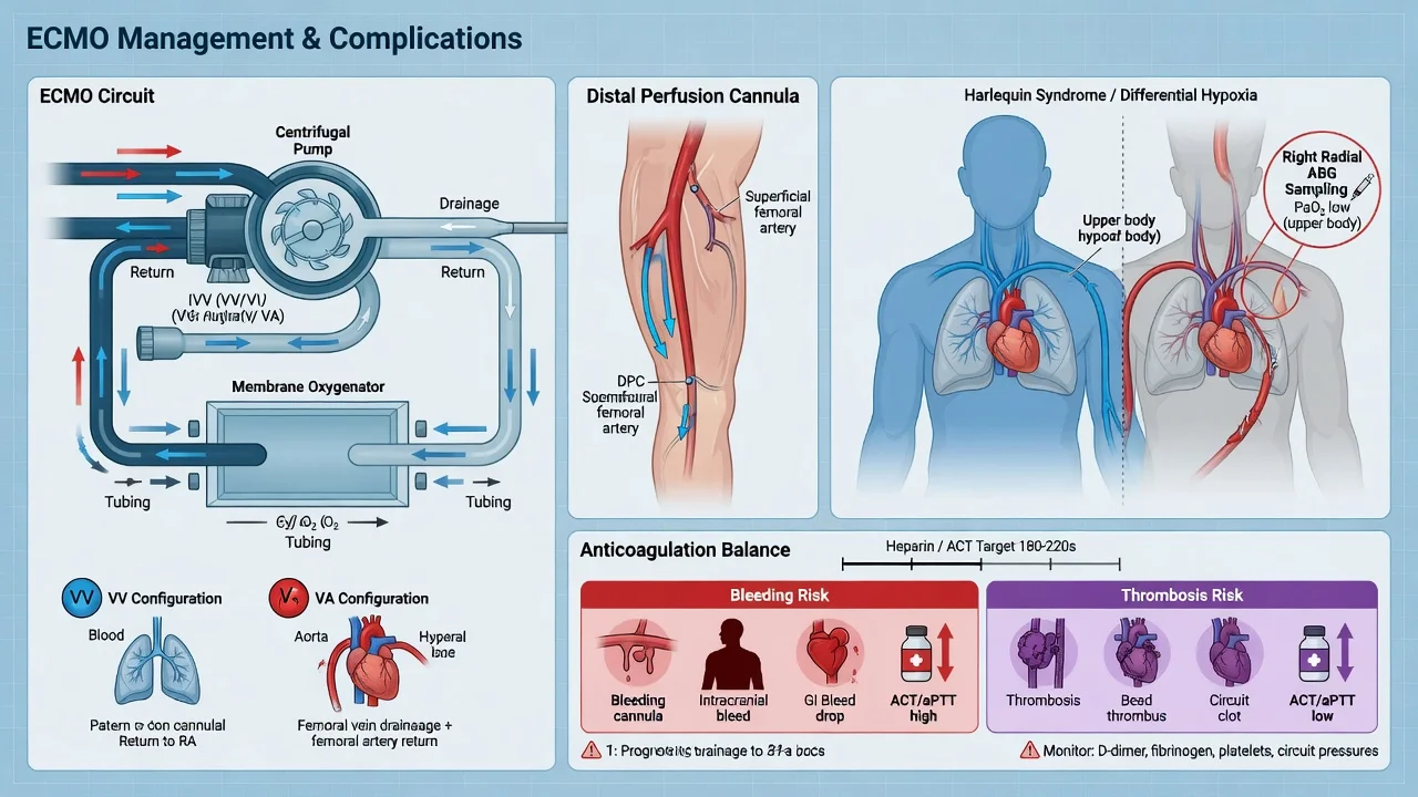
The ECMO circuit is a living organ — it must be inspected, measured, and maintained. The bedside specialist performs a structured hourly circuit check:[1][1]
- Flow and RPM — centrifugal pumps run at a set RPM and deliver flow dependent on preload and afterload; a fall in flow at constant RPM signals hypovolaemia (most common), a kinked drainage cannula, or poor venous drainage. Match flow to drainage — never "chase" flow by increasing RPM into a cavitating drainage limb.
- Pressures — pre-membrane (pre-oxygenator) pressure, post-membrane pressure, and the delta (trans-oxygenator) pressure gradient. A rising pre-membrane pressure with a rising delta = oxygenator clotting; a falling flow with a low delta = a drainage/preload problem, not clot.
- Gas transfer — the sweep gas flow (O₂/air blend) set on the oxygenator controls CO₂ clearance; FiO₂ of the sweep controls oxygenation (in VV-ECMO). A falling post-membrane PaO₂ or rising PaCO₂ for unchanged sweep settings = failing oxygenator.
- Plasma-free haemoglobin — send at least daily (twice-daily on long runs); a rising trend is the earliest sign of pump/oxygenator trouble.
- Anticoagulation monitoring — ACT bedside as needed, anti-Xa daily, aPTT as cross-check, platelet count daily (watch for HIT).
- Visual inspection — fibrin strands on the oxygenator face-plate, clot in the pump head, kinks, air bubbles, leaks at connectors, cannula-site inspection.
- Circuit change criteria — rising trans-oxygenator gradient beyond unit threshold, falling gas transfer, gross clot, refractory haemolysis, or persistently positive blood cultures. Circuit changes are performed by a trained team with two clamps and a primed circuit, often without interrupting support (a "parallel" or "Y" technique) for unstable patients.[1]
VV-ECMO vs VA-ECMO — the distinctive complication profile
| Domain | VV-ECMO (respiratory) | VA-ECMO (cardiac / ECPR) |
|---|---|---|
| Indication | Severe ARDS, refractory hypoxaemia/hypercapnia | Cardiogenic shock, cardiac arrest (ECPR), failure to wean bypass |
| Cannulation | Drainage + return both venous (femoro-jugular or double-lumen) | Venous drainage + arterial return (femoral or central) |
| Cardiac support | None — heart must pump | Full — provides systemic flow |
| Oxygenation delivered to | Pulmonary circulation → LV → systemic (depends on native cardiac output) | Aorta retrograde (peripheral) or antegrade (central) |
| Limb ischaemia risk | Very low (no large arterial cannula) | High — needs a distal perfusion cannula |
| Harlequin syndrome | Not applicable | Yes — if lungs diseased and LV ejecting desaturated blood |
| LV distension | Not applicable | Yes — retrograde afterload; may need IABP/Impella venting |
| Intracranial haemorrhage | 2–4% | 5–10% (highest in ECPR) |
| Bleeding (any site) | ~30% | ~40% (more cannulation sites, more surgical) |
| Air embolism consequence | Lung-filtered (usually) | Direct to brain — catastrophic |
| Recirculation phenomenon | Yes (return blood re-drained) | No |
Bleeding vs thrombosis — the anticoagulation balance on ECMO
| Variable | Bleeding (over-anticoagulation / coagulopathy) | Thrombosis (under-anticoagulation / stasis) |
|---|---|---|
| Commonest site | Cannulation/surgical site; GI; intracranial | Oxygenator; pump head; venous drainage limb |
| Circuit signs | Falling haemoglobin; visible ooze; dropping platelets/fibrinogen | Rising trans-oxygenator ΔP; falling post-membrane PaO₂; visible fibrin strands |
| Lab signature | High aPTT/anti-Xa; low fibrinogen/platelets; prolonged TEG R / low MA | Low aPTT/anti-Xa; high plasma-free Hb if pump clot; high fibrinogen |
| Anticoagulation move | Reduce/hold heparin; reverse with protamine; factor replacement | Increase heparin to target; check antithrombin; consider circuit change |
| Mortality impact | ICH is the leading preventable cause of ECMO death | Embolic stroke, limb loss, oxygenator failure |
| Prevention | Lowest effective target; viscoelastic-guided factor replacement; meticulous surgical haemostasis | Adequate anticoagulation; avoid low-flow stasis; daily ΔP trend |
Management of major bleeding on ECMO
- RECOGNISE AND LOCALISE — (a) Define the source: surgical/cannulation site (compressible, suturable), GI (endoscopy), intracranial (urgent CT — reduce anticoagulation before transport), retroperitoneal (CT), pulmonary (bronchoscopy), occult (suspect with falling Hb and haemodynamic instability). (b) Severity: minor (ooze, no haemodynamic effect) vs major (drop >2 g/dL, transfusion need, haemodynamic compromise, ICH). Send cross-match, coagulation, fibrinogen, platelets, TEG/ROTEM.[1][6]
- SOURCE CONTROL — (a) Surgical site: direct pressure, additional sutures, topical haemostatic agents, re-explore if needed. (b) Cannulation site: hold pressure, suture, consider re-siting. (c) GI: endoscopy ± clip/inject, octreotide/PPI, transfuse. (d) ICH: neurosurgery consult, control ICP, reverse anticoagulation fully.[1]
- REDUCE OR HOLD ANTICOAGULATION — (a) Minor bleeding: reduce heparin to lowest effective target (or pause briefly). (b) Major bleeding: STOP heparin; reverse with protamine 1 mg per 100 units UFH given in the previous 2 hours (max 50 mg; titrate to aPTT/anti-Xa; beware hypotension and paradoxical over-anticoagulation from protamine excess). (c) For DTIs (bivalirudin/argatroban) there is no specific reversal — stop the infusion (bivalirudin half-life ~25 min; argatroban longer in hepatic dysfunction); consider haemofiltration for bivalirudin.[6][7]
- CORRECT THE COAGULOPATHY (viscoelastic-guided) — (a) Platelets to >50 (target >100 for ICH/neurosurgery). (b) Fibrinogen to >1.5–2.0 g/L with cryoprecipitate or fibrinogen concentrate (guided by FIBTEM). (c) Prothrombin complex concentrate or FFP to correct INR <1.5. (d) Tranexamic acid 1 g IV for hyperfibrinolysis (prolonged TEG LY30). (e) Red cells to maintain Hb >70 (or >80–90 if bleeding/ICH).[1]
- REASSESS THE ECMO INDICATION — (a) If bleeding is uncontrollable on full reversal, the patient cannot safely be anticoagulated: weigh weaning ECMO (if the underlying indication has resolved) or continuing without anticoagulation (accepting thrombosis risk for a defined window, e.g., 24–48 h) under close circuit surveillance. (b) For ICH, multidisciplinary discussion of continuation vs withdrawal of ECMO.[1][10]
Approach to a suspected circuit problem (rising ΔP, falling gas transfer, or haemolysis)
- CLINICAL CONTEXT — Is the patient deteriorating (falling SpO₂/Hb, rising K⁺, dark urine, new lactataemia) or is this an isolated circuit alarm? Stabilise the patient first (increase native FiO₂, increase sweep FiO₂, transfuse if anaemic).[1][9]
- CIRCUIT INSPECTION — (a) Trans-oxygenator ΔP trending up + post-membrane PaO₂ falling for unchanged sweep = oxygenator clotting. (b) Pump power rising at constant RPM/flow = pump thrombosis. (c) Visible fibrin strands on the oxygenator face-plate. (d) Plasma-free Hb rising (send urgent) + haptoglobin falling. (e) Drainage and return pressures for kinks/position.[9]
- IDENTIFY AND ADDRESS THE CAUSE — (a) Kinked/malpositioned cannula → reposition under imaging. (b) High negative inlet pressure (cavitation) → reduce flow, give volume, add a second drainage cannula. (c) Under-anticoagulation → check anti-Xa/ACT, titrate heparin up, check antithrombin. (d) Clot in oxygenator/pump → plan circuit exchange.[1]
- CIRCUIT/OXYGENATOR EXCHANGE — (a) Indicated for: ΔP beyond unit threshold, falling gas transfer refractory to increased sweep, gross clot, refractory haemolysis, persistently positive blood cultures. (b) Performed by a trained ECMO team with a primed circuit; unstable patients may be supported on a parallel circuit or briefly on maximal ventilatory/inotropic support during the swap. (c) Send the old circuit for culture if infection suspected.[1][9]
- PREVENT RECURRENCE — Re-establish adequate anticoagulation (confirm anti-Xa), review cannula position (imaging), ensure adequate flow/preload to avoid stasis, and intensify surveillance (ΔP and plasma-free Hb every 6 h).[1]
Prevention and management of limb ischaemia in peripheral VA-ECMO
- AT CANNULATION (PREVENTION) — (a) Use an ultrasound-guided femoral-arterial puncture. (b) Place a prophylactic distal perfusion cannula (DPC) — a 6–8 Fr antegrade sheath in the superficial femoral or posterior tibial artery, connected to the arterial return line side-port. This is now considered standard of care and reduces limb ischaemia from ~25% to <5%.[5]
- HOURLY LIMB MONITORING (first 24 h, then 2–4 hourly) — Temperature, colour, capillary refill, palpable pulses and Doppler signals, sensation and motor function, calf NIRS (compare to contralateral leg and to baseline). Document a baseline vascular exam and NIRS at cannulation.[5]
- RECOGNISE ISCHAEMIA EARLY — (a) Cool, pale, pulseless leg with delayed refill; pain (if awake); paraesthesia; paralysis (late). (b) Falling calf NIRS or a NIRS differential >15% vs the contralateral leg. (c) Rising creatine kinase/lactate from the limb (rhabdomyolysis). A tense, very tender calf with pain on passive stretch = compartment syndrome — emergency.[5]
- MANAGE — (a) Check and flush the DPC (the commonest cause of new ischaemia is a kinked/clotted DPC); if clotted, re-establish flow or re-site the DPC. (b) If no DPC was placed, insert one urgently (percutaneous antegrade, or retrograde from the contralateral femoral). (c) Fasciotomy if compartment syndrome is present or incipient. (d) Optimise perfusion: adequate ECMO flow, haemoglobin >80, mean arterial pressure >65. (e) Ensure the anticoagulation is at target (under-anticoagulation clots the DPC).[5]
- TRANSITION — As the patient recovers and VA-ECMO is weaned, the DPC is removed with the arterial cannula under controlled conditions with protamine and manual/ultrasound-guided compression; check distal pulses afterwards.[5]
SAQ — Bleeding and circuit thrombosis on VV-ECMO
10 minutes · 10 marks
A 45-year-old man is on day 4 of VV-ECMO for severe influenza A ARDS (PaO2/FiO2 was 60). He is on an unfractionated heparin infusion titrated to anti-Xa 0.3 IU/mL. The nurse reports 300 mL of frank blood from the orogastric tube and oozing from cannula sites. Haemoglobin has fallen from 95 to 70 g/L. The examiners ask you to manage the bleeding while protecting the oxygenator.
SAQ — Limb ischaemia and Harlequin syndrome in VA-ECMO
10 minutes · 10 marks
A 58-year-old man is on peripheral femoral VA-ECMO for refractory cardiogenic shock post-MI. Twelve hours after cannulation the perfusionist reports falling venous saturations and worsening blood gases from the venous line; simultaneously the right leg is cool and pulseless distal to the cannula, and a pulmonary artery sample shows SpO2 84% while the right radial artery reads 99%. The examiners ask you to interpret and manage these complications of peripheral VA-ECMO.
Clinical pearls
Other complications (briefly)
- Limb ischaemia (peripheral VA-ECMO — a distal perfusion cannula).
- LV distension and pulmonary oedema (VA-ECMO — the failing LV cannot eject).
- The Harlequin syndrome (peripheral VA-ECMO with lung failure).
- Air embolism (circuit disruption).
- Acute kidney injury (common — from the shock, the haemolysis, and the inflammation; often needs renal replacement therapy).
- Fluid overload and electrolyte disturbance.[1]
Red flags
Prognosis
ECMO evidence, complications, and outcomes
EOLIA (2018, NEJM): Combes et al. — VV-ECMO vs conventional lung-protective ventilation with proning for very severe ARDS (PaO₂/FiO₂ <50, or <80 with high PEEP). Stopped early for futility at interim (no significant 60-day mortality difference), but Bayesian re-analyses suggest a high probability of clinically meaningful benefit; crossover (28% of control group received rescue ECMO) diluted the effect. Cemented ECMO as rescue therapy.[2] Munshi/Munster meta-analysis (2020, Intensive Care Med): systematic review and individual-patient-data meta-analysis of VV-ECMO RCTs (EOLIA + CESAR-era data) — showed a significant mortality reduction for severe ARDS when rescue crossover was accounted for.[3] CESAR (2009, Lancet): Peek et al. — referral to an ECMO centre vs conventional ventilation for severe adult respiratory failure. Survival to 6 months without disability was higher with ECMO-centre referral (63% vs 47%). Established the principle of centralised ECMO retrieval; patients were not all cannulated.[4] ELSO registry (ongoing): the largest ECMO dataset — bleeding in 30–50%, infection in 20–30%, neurological complications in 5–15%, mechanical/circuit events in 10–20% of adult runs; intracranial haemorrhage highest in ECPR (5–10%).[1] Martucci et al. (2024, AJRCCM): multicentre VV-ECMO anticoagulation cohort — anti-Xa-guided heparin strategies were associated with less bleeding than ACT/aPTT-guided strategies without excess thrombosis; supports anti-Xa as the preferred monitoring assay.[6] Kiskaddon et al. (2023, Semin Thromb Hemost): direct thrombin inhibitors (bivalirudin, argatroban) in ECMO — effective in HIT and in heparin resistance; no specific reversal; monitor by aPTT; bivalirudin shorter half-life and partly removable by haemofiltration.[7] Marbach et al. (2022, Int J Cardiol): strategies to reduce limb ischaemia in peripheral VA-ECMO — prophylactic distal perfusion cannula reduces ischaemia from ~25% to <5%, now standard of care; NIRS surveillance detects occult ischaemia.[5] Ryu et al. (2026, Perfusion): acute brain injury in adult post-cardiotomy VA-ECMO — intracranial haemorrhage and ischaemic stroke remain the leading neurological events; early imaging and aggressive reversal of anticoagulation improve outcomes.[10] Balthazar et al. (2026, Interv Cardiol Clin): prevention and management of haemolysis on mechanical circulatory support — centrifugal-pump design, avoidance of negative-inlet cavitation and high-RPM/high-afterload running, and timely oxygenator/pump exchange are the keys.[9] Outcomes (ELSO): survival to discharge ~50–60% for VV-ECMO (severe ARDS) and ~40–50% for VA-ECMO (cardiogenic shock), lower for ECPR (20–40%). Complications independently predict mortality — intracranial haemorrhage, refractory sepsis, and oxygenator failure carry the worst prognoses.[1]
Integration — the ECMO bedside round
A daily ECMO round revisits six questions, each mapping to a complication category:[1][1]
- Is the patient bleeding? — Hb trend, surgical/drain sites, gastric aspirate/stool, platelet and fibrinogen trend, latest anti-Xa/aPTT, viscoelastic if abnormal. Bleeding found → reduce/hold anticoagulation, source-control, factor replacement, reassess ECMO need.
- Is the circuit clotting? — ΔP trend, pump power, plasma-free Hb, visible clot, gas transfer. Clot found → titrate anticoagulation up, address preload/stasis, plan elective exchange.
- Is the patient infected? — temperature, white-cell and lactate trend, cultures, line sites, chest. Infection found → cultures, targeted antibiotics, source control, exchange circuit if persistent bacteraemia; check antibiotic levels (TDM).
- Is the brain OK? — pupillary exam, motor response, sedation hold if safe, NIRS trends, cEEG (ECPR), latest imaging. Any change → CT.
- Is the limb OK? (VA-ECMO) — temperature, pulses, NIRS, DPC patency. Change → check/flush DPC, fasciotomy if needed.
- Is the upper body oxygenated? (VA-ECMO) — cerebral NIRS vs lower-limb NIRS, right-radial ABG vs femoral ABG. Discordant → suspect Harlequin; fix the lungs, raise flow, convert configuration. [1]
This six-point round converts the abstract "four categories" into a checklist that surfaces every preventable complication before it becomes an emergency, and it is the structure examiners expect when asked "how do you manage a patient on ECMO on your round?"[1][1][1]
References
- [1]Choubey U, Mehta K, et al. Extracorporeal membrane oxygenation in cardiogenic shock: evidence, limitations, and patient selection in the contemporary era Postgrad Med, 2026.PMID 42178728
- [2]Combes A, Hajage D, Capellier G, et al. (EOLIA Trial) Extracorporeal Membrane Oxygenation for Severe Acute Respiratory Distress Syndrome N Engl J Med, 2018.PMID 29791822
- [3]Munshi L, Walkey A, Goligher E, et al. ECMO for severe ARDS: systematic review and individual patient data meta-analysis Intensive Care Med, 2020.PMID 33021684
- [4]Peek GJ, Mugford M, Tiruvoipati R, et al. (CESAR trial) Efficacy and economic assessment of conventional ventilatory support versus extracorporeal membrane oxygenation for severe adult respiratory failure (CESAR): a multicentre randomised controlled trial Lancet, 2009.PMID 19762075
- [5]Marbach JA, Mehta P, Ai Y, et al. Strategies to reduce limb ischemia in peripheral venoarterial extracorporeal membrane oxygenation: A systematic review and Meta-analysis Int J Cardiol, 2022.PMID 35523371
- [6]Martucci G, Grasselli G, Tanaka K, et al. Anticoagulation and Bleeding during Veno-Venous Extracorporeal Membrane Oxygenation: Insights from the PROTECMO Study Am J Respir Crit Care Med, 2024.PMID 37943110
- [7]Kiskaddon AL, Rizkalla N, Pollak U, et al. Anticoagulation with Intravenous Direct Thrombin Inhibitors in Pediatric Extracorporeal Membrane Oxygenation: A Systematic Review of the Literature Semin Thromb Hemost, 2023.PMID 37643746
- [8]Karam C, et al. Harlequin syndrome during peripheral cardiopulmonary bypass in a patient with an obstructing tracheal schwannoma: A case report Clin Case Rep, 2023.PMID 37323276
- [9]Balthazar T, et al. Prevention and Management of Hemolysis when Utilizing Mechanical Circulatory Support Interv Cardiol Clin, 2026.PMID 42209077
- [10]Ryu J, et al. Acute brain injury in adult post-cardiotomy cardiogenic shock patients receiving central vs. peripheral VA-ECMO: A single-center experience and systematic review with meta-analysis Perfusion, 2026.PMID 42306914