ICU · Respiratory / ventilation
High-Frequency Oscillatory Ventilation (HFOV)
Also known as HFOV · High-frequency oscillatory ventilation · High-frequency ventilation · OSCILLATE trial · OSCAR trial · MOAT trial · Mean airway pressure · mPaw · Pendelluft · Taylor dispersion · Bias flow · Power (amplitude) · Open-lung strategy
High-frequency oscillatory ventilation (HFOV) delivers a very high respiratory rate (3-15 Hz, 180-900 breaths/min) with a very small tidal volume (1-3 mL/kg, below the anatomic dead space) around a high continuous mean airway pressure (mPaw 25-35 cmH2O) — an open-lung, lung-protective concept. Gas exchange occurs not by bulk tidal flow but by five combined mechanisms (direct alveolar ventilation, Taylor/convective dispersion, Pendelluft, cardiogenic oscillation, and molecular diffusion; Chang 1984). CO2 removal is governed by amplitude/power and frequency in a counterintuitive way (lower frequency = more CO2 clearance), and oxygenation by the mPaw. Despite elegant physiology, two landmark trials in adult ARDS were negative: the OSCILLATE trial (NEJM 2013) was stopped early because HFOV increased in-hospital mortality (47 vs 35 per cent), and the OSCAR trial (NEJM 2013) showed no benefit; a Cochrane review (2016) found no mortality advantage and possible harm. HFOV is therefore NOT recommended for routine adult ARDS — it is essentially abandoned in adults, with a residual role only in neonatal respiratory failure and as a rarely-used rescue for refractory hypoxaemia.
On this page & tools
Your progress
Saved locally on this device.
Target exams
Overview & definition
High-frequency oscillatory ventilation (HFOV) uses a very high respiratory rate (about 3-15 Hz, or 180-900 breaths a minute) with a very small tidal volume (below the anatomic dead space) delivered around a high continuous mean airway pressure (mPaw). The rationale was the open-lung concept: the high mPaw keeps alveoli recruited, while the tiny tidal volume avoids the overdistension and cyclic collapse of conventional ventilation. Despite this elegant physiology, two large trials in adult ARDS showed HFOV is not beneficial and may be harmful, and it is now essentially abandoned in adult practice.[1][2][1]
The defining contrast with conventional ventilation: in volume- or pressure-control, each breath moves a tidal volume (about 6 mL/kg) that exceeds the dead space and reaches the alveoli by bulk flow — ventilation is therefore a function of tidal volume × respiratory rate (minute ventilation). In HFOV the tidal volume (about 1-3 mL/kg) is less than the dead space, so no single oscillation should, in theory, deliver fresh gas to the alveoli at all. That gas exchange nonetheless occurs — and can maintain near-normal gas tensions — is the central physiological puzzle of HFOV, solved by the five gas-transport mechanisms described by Chang.[5][1]
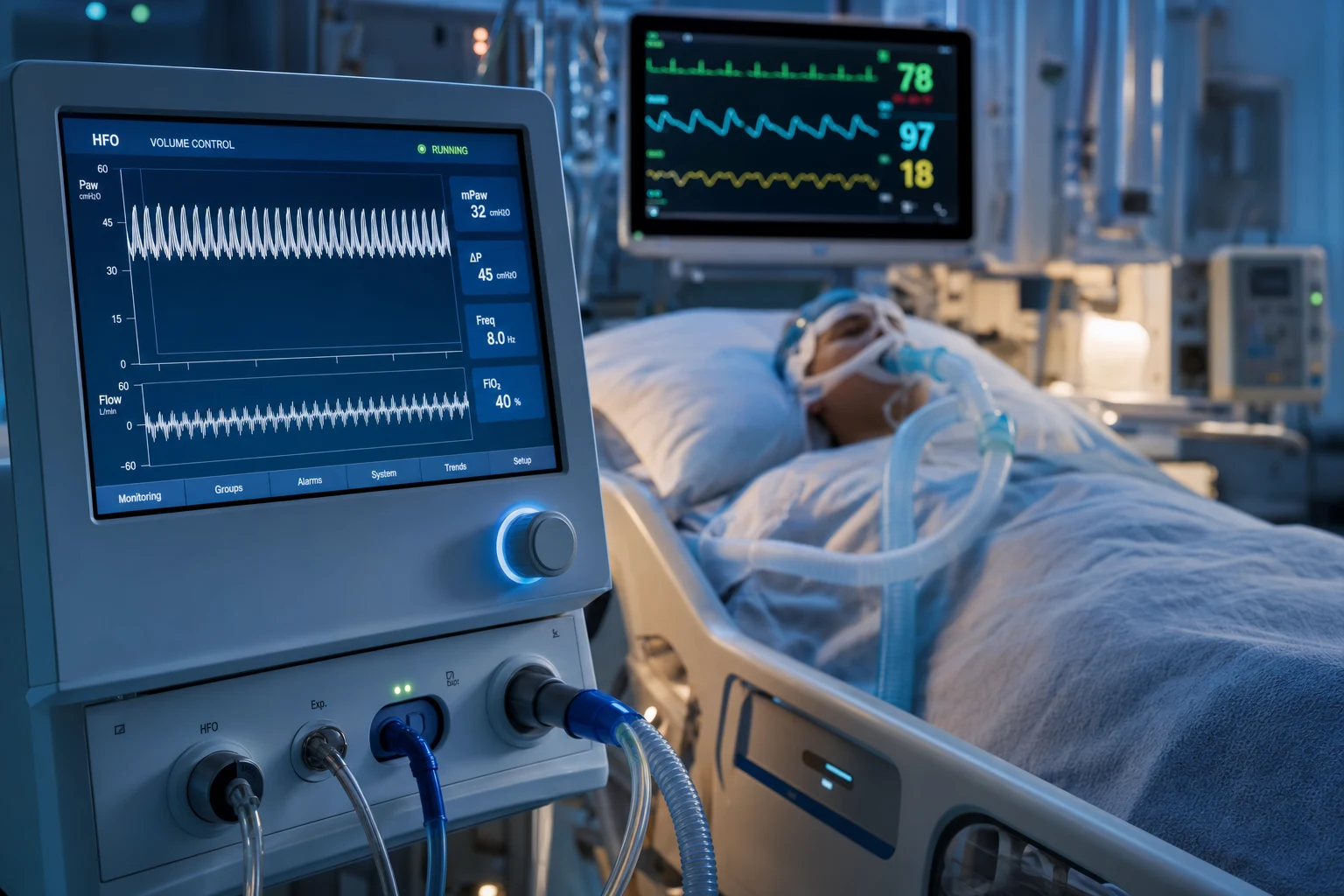
Mechanism and physiology
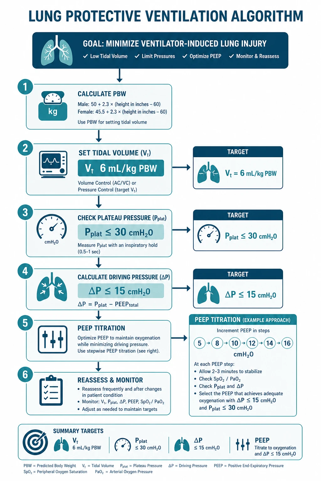
HFOV applies:[1]
- A continuous mean airway pressure (mPaw), set above the alveolar closing pressure to keep the lung open (the open-lung strategy).
- Small oscillations around the mPaw, generated by a piston or diaphragm, at a very high frequency (3-15 Hz).
- A tidal volume below the anatomic dead space — gas exchange occurs not by bulk tidal flow but by combined mechanisms: Pendelluft (gas mixing between lung units of differing time constants), convective mixing, Taylor dispersion, and molecular diffusion.
The intended advantages were sustained recruitment (oxygenation) with lung-protective tiny volumes (reduced VILI).[1]
How the oscillator generates the breath
A conventional ventilator is a flow generator: it pushes gas into the lung during inspiration and allows passive elastic expiration. A high-frequency oscillator is fundamentally different — it is an active push-pull device. An electrically driven piston, diaphragm (loudspeaker-style), or rotating valve oscillates the gas column in the breathing circuit at the set frequency, actively driving gas in on the forward stroke and actively pulling it back out on the return stroke. Because expiration is active (not passive), HFOV does not rely on elastic recoil to empty the lung — an advantage in theory, because it limits gas-trapping. A continuous bias flow of fresh gas (20-40 L/min in adults) is pushed across the top of the circuit (a T-piece), so the oscillating piston "jiggles" this fresh gas column; the mean pressure in the circuit is the mPaw, set by the resistance of the expiratory limb (a balloon valve or water column). Excess gas and CO2 leave via the expiratory limb.[1][4]
How the oscillator works — the push-pull story
A continuous bias flow sets the mean pressure
Fresh, humidified gas flows continuously into the circuit (20-40 L/min in adults) through a heated humidifier. The resistance of the expiratory limb holds this flow back, establishing the mean airway pressure (mPaw 25-35 cmH2O). This mPaw is the static recruitment force that keeps alveoli open throughout the cycle — there is no "expiration" that drops the pressure back to zero.
The piston oscillates the gas column
A motor-driven piston or diaphragm oscillates at the set frequency (3-15 Hz). On the forward stroke it pushes a small bolus of gas toward the patient; on the return stroke it actively withdraws gas back. The excursion of the piston (the amplitude, or "power") sets how big each oscillation is — this is the CO2-clearance lever.
Expiration is active, not passive
Unlike conventional ventilation (where expiration is passive elastic recoil), the oscillator actively pulls gas out. This is the theoretical basis for the claim that HFOV limits gas-trapping — but in obstructed airways the active pull can still stack gas, and air-trapping remains a real risk.
The tidal volume is below dead space — yet gas exchange occurs
Each oscillation moves only about 1-3 mL/kg, less than the ~2 mL/kg anatomic dead space. Fresh gas nonetheless reaches the alveoli by five simultaneous mechanisms (direct alveolar ventilation, Taylor/convective dispersion, Pendelluft, cardiogenic oscillation, and molecular diffusion). PaCO2 and PaO2 can be maintained near normal despite no single "breath" reaching the gas-exchange surface by bulk flow.
A continuous aerosol of tiny droplets of fresh gas is delivered
The net effect is that the dead space is "washed out" and refreshed continuously by the oscillating bias flow, while the high mPaw holds the recruited lung open. Oxygenation is a function of mPaw and FiO2; ventilation (CO2 clearance) is a function of amplitude and frequency.
The five gas-transport mechanisms (Chang, 1984)
Because no single oscillation delivers a tidal volume that reaches the alveoli, gas exchange in HFOV must rely on mechanisms beyond bulk convective flow. Chang's classic 1984 analysis identified five coexisting mechanisms that together explain how the alveolus is ventilated when the tidal volume is smaller than the dead space:[5]
The five gas-transport mechanisms of HFOV (Chang 1984)
| Mechanism | What it is | Where it dominates |
|---|---|---|
| 1. Direct alveolar ventilation (bulk flow) | The small fraction of tidal volume that reaches the proximal, short-pathway alveoli directly | Large airways / proximal alveoli with very short dead space |
| 2. Convective / Taylor dispersion (asymmetric velocity profile) | The oscillatory flow has a parabolic profile (faster in the centre). On forward and reverse strokes, fresh and residual gas interdigitate along the airway, greatly enhancing longitudinal mixing beyond what the tidal volume alone could achieve | Conducting airways (bronchi, bronchioles) — the major contributor |
| 3. Pendelluft (interregional mixing) | Adjacent lung units have different time constants and fill/empty out of phase. Gas therefore "sloshes" between units — moving from one alveolus to another across the alveolar septa and common airways without ever leaving the lung | Heterogeneous lung (ARDS) — regions of differing compliance/resistance |
| 4. Cardiogenic oscillation | Each heartbeat physically agitates the gas in the lung (especially the left lower lobe, adjacent to the heart), contributing measurable gas mixing | Near the heart; small but non-trivial contribution |
| 5. Molecular diffusion | Brownian motion of gas molecules across the alveolar-capillary membrane (normal Fick diffusion) — the final common step for all ventilation | Alveolar ducts and alveoli |
The practical point: in the injured, heterogeneous ARDS lung, Pendelluft and Taylor dispersion do most of the work. This is also why HFOV is conceptually attractive for ARDS — it is designed for exactly the heterogeneous, low-compliance, recruitable lung where these mechanisms operate. It is equally why HFOV was hypothesised to be lung-protective: with such small excursions, volutrauma and atelectrauma should be minimised.[5][1]
Settings
- mPaw — the mean airway pressure (the recruitment/oxygenation lever).
- Frequency (Hz) — the oscillation rate; a lower frequency gives a larger tidal volume.
- Amplitude (the power, ΔP) — the size of the oscillations (the CO2-clearance lever).
- Bias flow and FiO2.[1]
Initial adult settings (the Fessler roundtable protocol)
The most widely cited adult HFOV protocol is the roundtable consensus (Fessler 2007), which gives concrete starting values:[4]
HFOV initial adult settings (Fessler 2007 roundtable protocol)
| Parameter | Starting value | What it controls | How to titrate |
|---|---|---|---|
| mPaw (mean airway pressure) | 25-30 cmH2O (start ~3-5 cmH2O above the previous conventional Pplat; cap ~30-35 unless recruitment manoeuvre) | Oxygenation — recruits and holds alveoli open | Increase by 1-2 cmH2O for hypoxaemia (watch haemodynamics); wean by 1-2 cmH2O as FiO2 falls below 0.4-0.5 |
| Frequency | 4-8 Hz in adults (lower than neonates, who use 8-15 Hz) | CO2 clearance — counterintuitively, LOWER frequency = MORE CO2 clearance (see below) | Decrease Hz (e.g. 8→6→5→4) to raise PaCO2-correcting tidal volume if hypercapnia; increase Hz to lower PaCO2 if too much ventilation / air-trapping |
| Amplitude / power (ΔP) | 60-90 cmH2O initially; set by the "chest wiggle" sign (oscillation visible down to the umbilicus / pelvis) | CO2 clearance — the size of each oscillation | Increase power for rising PaCO2 (more wiggle); decrease if wiggle is excessive or causing haemodynamic perturbation |
| Bias flow | 20-40 L/min (adult 30-40) | Delivers fresh gas; sets the floor for CO2 washout | Increase if PaCO2 high despite max amplitude; do not reduce below 20 L/min (risks rebreathing and unstable mPaw) |
| FiO2 | Per oxygenation target (start 1.0 in severe ARDS, wean to SpO2 88-95%) | Oxygen content of delivered gas | Wean first as oxygenation improves; mPaw is weaned only once FiO2 is low |
| I:E time | Usually 1:1 (fixed on most devices) | Symmetric forward/reverse stroke | Rarely adjusted; some devices allow 1:2 (longer expiratory stroke) in obstruction |
Oxygenation versus ventilation — two independent levers
A central HFOV concept that examiners love: oxygenation and CO2 clearance are controlled by entirely separate settings.[4][1]
- Oxygenation is governed by mPaw and FiO2. Raising mPaw recruits more alveolus (improves the V/Q matching, reduces shunt) and so improves PaO2 — exactly like raising mean airway pressure in any recruitment strategy. The risk of raising mPaw is reduced venous return and cardiac output.
- CO2 clearance is governed by amplitude (power) and frequency. This is where HFOV is counterintuitive (see next section). [1]
The counterintuitive lever: lower frequency = more CO2 clearance
In conventional ventilation, raising the respiratory rate raises minute ventilation and so lowers PaCO2 — straightforward. In HFOV the opposite is partly true, because the delivered tidal volume increases as frequency falls (the piston has more time to travel on each stroke, so the excursion volume is larger). Because CO2 clearance is proportional to (frequency × tidal volume²), and tidal volume rises as frequency drops, reducing the frequency from 8 Hz to 5 Hz typically increases CO2 clearance — the larger tidal volume outweighs the fewer cycles per minute.[4][1]
- PaCO2 rising? → Decrease frequency (8→6→5→4 Hz) and/or increase amplitude/power.
- PaCO2 too low / air-trapping? → Increase frequency.
- Never assume "more breaths = more ventilation" in HFOV — this is the single most-tested HFOV concept. [1]
Titrating HFOV — step by step
Set the mPaw from the prior plateau pressure
Start mPaw 3-5 cmH2O above the conventional ventilation plateau pressure (typically 25-30 cmH2O). The goal is to maintain recruitment without causing haemodynamic compromise. Do not exceed ~35 cmH2O routinely. A recruitment manoeuvre (sustained high pressure 40-45 cmH2O for 40 s) may precede the set mPaw in centres that use them, though aggressive recruitment (ART trial) is itself harmful.
Set frequency 5-6 Hz (adult)
Begin at the upper-middle of the adult range (4-8 Hz). Lower frequencies give larger tidal volumes and better CO2 clearance; higher frequencies are used if air-trapping or if PaCO2 is driven too low. Neonates use 8-15 Hz.
Set the amplitude (power) by the chest wiggle sign
Start at ~60 cmH2O and increase until the oscillation is visible on the chest wall down to the umbilicus/pelvis (the "chest wiggle" sign — a clinical surrogate that the oscillation is reaching the alveoli). Typical adult power 60-90 cmH2O. This sets CO2 clearance.
Set bias flow 30-40 L/min and FiO2 to target
Adult bias flow 30-40 L/min. FiO2 usually starts at 1.0 in the severe-ARDS rescue setting and is weaned to maintain SpO2 88-95% (PaO2 55-80 mmHg).
Check a blood gas at 30-60 minutes
HFOV changes are slow to equilibrate — allow 30-60 min before rechecking the ABG. PaCO2 high → decrease Hz and/or increase power. PaO2 low → increase mPaw by 1-2 cmH2O and/or FiO2 (check blood pressure and lactate — watch for haemodynamic compromise).
Monitor for the four HFOV-specific problems
(a) Haemodynamic compromise (high mPaw → reduced venous return — check BP, lactate, vasopressor requirement). (b) Air-trapping / dynamic hyperinflation (increased mPaw with falling power effect, falling SpO2, rising Paw baseline). (c) Mucous plugging (sudden rise in PaCO2, falling chest wiggle — suction via in-line catheter; oscillators cannot generate a true "sigh"). (d) Pneumothorax (sudden rise in mPaw, falling SpO2, haemodynamic collapse — confirm with ultrasound/CXR, decompress).
Wean by a structured ladder
Wean FiO2 first (down to 0.4-0.5), then mPaw (down 1-2 cmH2O at a time to ~20-22). Only when both are low does transition back to conventional ventilation make sense (the lung is recruited and oxygenating on low pressure/oxygen). The transition itself is a vulnerable moment — set conventional ventilation with adequate PEEP to maintain the recruitment that HFOV established.
HFOV in context — versus the other rescue strategies
HFOV vs conventional lung-protective ventilation vs APRV vs VV-ECMO
| Feature | Conventional (VC/PC, ARDSNet) | HFOV | APRV | VV-ECMO |
|---|---|---|---|---|
| Mechanism | Volume/pressure controlled, cycled, 6 mL/kg | Oscillating diaphragm at 3-15 Hz around a high mPaw, Vt 1-3 mL/kg | Two CPAP levels (Phigh long, Plow brief), spontaneous breathing | Extracorporeal membrane lung drains venous blood, oxygenates, returns to RA |
| Oxygenation lever | FiO2 + PEEP | FiO2 + mPaw | Phigh (mean airway pressure) | Sweep gas + membrane FiO2 |
| CO2 lever | Vt × RR (minute ventilation) | Amplitude/power and frequency (lower Hz = more CO2) | Release phase (Tlow, frequency) | Sweep gas flow |
| Sedation/paralysis | Moderate | Heavy — almost always needs paralysis | Light (spontaneous breathing) | Moderate |
| Haemodynamic effect | Modest (PEEP-dependent) | Significant (high mPaw → reduced venous return) | Favourable (spontaneous breathing preserves venous return) | Favourable (rests the lung; low ventilating pressures) |
| Evidence in adult ARDS | Mortality benefit (ARDSNet)[9] | No benefit, possible harm (OSCILLATE, OSCAR)[1][2] | Oxygenation benefit, no mortality benefit | Refractory rescue (EOLIA neutral on primary; selected use)[8] |
| Current role | Standard for all ARDS | Rescue only, rarely used | Selected refractory hypoxaemia | Refractory hypoxaemia (P/F <50-80) |
The evidence — HFOV failed in adult ARDS
Two landmark trials in 2013 changed HFOV practice:[1][2]
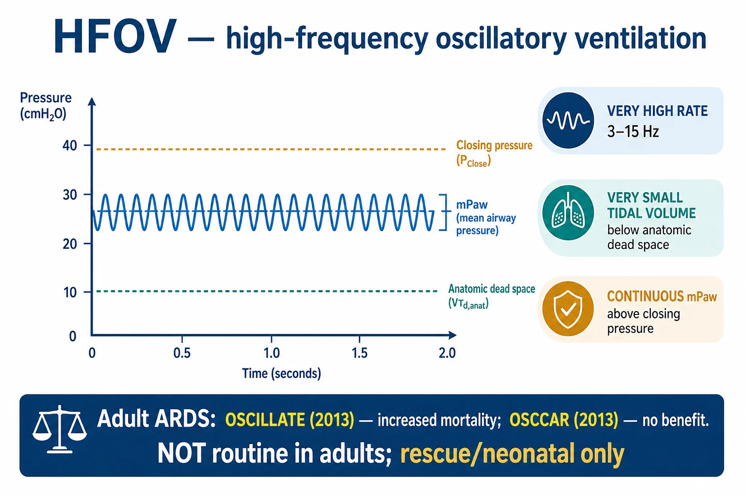
- The OSCILLATE trial (Ferguson, NEJM 2013) randomised adults with early moderate-severe ARDS to HFOV versus a low-tidal-volume control. It was stopped early for harm: HFOV increased in-hospital mortality (47 vs 35 per cent), with more sedation, more neuromuscular blockade, and more haemodynamic compromise (the high mPaw required heavy sedation and paralysed ventilation, and the high intrathoracic pressure dropped venous return).[1]
- The OSCAR trial (Young, NEJM 2013) found no difference in 30-day mortality between HFOV and conventional ventilation in adult ARDS.[2]
Together, these trials removed HFOV from routine adult ARDS practice.[1][2]
Earlier evidence — the MOAT trial
Before OSCILLATE and OSCAR, the MOAT trial (Multicentre Oscillatory Ventilation ARDS Trial, Derdak 2002) randomised 148 adults with ARDS to HFOV versus conventional ventilation. It found no difference in 30-day mortality (the primary outcome), though oxygenation improved in the first 24 hours and there was a trend to fewer barotrauma-related deaths with HFOV. MOAT was small, used a higher control-arm tidal volume than modern lung-protective ventilation, and was the basis for cautious optimism that HFOV might be lung-protective — optimism overturned by the larger, modern OSCILLATE/OSCAR trials.[3]
OSCILLATE
NEJM 2013
548 adults with early moderate-severe ARDS (P/F ≤200, on PEEP ≥10) — HFOV vs conventional low-tidal-volume ventilation
Key finding
STOPPED EARLY for harm. In-hospital mortality 47% (HFOV) vs 35% (control), RR 1.33. More sedation, more neuromuscular blockade, more vasopressors, higher mean airway pressure with HFOV.
Practice change
HFOV INCREASES mortality in adult ARDS — do not use routinely. The harm is attributed to haemodynamic compromise and the sedation/paralysis burden of the high mean airway pressure.
OSCAR
NEJM 2013
795 adults with acute respiratory distress syndrome (P/F ≤200) — HFOV vs conventional ventilation (UK, pragmatic)
Key finding
NO difference in 30-day mortality (41.7% HFOV vs 41.1% control). No benefit on any secondary outcome.
Practice change
HFOV does not improve survival in adult ARDS. Combined with OSCILLATE harm, HFOV has no routine place in adult ARDS.
MOAT
Am J Respir Crit Care Med 2002
148 adults with ARDS — HFOV vs conventional ventilation (early, pre-ARDSNet-standard control)
Key finding
No difference in 30-day mortality. Improved oxygenation in first 24 h with HFOV; trend to fewer barotrauma deaths. Small study, higher control-arm Vt than modern standard.
Practice change
Suggested HFOV was feasible and possibly lung-protective — the basis for the later OSCILLATE/OSCAR trials, which overturned this optimism.
Sud Cochrane meta-analysis
Cochrane Database Syst Rev 2016
Systematic review of RCTs comparing HFOV with conventional ventilation in ARDS (incl. OSCILLATE, OSCAR, MOAT)
Key finding
No statistically significant mortality benefit with HFOV; possible harm when OSCILLATE is included. Heterogeneity in protocols and control-arm ventilation limits pooled estimates.
Practice change
Confirms HFOV should not be used routinely in adult ARDS; reserve for rescue if used at all.
Why HFOV harmed in adults
Several mechanisms explain the OSCILLATE harm:[1][1]
- The high mean airway pressure required heavy sedation and neuromuscular blockade, with their own harms (prolonged ventilation, critical-illness myopathy).
- The high intrathoracic pressure reduced venous return and cardiac output (haemodynamic compromise).
- The small tidal volume and the difficulty monitoring it may have under- or over-ventilated the patient.
- The hemodynamic and sedation burden outweighed the lung-protective benefit.[1]
Why OSCILLATE showed harm — mechanism by mechanism
| Source of harm | What happens | How OSCILLATE showed it |
|---|---|---|
| Haemodynamic compromise | The high mPaw (mean ~30 cmH2O, often higher) raises intrathoracic pressure → reduces venous return and RV preload; can also raise RV afterload (pulmonary vascular compression) → falls in cardiac output and BP | More vasopressor use and higher mean airway pressure in the HFOV arm; likely mediator of the excess deaths |
| Sedation and paralysis burden | Patients fight the oscillator, so deep sedation and routine neuromuscular blockade are near-mandatory → prolonged ventilation, critical-illness polyneuromyopathy, loss of spontaneous venous return benefit | Significantly more sedation and NMBA use in the HFOV arm |
| Unseen tidal volume / overdistension | The actual delivered tidal volume is hard to measure at the bedside; an "ultra-protective" setting may in fact be delivering injurious transalveolar pressures in some regions | Cannot be excluded as a contributor to occult volutrauma |
| Late application / patient selection | In OSCILLATE, HFOV was applied relatively late and to patients already at high risk; a rescue mode that requires deep paralysis and high pressure has a narrow therapeutic window | Reflects real-world difficulty of doing HFOV safely even in expert centres |
Indications — rescue only
After OSCILLATE and OSCAR, HFOV has no routine indication in adult ARDS. If it is used at all, it is as a last-resort rescue for refractory hypoxaemia (P/F <100, often <80) that has failed the full conventional ladder, and only when ECMO is unavailable, contraindicated, or as a bridge to it.[1][2][1]
The rescue pathway for refractory hypoxaemia — where HFOV sits
Optimise conventional ventilation first (the standard of care)
Treat the cause. Lung-protective ventilation: Vt 4-6 mL/kg PBW, plateau pressure <30, driving pressure <15, permissive hypercapnia (pH >7.20). Titrate PEEP/FiO2 (ARDSNet table). This alone reduced mortality from 40% to 31% (ARDSNet 2000).
Add prone positioning for severe ARDS
PROSEVA: prone ≥16 h/day for P/F <150 reduces 28-day mortality from 32.8% to 16.0% — the single most effective adjunct in severe ARDS and the standard of care. Do this BEFORE reaching for HFOV.
Consider adjuncts: conservative fluids, inhaled pulmonary vasodilators, optimised haemodynamics
FACTT conservative fluid strategy; inhaled nitric oxide / prostacyclin as a physiological test (oxygenation improves but no mortality benefit); treat the cause (antibiotics, source control).
Refer for VV-ECMO early
For P/F <80 for >6 h (or <50 for >3 h), or pH <7.25 with PaCO2 ≥60 for >6 h, despite optimisation — refer to a regional ECMO centre (EOLIA). VV-ECMO rests the lung and is the modern rescue of choice for refractory hypoxaemia.
HFOV as a rarely-used rescue — the narrow niche
HFOV may be considered ONLY when the above have been applied or addressed AND ECMO is not immediately available (bridge to ECMO, ECMO contraindicated, or centre with no ECMO capability). It is NOT a substitute for any of steps 1-4. Expect heavy sedation and paralysis, and monitor haemodynamics closely (the OSCILLATE harm). Use it for as short a time as possible and transition to conventional ventilation or ECMO as soon as feasible.
Neonatal and paediatric role
HFOV retains a genuine role in neonatal and paediatric respiratory failure, where the physiology (small, highly recruitable lungs) and the evidence differ. In neonates with severe respiratory distress syndrome, persistent pulmonary hypertension of the newborn, meconium aspiration, and congenital diaphragmatic hernia, HFOV (often combined with inhaled nitric oxide) is an accepted strategy, particularly when conventional ventilation fails to oxygenate or generates injurious pressures. In paediatric ARDS, HFOV is used more liberally than in adults, though recent data favour lung-protective conventional ventilation first. The adult trials do not translate directly because neonatal/paediatric lung mechanics, the oscillator settings (higher frequency, 8-15 Hz), and the comparators differ.[1][6]
Contraindications
HFOV's high mean airway pressure, need for deep sedation/paralysis, and active exhalation make it unsuitable in several settings:[1][4]
Contraindications to HFOV — and why
| Contraindication | Reason |
|---|---|
| Severe obstructive airways disease (asthma, COPD) | The high mean airway pressure and active exhalation risk dynamic hyperinflation and air-trapping → worse hypercapnia, haemodynamic compromise. HFOV is a recruitment mode for a stiff, recruitable lung, not for obstructed airways. |
| Shock / haemodynamic instability / severe RV failure | The high intrathoracic pressure of mPaw 25-35 cmH2O reduces venous return and raises RV afterload — in a shocked patient this can precipitate cardiovascular collapse (a key driver of OSCILLATE harm). Stabilise haemodynamics before any consideration of HFOV. |
| Raised intracranial pressure (TBI, intracranial hypertension) | The high mean intrathoracic pressure impairs cerebral venous return and can raise ICP; the heavy sedation/paralysis and difficulty monitoring PaCO2 tightly add risk in a brain-injured patient who needs careful CO2 and perfusion control. |
| Inability to tolerate deep sedation / paralysis | HFOV nearly always requires neuromuscular blockade. Patients in whom paralysis is contraindicated (e.g. high risk of critical-illness myopathy, on drugs that interact with NMBA) are poor candidates. |
| Severe unilateral lung disease / bronchopleural fistula | HFOV applies pressure to both lungs equally; an unrecruitable unilateral process or an active air leak is worsened by sustained high pressure (continuous air leak, failure to oxygenate). |
| Unstable pneumothorax / large air leak | The continuous high pressure and active oscillation enlarge the leak and prevent resolution. A chest drain must be in place and the leak controlled first. |
Complications and monitoring
HFOV has a distinctive complication profile, driven by the high mPaw, the tiny tidal volume, and the near-mandatory paralysis:[1][1][4]
- Mucous plugging and inadequate secretion clearance — the oscillating humidified gas, small tidal volume, and inability to cough (paralysis) favour sticky secretions; a sudden rise in PaCO2 with a fading "chest wiggle" suggests a blocked tube or plug. Manage with regular in-line suction, adequate humidification, and saline instillation if needed; rarely requires bronchoscopy (which pauses oscillation).
- Haemodynamic compromise — high mPaw reduces venous return and raises RV afterload; presents as hypotension, rising lactate, rising vasopressor requirement. Manage by reducing mPaw (after ensuring recruitment is adequate), giving fluid/vasopressors, and excluding tension pneumothorax.
- Pneumothorax / air leak — sustained high pressure plus the heterogeneous ARDS lung predisposes to barotrauma. Presents as sudden rise in mPaw, falling SpO2, haemodynamic collapse. Confirm with ultrasound or CXR and decompress immediately (chest drain). HFOV can be continued across an in-situ chest drain if the leak is small.
- Dynamic hyperinflation / occult auto-PEEP — although expiration is active, gas-trapping still occurs, especially with high mPaw and high power; monitor the baseline Paw and the chest wiggle.
- Critical-illness polyneuromyopathy — the near-mandatory prolonged paralysis is a major contributor to long-term weakness; minimise NMBA duration.
- Inadequate humidification → airway injury — the high bias flow dries secretions; heated humidification is mandatory.
- Sedation excess and delirium — the deep sedation required adds to ICU-acquired weakness and delirium burden. [1]
Troubleshooting HFOV — the bedside response
PaO2 falling (hypoxaemia)
Increase FiO2 first. If insufficient, increase mPaw by 1-2 cmH2O (watch blood pressure and lactate — haemodynamic compromise is the price). Consider a recruitment manoeuvre. Exclude endotracheal tube obstruction, pneumothorax, and mucus plugging. Reassess the cause of ARDS (is it worsening?).
PaCO2 rising (hypercapnia)
Decrease frequency (e.g. 8→6→5 Hz — recall lower frequency = more CO2 clearance) and/or increase amplitude/power (more chest wiggle). Increase bias flow if already at max amplitude. Check tube patency (mucus plug?) and exclude air-trapping (compare baseline mPaw with baseline).
Sudden haemodynamic collapse on HFOV
Disconnect the circuit transiently (brief return to hand ventilation / bagging) to release intrathoracic pressure and assess. If BP recovers, the high mPaw was the cause — reduce it. If not, exclude tension pneumothorax (ultrasound) and decompress. Check for tube migration / mainstem intubation.
Falling chest wiggle
Loss of the visible oscillation down the chest wall indicates the oscillation is no longer reaching the alveoli — most often endotracheal tube obstruction (kink, mucus plug, bite), mucous plugging in the airway, or a circuit leak/disconnection. Suction the tube, check the circuit, reconfirm ETT position.
Air leak / pneumothorax
Sudden rise in mPaw with falling SpO2 and haemodynamic deterioration → pneumothorax until proven otherwise. Confirm with ultrasound/eFAST or CXR, insert a chest drain. Do not clamp a bubbling chest drain. HFOV may continue if the leak is tolerated; large leaks mandate a lower mPaw or conversion to conventional ventilation.
Current role
- Adult ARDS: HFOV is not recommended for routine use; it is essentially abandoned after OSCILLATE and OSCAR. It may be considered only as a rarely-used rescue in refractory hypoxaemia when all else (proning, optimised ventilation, ECMO consideration) has been addressed.[1][2][1]
- Neonatal and paediatric: HFOV retains a role in neonatal respiratory distress syndrome and selected paediatric lung disease, where the physiology and the evidence differ.[1]
SAQ — HFOV physiology and the counterintuitive frequency–CO2 relationship
10 minutes · 10 marks
A fellowship candidate is asked to explain, at the viva, how high-frequency oscillatory ventilation achieves gas exchange at tidal volumes below the anatomic dead space, and to justify the observation that reducing the frequency improves CO2 clearance. The examiners then ask how this physiology informed the design of the adult ARDS trials.
SAQ — The abandoned therapy: critical appraisal of HFOV in adult ARDS
10 minutes · 10 marks
A 45-year-old man with severe ARDS (PaO2/FiO2 70) is on lung-protective ventilation, has been proned for 16 hours, and is being considered for VV-ECMO. A junior colleague suggests 'trying HFOV first.' The examiners ask you to critically appraise this suggestion using the trial evidence and to outline your current management of refractory hypoxaemia.
Red flags
Clinical pearls
[1]HFOV by the numbers
References
- [1]Ferguson ND, Cook DJ, Guyatt GH, et al.; OSCILLATE Trial Investigators; Canadian Critical Care Trials Group. High-frequency oscillation in early acute respiratory distress syndrome N Engl J Med, 2013.PMID 23339639
- [2]Young D, Lamb SE, Shah S, et al.; OSCAR Study Group. High-frequency oscillation for acute respiratory distress syndrome N Engl J Med, 2013.PMID 23339638
- [3]Derdak S, Mehta S, Stewart TE, et al.; MOAT Study Investigators. High-frequency oscillatory ventilation for acute respiratory distress syndrome in adults: a randomized, controlled trial Am J Respir Crit Care Med, 2002.PMID 12231488
- [4]Fessler HE, Derdak S, Ferguson ND, et al. A protocol for high-frequency oscillatory ventilation in adults: results from a roundtable discussion Crit Care Med, 2007.PMID 17522576
- [5]Chang HK. Mechanisms of gas transport during ventilation by high-frequency oscillation J Appl Physiol Respir Environ Exerc Physiol, 1984.PMID 6368498
- [6]Sud S, Sud M, Friedrich JO, et al. High-frequency oscillatory ventilation versus conventional ventilation for acute respiratory distress syndrome Cochrane Database Syst Rev, 2016.PMID 27043185
- [7]Guérin C, Reignier J, Richard JC, et al.; PROSEVA Trial Investigators. Prone positioning in severe acute respiratory distress syndrome N Engl J Med, 2013.PMID 23688302
- [8]Combes A, Hajage D, Capellier G, et al.; EOLIA Trial Investigators. Extracorporeal Membrane Oxygenation for Severe Acute Respiratory Distress Syndrome N Engl J Med, 2018.PMID 29791822
- [9]Acute Respiratory Distress Syndrome Network (ARDSNet); Brower RG, et al. Ventilation with lower tidal volumes as compared with traditional tidal volumes for acute lung injury and the acute respiratory distress syndrome N Engl J Med, 2000.PMID 10793162