ICU · respiratory
Hypoxaemia Mechanisms — Comprehensive (5 Causes, A-a Gradient, Shunt vs V/Q Mismatch)
Also known as Hypoxaemia · Mechanisms of hypoxaemia · Five causes of hypoxaemia · V/Q mismatch · Shunt · Venous admixture · Diffusion impairment · Hypoventilation · Type 1 respiratory failure · Type 2 respiratory failure · A-a gradient · Alveolar gas equation · Shunt equation · P/F ratio · Berlin ARDS definition · Oxygenation index
There are exactly FIVE mechanisms of hypoxaemia: (1) ventilation-perfusion (V/Q) MISMATCH — the commonest cause, which RESPONDS to oxygen; (2) SHUNT — blood bypasses ventilated alveoli, which does NOT respond to oxygen and needs PEEP; (3) DIFFUSION IMPAIRMENT — a thickened blood-gas barrier, which responds to oxygen and worsens with exercise; (4) HYPOVENTILATION — type 2 failure with a NORMAL A-a gradient; and (5) LOW INSPIRED PO2 — altitude. Two bedside discriminators separate them. The ALVEOLAR GAS EQUATION gives PAO2 = FiO2(Patm − PH2O) − PaCO2/RQ (~100 mmHg on room air), so the A-a GRADIENT (PAO2 − PaO2) is normal (<15 young, <25 elderly) in hypoventilation and low inspired PO2, and ELEVATED in V/Q mismatch, shunt, and diffusion impairment. The SHUNT EQUATION Qs/Qt = (CcO2 − CaO2)/(CcO2 − CvO2) is normally <5% and 10% is significant. The definitive V/Q-mismatch-vs-shunt test is 100% FiO2 for 15 minutes: if PaO2 rises above ~350 mmHg the problem is V/Q mismatch (correctable), if it stays below ~350 mmHg there is a significant SHUNT (needs PEEP, proning, ECMO). The P/F RATIO (PaO2/FiO2) is the Berlin ARDS yardstick: <300 mild, <200 moderate, <100 severe. Oxygenation indices rank severity: P/F ratio, A-a gradient, and the Oxygenation Index (OI = FiO2 × MAP × 100 / PaO2). The clinical approach is stepwise: (1) calculate the A-a gradient; (2) if elevated, get a chest X-ray and echocardiogram to localise the lung or heart problem; (3) treat the cause — oxygen for V/Q mismatch, PEEP/recruitment/proning for shunt, ventilation for hypoventilation.
On this page & tools
Your progress
Saved locally on this device.
Target exams
Red flags
Overview
Definitions — hypoxaemia vs hypoxia vs type 1 vs type 2
| Term | Definition | Key point |
|---|---|---|
| Hypoxaemia | Low arterial oxygen TENSION — PaO2 below the normal range for age | A blood finding, measured by ABG. Normal PaO2 falls with age (approx 100 − age/3 at rest) |
| Hypoxia | Low oxygen DELIVERY / utilisation at the tissue level | A tissue problem. Can occur with a normal PaO2 (anaemia, low cardiac output, cyanide) |
| Type 1 respiratory failure | PaO2 <60 mmHg (SpO2 <90%) with a normal or low PaCO2 | Hypoxaemic failure — V/Q mismatch, shunt, diffusion. The commonest pattern in the ICU |
| Type 2 respiratory failure | PaO2 <60 mmHg with a high PaCO2 (>45 mmHg) | Ventilatory (hypercapnic) failure — hypoventilation. The A-a gradient is NORMAL |
Note that hypercapnia is not one of the five mechanisms of hypoxaemia — it is type 2 (ventilatory) failure. It causes hypoxaemia only indirectly: through the alveolar gas equation, a rising PaCO2 lowers the PAO2, with a normal A-a gradient. The treatment is ventilation, not oxygen.[1]
The oxygen cascade and the alveolar gas equation
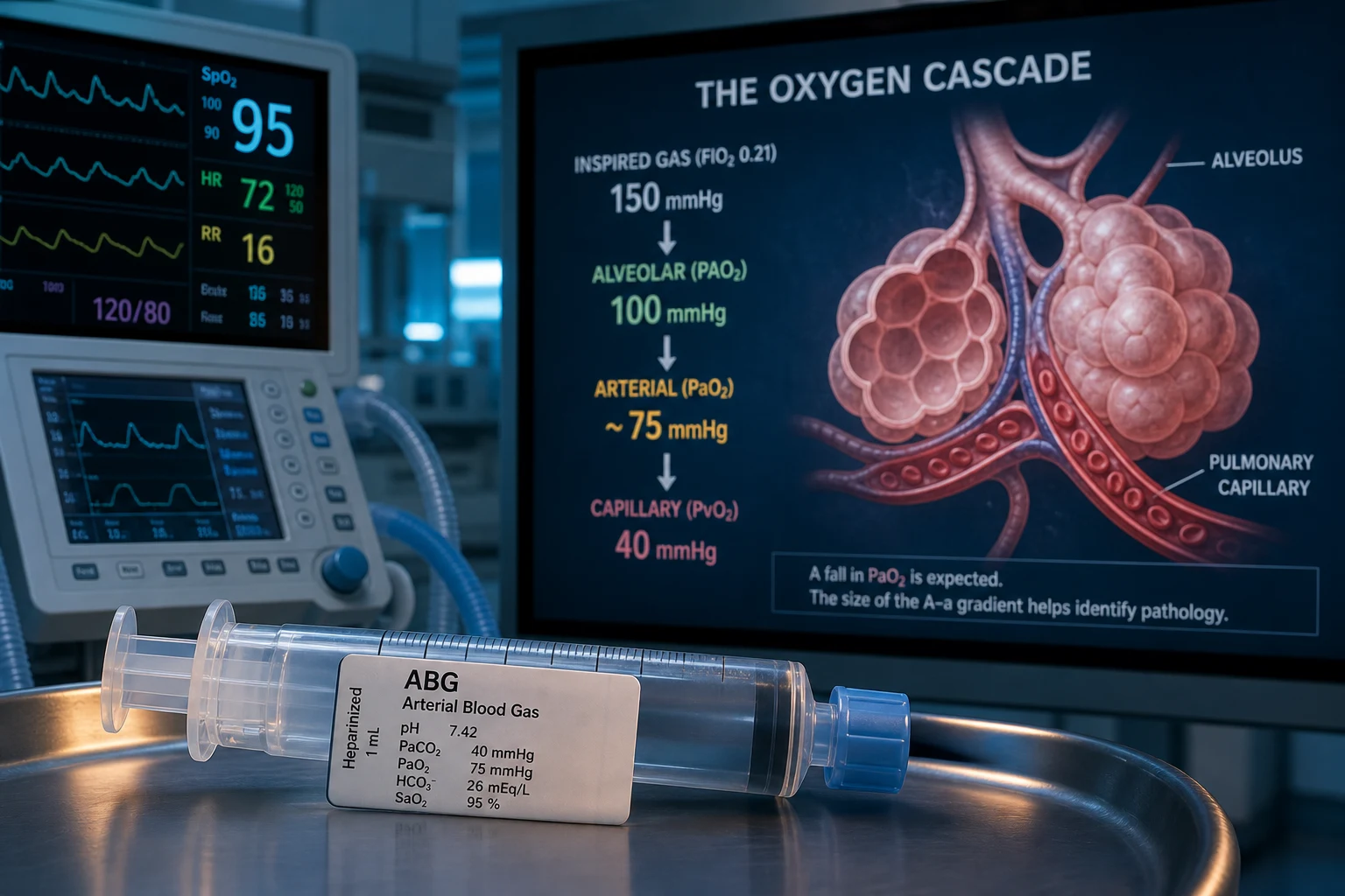
Oxygen partial pressure falls in a series of steps from the dry inspired gas to the mitochondrion. Understanding each step explains both normal gas exchange and where each mechanism of hypoxaemia acts.[2]
The oxygen cascade — each step reduces PO2
| Location | PO2 (mmHg) | Mechanism of the reduction |
|---|---|---|
| Dry atmosphere | 159 | FiO2 0.21 × Patm 760 mmHg |
| Trachea (humidified) | 149 | Subtract PH2O 47 mmHg → FiO2 × (760 − 47) = 0.21 × 713 |
| Alveolus (PAO2) | ~100 | Subtract PaCO2/RQ (40/0.8 = 50) → the alveolar gas equation |
| Artery (PaO2) | ~95 | Shunt + V/Q mismatch: the A-a gradient (normal <15 young, <25 elderly) |
| Systemic capillary | ~40 | Tissue oxygen extraction |
| Mitochondrion | 1–3 | Critical for oxidative phosphorylation; if <1 → anaerobic metabolism → lactate |
| Mixed venous (PvO2) | ~40 | SvO2 ~75% — the balance between DO2 and VO2 |
The five mechanisms of hypoxaemia
The five mechanisms — A-a gradient, response to oxygen, examples, and management
| Mechanism | A-a gradient | Response to 100% O2 | Typical causes | Management |
|---|---|---|---|---|
| V/Q mismatch (commonest) | Elevated | CORRECTS (PaO2 >350 on FiO2 1.0) | Pneumonia, COPD, asthma, pulmonary oedema, pulmonary embolism | Increase FiO2 + treat the cause |
| Shunt | Elevated | DOES NOT CORRECT (PaO2 stays <350 on FiO2 1.0) | ARDS, dense consolidation, complete atelectasis, PFO, pulmonary AVM, hepatopulmonary syndrome | PEEP (recruit), proning, ECMO; treat the anatomical shunt |
| Diffusion impairment | Elevated | CORRECTS (raises the diffusion gradient) | Interstitial lung disease, pulmonary fibrosis, early pulmonary oedema, emphysema | Increase FiO2; worsens with exercise |
| Hypoventilation | NORMAL | Improves PaO2 (but CO2 keeps rising) | Opioid/sedative overdose, neuromuscular disease (GBS, MG), CNS depression, chest-wall disorder | VENTILATE (NIV or intubation) — oxygen alone is insufficient |
| Low inspired PO2 | NORMAL | Improves | High altitude, a hypoxic gas mixture, a low-delivered FiO2 | Increase FiO2 (or descend) |
1. Ventilation-perfusion (V/Q) mismatch — the commonest cause
In a perfectly matched lung every alveolus receives ventilation in proportion to its blood flow (V/Q ≈ 1). In disease that matching breaks down. Low-V/Q units — perfused but under-ventilated (consolidation, oedema, atelectasis, bronchospasm) — behave as a partial shunt and are the dominant reason PaO2 falls. High-V/Q units — ventilated but under-perfused (embolism, emphysema) — behave as dead space; they cannot compensate for the low-V/Q units because blood leaving them is already nearly saturated, so extra oxygen barely raises the total CaO2. The net effect in almost all lung disease is dominated by the low-V/Q units that drag the PaO2 down.[1][2]
It is the commonest mechanism of hypoxaemia in the ICU (pneumonia, asthma, COPD, pulmonary oedema, pulmonary embolism), and it is the most treatable: because even poorly ventilated units are still open to the alveolus, raising the inspired FiO2 raises the PAO2 in those units and oxygenates their blood. V/Q mismatch corrects well with oxygen. Hypoxic pulmonary vasoconstriction (HPV) is the lung's own defence — it diverts blood away from poorly ventilated units — and anything that abolishes it (vasodilators, sepsis, cirrhosis) worsens the mismatch.[2]
2. Shunt — does NOT correct with 100% oxygen
In shunt, blood bypasses ventilated alveoli entirely, so it never equilibrates with alveolar gas and contributes deoxygenated blood directly to the arterial circulation. Because the shunted blood never contacts alveolar gas at all, raising the inspired FiO2 cannot reach it — the defining feature of true shunt. This is the single most important physiological distinction in the whole topic.[1][4]
Shunt is either anatomical (blood bypasses the lungs — a patent foramen ovale, pulmonary arteriovenous malformation, hepatopulmonary syndrome) or physiological (perfusion of completely unventilated lung — severe ARDS, dense consolidation, complete atelectasis, or a mucus plug occluding a lobe). In ARDS the physiological shunt fraction often exceeds 30%, which is why these patients are so hard to oxygenate. The landmark Mancini/Rodriguez-Roisin study used the multiple inert gas elimination technique (MIGET) to show that the improvement in oxygenation from a lung-protective (recruitment) strategy came almost entirely from a fall in intrapulmonary shunt as collapsed alveoli were recruited — PEEP works by reducing shunt, not by adding oxygen.[4]
Because FiO2 fails, the treatment of shunt is PEEP and alveolar recruitment (reopen collapsed units so blood can reach them), prone positioning (redistributes perfusion to dorsal, previously shunted lung), inhaled pulmonary vasodilators (redirect blood to ventilated units), and ultimately VV-ECMO for refractory shunt. [1]
3. Diffusion impairment
A thickened alveolar-capillary membrane slows oxygen transfer so that venous blood does not fully equilibrate with alveolar gas before it leaves the capillary. Causes include interstitial lung disease, pulmonary fibrosis, and the early exudative phase of pulmonary oedema. It responds to oxygen (raising FiO2 steepens the diffusion gradient) and characteristically worsens with exercise — because exercise raises cardiac output and shortens capillary transit time, so blood has less time to equilibrate. In healthy trained athletes exercise can itself cause a mild diffusion limitation and widen the A-a gradient — the phenomenon of exercise-induced arterial hypoxaemia.[3]
4. Hypoventilation (type 2 respiratory failure) — NORMAL A-a gradient
Alveolar ventilation is inadequate, so CO2 accumulates and, through the alveolar gas equation, PAO2 falls (as PaCO2 rises, PAO2 falls). The A-a gradient is normal — the alveoli themselves are fine; the problem is moving gas in and out. Causes are opioid or sedative overdose, neuromuscular disease (Guillain-Barré, myasthenia, ALS), CNS depression, and chest-wall disorders (kyphoscoliosis, obesity-hypoventilation). Oxygen improves PaO2 transiently but does not stop CO2 from rising; the treatment is ventilation (non-invasive or invasive) and reversal of the cause.[1]
5. Low inspired PO2 (altitude)
The inspired gas itself carries less oxygen — at altitude the Patm (and therefore the PiO2) is lower, so the whole cascade shifts down. The A-a gradient is normal. Corrected by increasing the inspired oxygen (or by descending). The classic example is high-altitude pulmonary oedema, where hypoxic pulmonary vasoconstriction raises capillary pressures and causes a non-cardiogenic oedema — but the primary mechanism at altitude is simply a low inspired PO2 with a normal gradient.[2]
The A-a gradient — calculation and interpretation
The alveolar-arterial oxygen gradient (A-a gradient) is the most useful single calculation for working out the cause of hypoxaemia. It compares the PAO2 the lung should generate (from the alveolar gas equation) with the PaO2 it actually achieves (from the ABG).[1][2]
Interpreting the A-a gradient — the key fork in the road
| A-a gradient | Meaning | Mechanism | Clue | Treatment |
|---|---|---|---|---|
| NORMAL (<15 young; <25 elderly; rises ~3 mmHg per decade) | Gas exchange is intact — the alveoli are fine | Hypoventilation or low inspired PO2 | PaCO2 is elevated (hypoventilation) or the patient is at altitude | Ventilate (hypoventilation); increase FiO2 / descend (altitude) |
| ELEVATED (>15–25) | The lung itself is the problem | V/Q mismatch, shunt, or diffusion impairment | PaCO2 is normal or low (hyperventilation offsetting the hypoxaemia) | Oxygen + treat the cause; PEEP/recruitment if shunt |
Worked example 1 — hypoventilation. A post-op patient on an opioid PCA: PaO2 55, PaCO2 70, on room air. PAO2 = 0.21 × 713 − 70/0.8 = 150 − 87.5 = 62. A-a gradient = 62 − 55 = 7 mmHg (normal). The low PaO2 is entirely explained by the high CO2 (hypoventilation). Treatment is ventilation/reversal, not oxygen. [1]
Worked example 2 — V/Q mismatch (pneumonia). PaO2 55, PaCO2 32, on room air. PAO2 = 0.21 × 713 − 32/0.8 = 150 − 40 = 110. A-a gradient = 110 − 55 = 55 mmHg (markedly elevated). The lung is the problem — V/Q mismatch from consolidation. Oxygen will help; treat the pneumonia. [1]
Worked example 3 — severe shunt (ARDS). Ventilated patient on FiO2 0.8: PaO2 60, PaCO2 40. PAO2 = 0.8 × 713 − 40/0.8 = 570 − 50 = 520. A-a gradient = 520 − 60 = 460 mmHg (massive). The gradient is enormous and the patient is refractory to a high FiO2 — this is shunt, needing PEEP, proning, and possibly ECMO.[4][6]
The age correction matters. The normal A-a gradient rises about 3 mmHg per decade (a common bedside formula is age/4 + 4). A gradient of 25 mmHg is reassuring in an 80-year-old but pathological in a 20-year-old — always interpret the number in the context of age.[1]
The shunt equation and venous admixture
The shunt equation quantifies the fraction of cardiac output that perfuses non-ventilated alveoli. It is the formal definition of "how much shunt is there?" [1]
Qs/Qt = (CcO2 − CaO2) / (CcO2 − CvO2) [1]
Where:
- Qs/Qt = shunt fraction (shunted flow / total cardiac output)
- CcO2 = end-capillary oxygen content (the theoretical maximum — calculated from the PAO2)
- CaO2 = arterial oxygen content (measured)
- CvO2 = mixed venous oxygen content (measured from a pulmonary artery catheter) [1]
Normal Qs/Qt is <5%; >10% is significant; in severe ARDS it can exceed 30–50%. Because it requires a mixed venous sample from a PA catheter, it is rarely calculated at the bedside — but the concept is examined and the bedside 100% oxygen test (below) is its practical surrogate.[2]
Venous admixture — the unifying concept
Venous admixture is the total amount of mixed venous blood that effectively "mixes" with oxygenated end-capillary blood and drags down the arterial content. It has two components:
- True shunt (V/Q = 0) — blood perfusing completely unventilated alveoli.
- Low-V/Q units — blood perfusing poorly ventilated alveoli that are not fully equilibrated. [1]
Venous admixture therefore captures both shunt and severe V/Q mismatch. The crucial practical point: venous admixture rises when mixed venous oxygen falls (low cardiac output, high extraction, fever, shivering). A falling CvO2 magnifies the effect of any existing shunt, because the venous blood dumped into the arterial circulation is even more desaturated. This is why improving cardiac output, haemoglobin, and oxygen demand (analgesia, sedation, paralysis, fever control) can improve PaO2 in a shunt patient without touching the lung — you raise the CvO2 and so dilute the shunted blood. Conversely, the same shunt looks far worse in a shocked, extracting patient.[2][4]
V/Q mismatch vs shunt — the 100% oxygen test
The definitive bedside discriminator between V/Q mismatch and true shunt is the response to 100% oxygen. Give FiO2 1.0 for ~15 minutes (long enough to wash out nitrogen from even poorly ventilated units), then re-measure the PaO2.[1][2]
The 100% oxygen test — distinguishing V/Q mismatch from shunt
| After 15 min on FiO2 1.0 | Interpretation | Why |
|---|---|---|
| PaO2 rises above ~350 mmHg | V/Q mismatch — correctable | Even poorly ventilated units are still open to alveolus; 100% O2 raises their PAO2 and oxygenates their blood |
| PaO2 stays below ~350 mmHg | Significant SHUNT — not correctable by FiO2 | The shunted blood never contacts alveolar gas, so FiO2 cannot reach it. The ceiling is set by how much blood is being shunted |
The ~350 mmHg threshold is pragmatic: on pure oxygen, a lung with only V/Q mismatch (no shunt) should achieve a PaO2 well above 350 mmHg, because every perfused alveolus — even a poorly ventilated one — is now filled with 100% oxygen. A PaO2 stuck below 350 mmHg means a meaningful fraction of the cardiac output is bypassing ventilated alveoli altogether. This single test redirects the whole management: a correctable patient gets oxygen and treatment of the cause; an uncorrectable patient gets PEEP, recruitment, proning, and consideration of ECMO. Do not keep escalating FiO2 in a patient who fails the 100% test — that is the definition of refractory shunt.[1]
The P/F ratio and the Berlin ARDS definition
The P/F ratio (PaO2/FiO2) is the simplest oxygenation index and the backbone of the Berlin ARDS definition. It is measured on a minimum of 5 cmH2O PEEP (or CPAP).[6]
Berlin ARDS severity — the P/F ratio (on PEEP/CPAP >=5 cmH2O)
| Berlin category | P/F ratio (mmHg) | Clinical implication |
|---|---|---|
| Mild | 200–300 | Close monitoring; HFNC/NIV often sufficient; lung-protective ventilation if intubated |
| Moderate | 100–200 | Usually intubated; lung-protective ventilation + PEEP titration; consider proning if worsening |
| Severe | <100 | Full ARDS bundle: low Vt, high PEEP strategy, proning >16 h/day (PROSEVA), consider cisatracurium and VV-ECMO referral |
The P/F ratio is not perfect — it depends on FiO2 (the ratio improves then worsens as FiO2 rises, because of the shunt effect), on PEEP, and on chest-wall compliance — but it is universally available and is the entry criterion for virtually every ARDS trial. It frames the mechanism: a low P/F on a high FiO2 is shunt-dominant disease, which is exactly why ARDS management is built around PEEP and recruitment rather than FiO2.[6]
Oxygenation indices — P/F, A-a gradient, and the Oxygenation Index
Three indices rank the severity of oxygenation failure. Each captures something slightly different.[6]
Oxygenation indices — what each measures and when to use it
| Index | Formula | Normal / threshold | Strengths and weaknesses |
|---|---|---|---|
| P/F ratio | PaO2 / FiO2 | Normal >400; ARDS <300 (mild/moderate/severe as above) | Simplest, universal, trial entry criterion. Conflates FiO2 and PEEP effects; not valid on different PEEP |
| A-a gradient | PAO2 − PaO2 (PAO2 from the alveolar gas equation) | <15 young, <25 elderly | The best diagnostic index — separates hypoventilation (normal) from a lung problem (high). Requires the full alveolar gas equation |
| Oxygenation Index (OI) | (FiO2 × MAP × 100) / PaO2 | Normal <5; >5 abnormal; >40 = severe (used in paediatric ARDS and ECMO criteria) | Incorporates mean airway pressure (MAP), so it rewards lung-protective low-pressure strategies and is the best severity index for tracking progress on the ventilator. Higher = worse |
The Oxygenation Index (OI) is underused in adult practice but is the gold-standard severity marker in paediatric ARDS and is part of many ECMO referral criteria: OI >40 for >4 hours is a classic VV-ECMO trigger in children. In adults, the simpler P/F ratio and the P(ECO2)/PaO2 (the "oxygenation index" without MAP) are more common, but the principle is the same — the more pressure and oxygen you must apply to achieve a given PaO2, the sicker the lung.[6]
Clinical approach to the hypoxaemic patient
The three-step approach to hypoxaemia — A-a gradient, image, treat
Step 1 — Calculate the A-a gradient from an ABG
Take an arterial blood gas on a known FiO2. Compute PAO2 = FiO2 × (760 − 47) − PaCO2/0.8, then the A-a gradient (PAO2 − PaO2). NORMAL gradient (<15 young, <25 elderly, age/4 + 4) → hypoventilation (high PaCO2) or low inspired PO2 (altitude) — the alveoli are fine; VENTILATE or raise FiO2. ELEVATED gradient → a lung (or heart) problem — go to Step 2.
Step 2 — If the gradient is elevated, localise with a chest X-ray and echocardiogram
The CXR separates diffuse (pulmonary oedema, ARDS, pneumonia) from focal (consolidation, atelectasis, effusion, pneumothorax) disease. The echocardiogram separates a cardiac cause (acute pulmonary oedema from LV failure, a right-to-left shunt through a PFO, pulmonary hypertension) from a primary lung problem. Bedside lung ultrasound adds rapid pleural and B-line information. If the CXR is clear and the echo is normal, think pulmonary embolism (V/Q scan or CTPA), early interstitial disease, pulmonary vasculitis, or a shunt (hepatopulmonary syndrome, pulmonary AVM).
Step 3 — Treat the mechanism, not the number
V/Q mismatch and diffusion impairment → OXYGEN and treat the cause (antibiotics, bronchodilators, diurese the oedema). HYPOVENTILATION → VENTILATION (NIV or intubation) and reverse the cause — oxygen buys time but will not stop CO2 rising. SHUNT (fails the 100% oxygen test, low P/F on high FiO2) → PEEP and alveolar recruitment, lung-protective ventilation, prone positioning, inhaled vasodilators, and VV-ECMO for refractory disease. In parallel, optimise the non-pulmonary determinants of arterial oxygenation: haemoglobin (transfuse), cardiac output (inotrope/fluid), oxygen demand (analgesia, sedation, fever control) — raising mixed venous oxygen dilutes any shunt.
The three steps collapse the five mechanisms into a single pathway: the gradient tells you whether to ventilate or image; the image tells you where the problem is; the mechanism (and the 100% oxygen test) tells you whether the patient needs oxygen or PEEP. The whole framework is designed to stop the commonest error in hypoxaemia — escalating FiO2 in a shunt patient who needs PEEP, or chasing a lung problem in a hypoventilating patient who needs ventilation.[1][2]
Clinical pearls
Red flags
Prognosis
Outcomes by mechanism and oxygenation index — the evidence
| Scenario | Prognostic marker | Outcome implication |
|---|---|---|
| ARDS severity (Berlin) | P/F <100 (severe) | Hospital mortality ~45%; drives the full bundle (proning, ECMO). P/F 200–300 (mild) mortality ~27%[6] |
| Paediatric / refractory ARDS | Oxygenation Index (OI) >40 sustained | Classic VV-ECMO trigger; high mortality without advanced support |
| Refractory shunt on 100% O2 | PaO2 <350 on FiO2 1.0 | Indicates significant shunt; FiO2 escalation futile — needs PEEP, proning, ECMO |
| Hypoventilation | Normal A-a gradient | Excellent once ventilation is restored — the lung is structurally intact |
| Altitude / low inspired PO2 | Normal A-a gradient, corrects with O2 | Excellent with oxygen or descent; watch for high-altitude pulmonary oedema |
Key trials and evidence
ARDS Definition Task Force — the Berlin Definition (PMID 22797452)
Study design
Consensus panel + meta-analysis of 4188 patients across 4 multicentre datasets
Population
Acute hypoxaemic respiratory failure within 1 week of a known insult, bilateral imaging not fully explained by cardiac failure
Key output
P/F ratio on PEEP/CPAP >=5 cmH2O grades severity: mild 200–300, moderate 100–200, severe <100
Mortality by grade
Mild 27%, moderate 32%, severe 45% — severity predicted mortality and ventilator-free days
Clinical bottom line
The P/F ratio is the universal ARDS entry and severity criterion — and a low P/F on a high FiO2 signals a shunt-dominant lung needing PEEP and recruitment, not more oxygen
Mancini, Rodriguez-Roisin et al. — why PEEP works in ARDS (PMID 11704594)
Study design
Prospective physiological study — 8 patients with early ARDS, MIGET (multiple inert gas elimination technique)
Intervention
Protective ventilatory strategy (low Vt, PEEP set 2 cmH2O above the lower inflection point) vs baseline conventional ventilation, at the same FiO2
Key finding
PaO2 rose from 93 to 166 mmHg, and intrapulmonary shunt fell from 39% to 31% — the oxygenation improvement tracked the fall in shunt, driven by alveolar recruitment
Clinical bottom line
The mechanism by which PEEP improves oxygenation in ARDS is REDUCTION OF SHUNT (recruiting collapsed alveoli), not addition of oxygen — the physiological proof that shunt is treated by recruitment, not by FiO2
Petersson & Glenny — Gas Exchange in the Lung (PMID 37816345)
Article type
Comprehensive review — Seminars in Respiratory and Critical Care Medicine, 2023
Scope
The principles of lung gas exchange and the mechanisms of hypoxaemia and hypercapnia: V/Q mismatch, shunt, diffusion limitation, and the metrics (A-a gradient, shunt equation, dead space) that quantify the deviation from ideal gas exchange
Clinical bottom line
The modern authoritative reference for the five-mechanism framework and the oxygenation metrics used at the bedside — the basis for the A-a gradient, shunt equation, and 100% oxygen test discussed throughout this topic
Exam practice — SAQ
SAQ — Hypoxaemia mechanisms comprehensive — five causes, A-a, shunt vs V/Q
10 minutes · 10 marks
You are the ICU consultant reviewing a complex case that hinges on hypoxaemia mechanisms comprehensive — five causes, a-a, shunt vs v/q. Apply fellowship-level reasoning.
References
- [1]Sarkar M, Niranjan N, Banyal PK Mechanisms of hypoxemia Lung India, 2017.PMID 28144061
- [2]Petersson J, Glenny RW Gas Exchange in the Lung Semin Respir Crit Care Med, 2023.PMID 37816345
- [3]Hopkins SR Exercise induced arterial hypoxemia: the role of ventilation-perfusion inequality and pulmonary diffusion limitation Adv Exp Med Biol, 2006.PMID 17089876
- [4]Mancini M, Zavala E, Mancebo J, Fernandez C, Barbera JA, Rossi A, Roca J, Rodriguez-Roisin R Mechanisms of pulmonary gas exchange improvement during a protective ventilatory strategy in acute respiratory distress syndrome Am J Respir Crit Care Med, 2001.PMID 11704594
- [5]Petersson J, Glenny RW Imaging regional PAO2 and gas exchange J Appl Physiol (1985), 2012.PMID 22604886
- [6]ARDS Definition Task Force; Ranieri VM, Rubenfeld GD, Thompson BT, et al Acute respiratory distress syndrome: the Berlin Definition JAMA, 2012.PMID 22797452