ICU · Respiratory
Lung abscess and aspiration pneumonia in ICU
Also known as Lung abscess · Pulmonary abscess · Aspiration pneumonia · Necrotising pneumonia · Empyema · Mendelson syndrome
Lung abscess: localised collection of pus in lung parenchyma (cavity with air-fluid level). Primary abscess arises from aspiration of oropharyngeal contents (anaerobes, S. milleri) in a host with impaired consciousness or swallow — alcoholism, seizure, stroke, poor dentition. Secondary abscess follows bacteraemic seeding — right-sided endocarditis (S. aureus in IVDU), septic thrombophlebitis (Lemierre, Fusobacterium), infected lines — or bronchial obstruction (cancer, foreign body). Necrotising pneumonia is a severe, confluent tissue-destructive process (PVL-producing S. aureus, Klebsiella, type 3 pneumococcus). Aspiration PNEUMONIA (bacterial infection after macro-aspiration, 24-48 h onset, dependent segments, anaerobes) is distinct from aspiration PNEUMONITIS (Mendelson syndrome — sterile chemical injury from acidic gastric contents, onset within hours, no routine antibiotics). Treatment: prolonged antibiotics 4-6 weeks (clindamycin or amoxycillin/clavulanate for aspiration; organism-directed for necrotising), percutaneous drainage if >6 cm or no response by 7-10 days, surgical resection for failure or massive haemoptysis, and treatment of the underlying cause plus an aspiration prevention bundle.
On this page & tools
Your progress
Saved locally on this device.
Target exams
Red flags
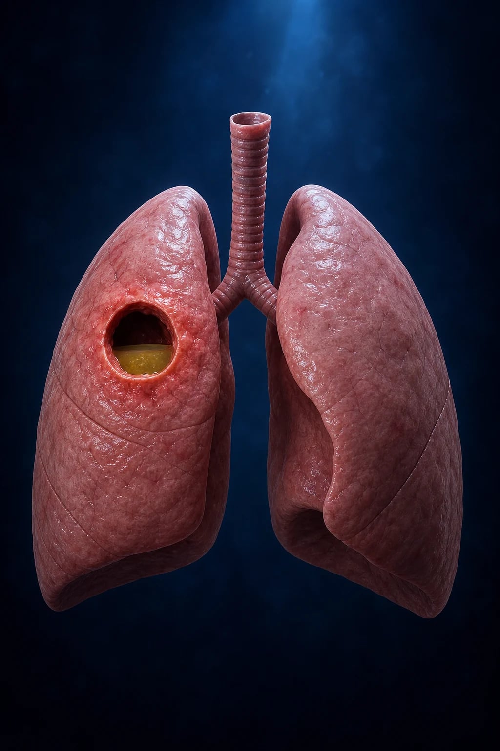
Management of lung abscess
- Recognise — fever, cough, foul-smelling sputum, weight loss. Chest X-ray/CT: cavity with air-fluid level, thick wall
- Identify cause — aspiration (alcoholism, seizure, stroke), necrotising pneumonia (S. aureus, Klebsiella), septic emboli (endocarditis — examine for murmur, IVDU), bronchial obstruction (cancer, foreign body), immunocompromise
- Antibiotics (prolonged) — IV initially, then oral. Aspiration/anaerobic: clindamycin OR amoxycillin/clavulanate. Necrotising: cover specific organism (flucloxacillin/vancomycin for S. aureus). Duration: 4-6 weeks (until cavity resolves or stable)
- Source control — percutaneous drainage (if >6 cm, not responding to antibiotics by 7-10 days, or deterioration). Surgical resection (if drainage fails, massive haemoptysis, malignancy)
- Treat underlying cause — dental hygiene (aspiration), alcohol rehabilitation, seizure control, stroke rehabilitation, endocarditis treatment, relief of obstruction
- Nutrition — these patients are often malnourished (alcoholism, chronic illness). Enteral nutrition, thiamine and B vitamins in alcohol use disorder
- Follow-up — repeat imaging (ensure cavity resolving). Bronchoscopy if not resolving (exclude malignancy, foreign body, obstruction)
SAQ — Cavitating lung lesion in a chronic alcoholic
10 minutes · 10 marks
A 58-year-old man with chronic alcohol use disorder is admitted from the ED with a two-week history of productive cough, fever, 8 kg weight loss and foul-smelling sputum. He has poor dentition. Chest X-ray shows a 5 cm cavitating lesion with an air-fluid level in the posterior segment of the right upper lobe. CRP 240, WCC 18.4,Albumin 28.
SAQ — Aspiration pneumonia versus aspiration pneumonitis (Mendelson syndrome)
10 minutes · 10 marks
A 72-year-old man is admitted to ICU three hours after a large witnessed aspiration of gastric contents during a generalised tonic-clonic seizure. He has a residual dense right hemiparesis from an ischaemic stroke four months ago. He is tachypnoeic, SpO2 88 per cent on room air, diffusely wheezy with crackles in the right lower lobe. CXR shows new bilateral basal infiltrates.
Clinical pearls
Red flags
Prognosis
Outcomes of lung abscess — what to expect
Modern case series and reviews:[3][1]
- Overall mortality: 5-10 per cent with prompt antibiotics and drainage (was 30-50 per cent in the pre-antibiotic era)
- Resolution with antibiotics alone: 80-90 per cent of primary abscesses (drainage not required)
- Percutaneous catheter drainage success: 80-90 per cent for large or non-responding abscesses
- Surgical resection: required in 5-10 per cent (drainage failure, massive haemoptysis, underlying malignancy)
- Median time to radiological resolution: 4-6 weeks; small stable residual cavities are acceptable
- Risk factors for poor outcome: abscess >6 cm, necrotising pneumonia, immunocompromise, delayed treatment, older age, large airway obstruction
Aspiration pneumonia mortality: 5-15 per cent (community) vs 20-30 per cent (hospital-acquired / ICU). Necrotising pneumonia mortality: 15-30 per cent, highest with PVL-producing S. aureus and hypervirulent Klebsiella.[4]
Lung abscess — pathology and classification
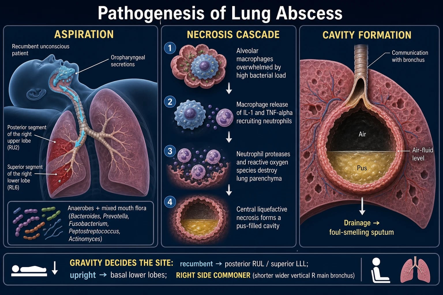
A lung abscess is a localised collection of pus within the lung parenchyma that has undergone central liquefactive necrosis, producing a cavity whose wall is formed by the surrounding inflamed lung. When the cavity communicates with a bronchus, the necrotic material is partially expectorated and air enters, producing the hallmark air-fluid level.[1]
Abscesses are classified by mechanism into primary and secondary, a distinction that determines both the microbiology and the source-control strategy.[3]
Primary lung abscess (aspiration)
A primary abscess arises in a previously normal lung from aspiration of infected oropharyngeal or gastric contents. A single large aspiration event, or repeated micro-aspiration, seeds bacteria into the dependent segments where mucociliary clearance is impaired by impaired consciousness or impaired swallow.[1]
Risk factors for primary aspiration abscess: [1]
- Impaired consciousness — alcohol use disorder (the classic cause), post-ictal state, drug overdose, anaesthesia, ICU sedation, head injury
- Neurological swallow failure — acute stroke (especially brainstem or large hemisphere), dementia, Parkinson's disease, motor neuron disease, bulbar weakness (myasthenia gravis, Guillain-Barré)
- Mechanical / anatomical — oesophageal disease (achalasia, Zenker's diverticulum, stricture, severe GERD), prolonged intubation, tracheostomy
- Increased bacterial inoculum — poor dentition and periodontal disease (the reservoir for anaerobes), poor oral hygiene, proton-pump inhibitor use (raises gastric pH and permits Gram-negative colonisation), nursing-home residence [1]
The microbiology of the primary abscess mirrors the oropharyngeal flora: anaerobes (Peptostreptococcus, Prevotella, Fusobacterium, Bacteroides, Porphyromonas, Actinomyces) mixed with the S. milleri group of streptococci.[2]
Secondary lung abscess
A secondary abscess arises by one of three non-aspiration mechanisms: [1]
- Haematogenous seeding — right-sided endocarditis (S. aureus in people who inject drugs), septic thrombophlebitis (Lemierre's syndrome, Fusobacterium necrophorum), infected central venous catheter, or septic embolism from any peripheral source. Lesions are typically multiple, peripheral, and cavitating.[16]
- Bronchial obstruction — bronchogenic carcinoma (squamous), bronchial carcinoid, foreign body aspiration, extrinsic compression by nodes. An abscess that develops distal to an obstruction (post-obstructive) will not resolve until the obstruction is relieved — this is the classic cause of a non-resolving abscess.[20]
- Direct extension — subphrenic or hepatic abscess (often Klebsiella in East Asian series, classically from pyogenic liver abscess), oesophageal perforation, or suppurative mediastinitis.
Secondary abscesses are caused by aerobic, often Gram-negative or nosocomial, organisms (S. aureus, Klebsiella, Pseudomonas, Enterobacteriaceae) rather than the mixed anaerobic flora of aspiration, and they carry a higher mortality.[3]
Primary vs secondary lung abscess — the exam answer side by side
| Feature | Primary lung abscess | Secondary lung abscess |
|---|---|---|
| Mechanism | Aspiration of oropharyngeal contents | Bacteraemic seeding, obstruction, or direct extension |
| Host | Impaired consciousness / swallow; poor dentition | IVDU, endocarditis, line infection, malignancy, immunocompromise |
| Number | Usually single | Often multiple (septic emboli) |
| Location | Dependent segments (posterior upper lobe, superior lower lobe) | Peripheral / wedge-shaped (septic emboli); distal to obstruction |
| Microbiology | Mixed anaerobes + S. milleri | S. aureus, Klebsiella, Pseudomonas, Enterobacteriaceae |
| Foul sputum | Often (anaerobes) | Uncommon |
| Resolution with antibiotics alone | 80-90 per cent | Lower — needs source control |
| Key management step | Prolonged anaerobic cover | Treat the source (endocarditis, line, obstruction) |
| Mortality | 5-10 per cent | 20-30 per cent |
The organisms that cavitate — and the clues that point to each
| Organism | Clue / setting | Distinguishing feature |
|---|---|---|
| Anaerobes + S. milleri (aspiration) | Alcoholism, post-ictal, stroke, poor dentition | Foul-smelling sputum; single dependent-segment cavity; subacute |
| PVL-producing S. aureus | Young, previously well; post-influenza; IVDU; skin/soft tissue infection | Rapidly progressive, multilobar necrosis, high mortality, leucopenia* |
| Klebsiella pneumoniae | Alcoholism, diabetes; East Asian (hypervirulent K1/K2) | "Bulging fissure" lobar consolidation; thickcreamy ("currant jelly") sputum; multi-focal abscesses |
| Type 3 S. pneumoniae | Young children; post-influenza | Bilateral necrotising pneumonia; effusion common |
| Pseudomonas aeruginosa | Nosocomial; bronchiectasis; structural lung disease | Bacteremic, bilateral, rapidly cavitating |
| Group A streptococcus | Post-viral; healthy young adults | Necrotising pneumonia + toxin-mediated shock |
| Septic emboli (S. aureus, Fusobacterium) | IVDU; right-sided endocarditis; Lemierre | Multiple peripheral "cannonball" cavities; feeding-vessel sign |
| Fungi (Aspergillus, Mucorales, Histoplasma) | Immunocompromise (neutropenia, transplantation, diabetes for Mucorales) | Halo and reverse-halo signs; nodular cavity wall |
*PVL-producing S. aureus necrotising pneumonia carries a reported mortality of over 30 per cent and frequently presents with leucopenia, haemoptysis and multilobar involvement.[4][17]
Aspiration pneumonia vs aspiration pneumonitis (Mendelson syndrome) — the core distinction
A single aspiration event can produce two entirely different syndromes, and the difference determines whether antibiotics are given. Confusing them is one of the commonest antibiotic-stewardship errors in the ICU.[5]
Aspiration pneumonitis (Mendelson syndrome)
Aspiration of sterile, acidic gastric contents (pH < 2.5) causes an acute chemical burn of the bronchial and alveolar epithelium. It is sterile at onset — the injuring agent is the acid, not bacteria. Onset is within minutes to hours, classically after anaesthesia, seizure, intoxication, or resuscitation (Curtis Mendelson's 1946 obstetric description in 66,016 pregnancies).[15] Features include abrupt dyspnoea, bronchospasm, frothy or pink sputum, hypoxaemia that is often out of proportion to the initial chest X-ray, and bilateral infiltrates that develop over hours. Roughly 10 per cent of massive aspirations progress to ARDS, and a minority develop secondary bacterial infection over the following days.[7]
Management is supportive: airway protection, supplemental oxygen, NIV or invasive ventilation for respiratory failure, judicious fluids. No routine antibiotics, no corticosteroids, no bronchoalveolar lavage. Antibiotics are added only if infection declares itself (fever beyond 48 h, purulent sputum, rising inflammatory markers, progressive infiltrates).[5]
The Mendelson cascade:[7]
- Acid reaches the alveoli and spreads throughout the bronchial tree within 12-18 seconds.
- A pH below 2.5 causes immediate coagulative necrosis of bronchial and alveolar epithelium.
- Alveolar-capillary disruption within minutes allows a protein-rich exudate to flood the alveoli — non-cardiogenic pulmonary oedema.
- Neutrophil infiltration (1-4 h) releases reactive oxygen species and proteases that amplify injury — acute inflammation, not infection.
- Surfactant inactivation causes atelectasis and a fall in compliance.
- V/Q mismatch and intrapulmonary shunt produce hypoxaemia, often disproportionate to the radiograph early on.
- By 24-36 h, around 10 per cent progress to ARDS; a subset develop secondary bacterial infection over subsequent days.
Aspiration pneumonia
Aspiration of bacteria-laden oropharyngeal or gastric secretions causes a bacterial pneumonia. It is more indolent, evolving over 24-48 h (or insidiously over days in elderly or demented patients with repeated small aspirations). It arises in patients with impaired swallow or chronic aspiration: stroke, dementia, Parkinson's, neuromuscular weakness, poor dentition, prolonged intubation. The infiltrate localises to dependent segments and may cavitate (anaerobes) or progress to abscess or empyema.[5]
Management requires antibiotics covering both aerobes and anaerobes.[2]
Aspiration pneumonitis vs aspiration pneumonia — the side-by-side answer
| Feature | Aspiration pneumonitis (Mendelson) | Aspiration pneumonia |
|---|---|---|
| Mechanism | Chemical injury — sterile gastric acid | Bacterial infection — oropharyngeal flora |
| pH of aspirate | < 2.5 (acidic) | Any (usually colonised, near-neutral) |
| Onset | Minutes to hours | 24-48 h (or insidious in elderly) |
| Typical setting | Anaesthesia, post-ictal, intoxication, resuscitation | Stroke, dementia, neuromuscular, poor dentition |
| Inoculum | Sterile gastric acid | Bacteria-laden secretions |
| Fever / purulent sputum | Absent initially | Present |
| Chest X-ray | Bilateral diffuse infiltrates ± ARDS | Focal dependent-segment consolidation ± cavitation |
| Dependent localisation | Less localised | Posterior upper lobe + superior lower lobe (supine); basal lower lobe (upright) |
| Anaerobes | — | Peptostreptococcus, Fusobacterium, Prevotella, Bacteroides |
| Procalcitonin | Low (< 0.1) | Elevated (> 0.25)[10] |
| Antibiotics | NO (unless infection develops) | YES — aerobes + anaerobes |
| Corticosteroids | NO | NO |
| Bronchoalveolar lavage | NO | NO |
| Duration of therapy | Supportive | 7 days (4-6 weeks if abscess or empyema) |
Why the distinction matters
Up to a third of inpatients labelled "aspiration pneumonia" actually have sterile pneumonitis for which antibiotics add nothing and cause harm — Clostridioides difficile colitis, antimicrobial resistance, adverse drug events, and cost. A 48-72 h observation window with procalcitonin and CRP trends and repeat imaging is the stewardship-friendly approach in equivocal cases.[8] Procalcitonin is low in pneumonitis (< 0.1 ng/mL) and elevated in bacterial aspiration pneumonia (> 0.25 ng/mL), helping to distinguish them.[10]
Microbiology of aspiration and abscess
The classic anaerobic + aerobic mix
Anaerobes (originate from gingival crevices and dental plaque; need anaerobic transport and are rarely recovered from expectorated sputum):[1][2]
- Peptostreptococcus species — the single most common anaerobe in lung abscess
- Fusobacterium nucleatum — necrotising; the organism of Lemierre's syndrome (F. necrophorum)
- Prevotella, Bacteroides (including the B. fragilis group in abscess)
- Porphyromonas, Actinomyces (chronic, indolent infection with sinus tracts) [1]
Aerobes: [1]
- Streptococcus pneumoniae, Haemophilus influenzae — community-acquired
- Staphylococcus aureus (including MRSA and PVL-producing strains)
- Enteric Gram-negative bacilli — Klebsiella, E. coli, Pseudomonas — more prominent in healthcare-associated aspiration, alcoholism, and structural lung disease [1]
High-yield exam pearl: Edentulous patients have markedly fewer anaerobes (no gingival crevices), so anaerobic coverage can often be narrowed, and aspiration in an edentulous patient is more likely to be aerobic (S. pneumoniae, H. influenzae, Gram-negative bacilli).[2]
Organisms by syndrome and setting — what to cover
| Setting | Predominant organisms | Empirical cover |
|---|---|---|
| Community aspiration, dentate | Anaerobes + S. pneumoniae, H. influenzae | Amox-clav OR clindamycin ± respiratory FQ |
| Community aspiration, edentulous | Aerobes (S. pneumo, H. flu, GNB) — few anaerobes | Ceftriaxone ± metronidazole (anaerobe cover optional) |
| Severe / hospitalised | Anaerobes + aerobes ± GNB | Pip-tazo OR clindamycin + ceftriaxone |
| Healthcare-associated / MDR risk | Anaerobes + Pseudomonas, MRSA, ESBL | Meropenem/cefepime + vanc/linezolid + metronidazole |
| Lung abscess / empyema | Anaerobes (necrotising) | Drain + prolonged anaerobe cover (4-6 weeks) |
| Necrotising pneumonia | PVL S. aureus, Klebsiella, type 3 pneumococcus | Linezolid/vancomycin + organism-directed cover |
| Pneumonitis (sterile) | None | None (antibiotics only if infection develops) |
Necrotising pneumonia
Necrotising pneumonia is a severe, confluent tissue-destructive process in which pneumonia progresses to liquefactive necrosis of the lung parenchyma, producing thin-walled cavities, pneumatoceles, and often an empyema or bronchopleural fistula.[4]
Classic organisms: [1]
- PVL-producing Staphylococcus aureus — community-acquired (CA-MRSA or MSSA); affects previously healthy young people, often after an influenza-like illness; rapidly progressive multilobar necrosis, haemoptysis, leucopenia, septic shock; mortality > 30 per cent.[4][17]
- Klebsiella pneumoniae — classically in alcoholics and diabetics; hypervirulent capsular serotype K1/K2 in East Asian series; "currant jelly" sputum, bulging fissure, multi-focal abscesses.[3]
- Type 3 Streptococcus pneumoniae — children and post-influenza adults.
- Pseudomonas aeruginosa — nosocomial; bacteremic, bilateral, rapidly cavitating.
- Group A streptococcus — post-viral; toxin-mediated shock.
Management combines aggressive organism-directed antibiotics (including anti-toxin activity — linezolid or clindamycin inhibit protein synthesis and hence toxin production in PVL staphylococcal disease), source control (drainage of abscess or empyema), and supportive care for the frequently associated septic shock and ARDS. Surgical resection is occasionally required for massive necrosis or uncontrolled bronchopleural fistula.[18]
Clinical features and radiology
- Subacute presentation is typical of primary abscess: 1-3 weeks of fever, productive cough, foul-smelling (putrid) sputum, pleuritic chest pain, weight loss, night sweats, and haemoptysis (occasionally massive).
- Acute presentation dominates in necrotising pneumonia and secondary (bacteraemic) abscess: high fever, rigors, septic shock, rapidly progressive respiratory failure.
- Crackles, bronchial breathing, and decreased breath sounds over the involved zone; a pleural rub or dullness if an effusion or empyema has developed. [1]
Radiographic distribution — gravity dictates the site
| Patient position | Involved segments |
|---|---|
| Supine / recumbent (most ICU patients) | Posterior segment of the upper lobes + superior segment of the lower lobes |
| Upright / ambulant | Basal segments of the lower lobes |
| Right > Left | The right main bronchus is wider, shorter and more vertical |
Imaging patterns
- Chest X-ray — round opacity with an air-fluid level (only if cavity communicates with a bronchus); surrounding consolidation; may show effusion or empyema.
- CT chest (definitive) — thick-walled cavity with air-fluid level; surrounding consolidation; greater sensitivity for cavitation, abscess, empyema, necrosis, gas-forming infection, and an aspirated foreign body; can guide percutaneous drainage.
- Pneumatoceles — thin-walled, air-filled cystic spaces seen in S. aureus (especially in children) and Pneumocystis pneumonia.
- Septic emboli — multiple peripheral wedge-shaped nodules/cavities with a feeding vessel ("cannonball" lesions). [1]
Cavitating lung lesion — the differential and the discriminating clues
| Lesion | Discriminating clue | Confirmatory test |
|---|---|---|
| Lung abscess (aspiration) | Dependent segment, foul sputum, fever, surrounding consolidation | CT (air-fluid level, thick wall); sputum/BAL |
| Cavitating lung cancer | Older smoker, weight loss, thick irregular wall (>15 mm), no air-fluid level, no surrounding consolidation | Bronchoscopic biopsy |
| Tuberculosis | Upper-lobe predominance, cough, weight loss, night sweats, immigration/exposure | Sputum AFB, NAAT, culture |
| Aspergilloma | Pre-existing cavity, "monod sign"/air crescent, immunocompromise | Serum galactomannan, CT |
| Septic emboli | Multiple peripheral cavities, IVDU, right-sided endocarditis | Blood cultures, echocardiography |
| Wegener's granulomatosis (GPA) | Multiple cavitating nodules, sinusitis, renal involvement, c-ANCA | Biopsy, ANCA |
| Hydatid cyst | Endemic area, "water lily" sign, intact cyst | Serology, CT |
Investigations
- Bloods — FBC (leucocytosis, or leucopenia in severe PVL disease), EUC, LFTs, CRP, procalcitonin (low in pneumonitis, high in bacterial infection)[10]
- ABG/VBG — quantify hypoxaemia, the A-a gradient, and acid-base status
- Chest X-ray on presentation and serially; CT chest if cavitation, abscess, empyema, obstruction, or a foreign body is suspected, or for poor-resolution cases
- Blood cultures before antibiotics (in pneumonia, sepsis, or suspected endocarditis)
- Sputum / endotracheal aspirate Gram stain and culture — aerobic; anaerobic culture of expectorated sputum is unhelpful because of contamination by oral flora
- Invasive sampling (bronchoscopic BAL, protected specimen brush) — for severe, healthcare-associated, immunocompromised, or non-responding cases; also for anaerobic culture (trans-tracheal or aspirate)
- Bronchoscopy — for retrieval of large particulate / food / foreign body aspiration; not for lavage of acidic liquid; to relieve obstruction and to biopsy a suspected malignancy
- Percutaneous or bronchoscopic aspirate of an abscess for anaerobes in complicated cases
- Echocardiography if septic emboli or right-sided endocarditis is suspected; Doppler of the internal jugular vein if Lemierre is suspected
- HIV serology if risk factors — S. aureus bacteraemia, pneumococcal necrotising pneumonia, and atypical organisms are more common in undiagnosed HIV
Management
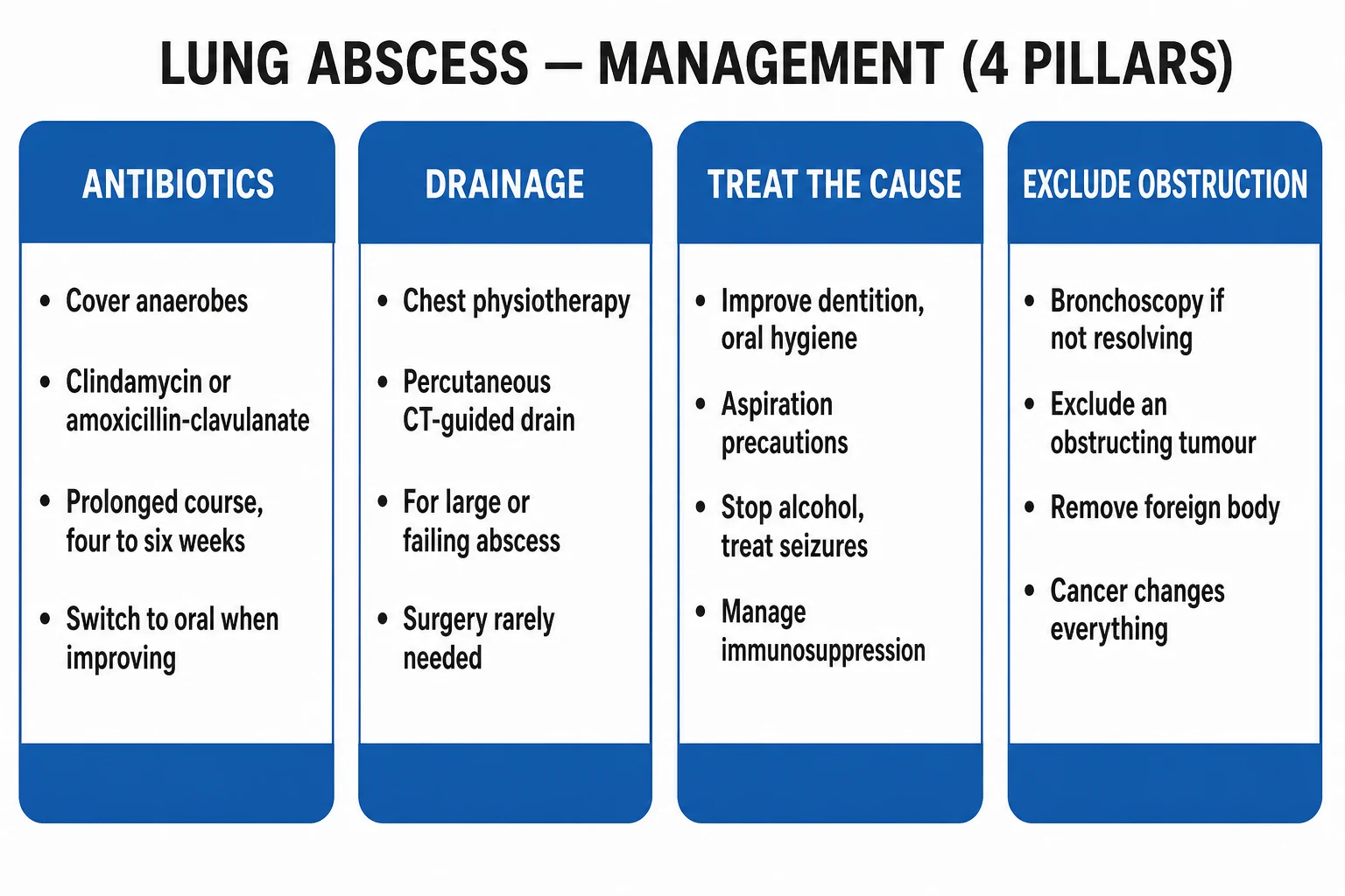
1. Immediate airway and respiratory support
- Airway — clear and protect the airway; suction the oropharynx; intubate if GCS < 8, unable to protect the airway, or severe hypoxaemia
- Oxygen — target SpO₂ 92-96 per cent (88-92 per cent if COPD/hypercapnia risk)
- Ventilatory support — NIV may avoid intubation in moderate pneumonitis (each intubation raises VAP risk); invasive ventilation for refractory hypoxaemia, ARDS, or necrotising pneumonia with respiratory failure
- Positioning — head of bed elevated; lateral with the affected side down to localise the process
- Fluids — judicious; capillary leak favours cautious resuscitation, but hypovolaemia worsens perfusion [1]
2. The antibiotic decision
Aspiration pneumonitis → NO empirical antibiotics. Reassess at 48-72 h and start only if infection develops (fever, purulent sputum, leucocytosis, progressive infiltrates), or if there is high baseline suspicion of bacterial co-infection (sepsis, immunocompromise).[5]
Aspiration pneumonia → antibiotics covering aerobes + anaerobes:[2][6]
- Community-acquired, mild to moderate — amoxicillin-clavulanate OR clindamycin (± a respiratory fluoroquinolone)
- Severe / hospitalised — piperacillin-tazobactam OR clindamycin + ceftriaxone (or cefepime + metronidazole)
- Healthcare-associated / MDR risk — extended-spectrum antipseudomonal β-lactam (pip-tazo, cefepime, meropenem) ± MRSA cover (vancomycin or linezolid)
- Abscess / empyema — drain + prolonged anaerobe cover (4-6 weeks)[1]
3. Source control — drainage and surgery
- Percutaneous catheter drainage (CT- or ultrasound-guided) for abscess > 6 cm, non-response at 7-10 days, deterioration, or coexisting empyema. Success 80-90 per cent. Complications: bronchopleural fistula, bleeding, pneumothorax.[14]
- Surgical resection (lobectomy or pneumonectomy) for drainage failure, massive haemoptysis, underlying malignancy, or extensive necrosis with bronchopleural fistula.[21]
- Empyema — chest drain (large bore); intrapleural tPA + DNase for loculated collections (Rahman 2011 NEJM showed DNase + tPA improved drainage; streptokinase alone did not — MIST1 trial); VATS for failure or multi-loculated disease.[12][13]
4. Therapies NOT recommended
- Corticosteroids — no benefit and possible harm across multiple RCTs and meta-analyses for aspiration pneumonitis.[5]
- Bronchoalveolar lavage to neutralise or remove acid — distributes the injury more widely and worsens outcomes.[7]
- Empirical antibiotics for sterile chemical pneumonitis — stewardship harm.[8]
5. Supportive and adjunctive
- Chest physiotherapy — postural drainage, percussion, breathing exercises once the patient is stable
- Bronchodilators for bronchospasm
- VAP and aspiration prevention bundle (see below)
- DVT and stress-ulcer prophylaxis per ICU protocol
- Early enteral nutrition via NG or NJ with swallow reassessment before any oral intake; thiamine (100 mg IV OD for 3-5 days) before glucose in suspected alcohol use disorder to prevent Wernicke encephalopathy
- Treat the underlying cause — alcohol rehabilitation, seizure control, stroke rehabilitation, dental care, endocarditis therapy, relief of bronchial obstruction, line removal [1]
Aspiration event in the ICU — the management algorithm
Immediate (0-10 min): airway and oxygen
Clear and suction the oropharynx; intubate if GCS < 8 or airway unprotected; oxygen to SpO2 92-96 per cent (88-92 per cent if COPD). Position head up, affected side down.
Assess: pneumonitis vs pneumonia
Timing (hours = pneumonitis; 24-48 h = pneumonia), setting (anaesthesia/intoxication vs stroke/dementia), purulence of sputum, fever, CXR pattern, procalcitonin (low vs high).
Decision: antibiotics or not
If pneumonitis → supportive care, NO antibiotics, observe 48-72 h. If pneumonia → antibiotics covering aerobes + anaerobes (pip-tazo OR clindamycin + ceftriaxone); send blood + sputum cultures first.
Look for an abscess
CXR ± CT chest for cavity/air-fluid level. If abscess present: prolonged antibiotics 4-6 weeks (clindamycin or amox-clav); drain if >6 cm or non-response at 7-10 days.
Supportive care
NIV/invasive ventilation for respiratory failure; judicious fluids; chest physiotherapy once stable; bronchodilators for wheeze; DVT and stress-ulcer prophylaxis; early enteral nutrition; thiamine in alcohol use disorder.
Reassess at 48-72 h
For pneumonitis: start antibiotics only if fever, purulent sputum, rising inflammatory markers, or progressive infiltrates appear. For pneumonia: narrow to culture results; duration 7 days (4-6 weeks if abscess or empyema).
Investigate non-resolution
If not improving at 7-14 days → bronchoscopy (exclude obstruction, foreign body, malignancy), repeat CT, consider resistant organism, undrained collection, or immunodeficiency.
Prevent recurrence (the bundle)
HOB 30-45 degrees; oral care with chlorhexidine; subglottic suction ETT; cuff pressure 20-30 cmH2O; minimise sedation; swallow assessment before oral intake; review PPI and feeding route.
Lung abscess — source-control decision ladder
Step 1 — antibiotics first
Prolonged IV then oral anaerobe cover (clindamycin 600 mg IV TDS or amox-clav 1.2 g IV TDS) plus organism-directed cover for necrotising organisms. Most primary abscesses (80-90%) resolve on antibiotics alone.
Step 2 — when to drain
Percutaneous catheter drainage (CT/US-guided) if: abscess >6 cm, no clinical/radiological response by 7-10 days, deterioration, rupture into pleura (empyema), or immunocompromise. Avoid routes crossing uninvolved lung.
Step 3 — bronchoscopy
If not resolving or if obstruction/foreign body suspected — diagnostic (biopsy, BAL) and therapeutic (foreign-body removal, relief of obstruction).
Step 4 — surgery
Reserved for: drainage failure, massive haemoptysis, underlying malignancy, bronchopleural fistula, extensive necrosis. Lobectomy or pneumonectomy.
Step 5 — treat the cause + follow-up
Dental care, alcohol rehab, seizure/stroke management, endocarditis therapy, line removal. Serial CXR/CT until the cavity resolves or is stable.
Antibiotic regimens for aspiration / abscess by severity and setting
| Setting | First-line regimen | Alternative | Notes |
|---|---|---|---|
| Community aspiration, mild (oral) | Amox-clav 875/125 mg PO BD | Clindamycin 300 mg PO QID ± moxifloxacin | Covers aerobes + anaerobes; edentulous may drop anaerobe cover |
| Community aspiration, severe (IV) | Clindamycin 600 mg IV TDS + ceftriaxone 2 g IV OD | Piperacillin-tazobactam 4.5 g IV TDS | Classic anaerobe + aerobic combination |
| Hospitalised, broad cover | Piperacillin-tazobactam 4.5 g IV TDS | Cefepime + metronidazole; meropenem | Single agent covers anaerobes + GNB |
| Healthcare-associated / MDR | Meropenem 1 g IV TDS + vancomycin (if MRSA) | Cefepime + metronidazole + linezolid | Add MRSA and Pseudomonas cover |
| PVL S. aureus necrotising | Linezolid + (vancomycin or anti-staphylococcal β-lactam) | Clindamycin (if susceptible) — inhibits toxin | Linezolid/clindamycin inhibit PVL toxin synthesis |
| Abscess / empyema | Drainage + prolonged anaerobe cover (4-6 wk) | Clindamycin or amox-clav IV→PO | Drainage is essential; antibiotics alone fail |
Special situations
Septic emboli, right-sided endocarditis and Lemierre's syndrome
- Right-sided endocarditis — classically in people who inject drugs; S. aureus; septic pulmonary emboli produce multiple peripheral cavitating nodules. Listen for a tricuspid regurgitation murmur; obtain blood cultures and an echocardiogram; remove any infected access device.
- Lemierre's syndrome — Fusobacterium necrophorum infection of the palatine tonsil spreads to the internal jugular vein causing septic thrombophlebitis, bacteraemia, and septic emboli to the lungs (cavitating nodules, abscesses, empyema) and joints. Suspect in a previously well young adult with tonsillitis that fails to improve, neck pain, and pulmonary cavities. Treat with prolonged β-lactam (e.g., piperacillin-tazobactam or ceftriaxone); anticoagulation is contentious.[16]
- Infected central venous catheter — S. aureus or Candida; remove the line, organism-directed therapy.
Post-obstructive abscess
An abscess developing distal to a bronchial obstruction (squamous carcinoma, carcinoid, foreign body, nodal compression) will not resolve with antibiotics alone. Any non-resolving or recurrent abscess in the same location, particularly in an older smoker, mandates bronchoscopy with biopsy to exclude malignancy.[20]
Immunocompromise
- Neutropenia — Gram-negative bacilli, S. aureus, and moulds (Aspergillus, Mucorales).
- Transplantation / high-dose corticosteroids — consider CMV, Pneumocystis, Nocardia, and endemic fungi; obtain BAL early; serology/galactomannan/beta-D-glucan.
- HIV — pneumococcal and S. aureus necrotising pneumonia, TB, and atypical mycobacteria are all more common. [1]
Aspiration prevention bundle (the ICU bundle)
All prevention strategies target the two convergent mechanisms: impaired airway protection and increased bacterial inoculum.[5][22]
| Domain | Intervention | Rationale |
|---|---|---|
| Position | Head of bed elevated 30-45° at all times (unless contraindicated) | Reduces gastro-oesophageal reflux and passive aspiration |
| Sedation | Daily sedation interruption (SAT) + spontaneous breathing trials (SBT); minimise benzodiazepines | Reduces time on the ventilator and periods of ↓GCS |
| Airway | Maintain cuff pressure 20-30 cmH₂O; subglottic secretion drainage ETT; minimise reintubation | Prevents micro-aspiration around the cuff |
| Oral care | Toothbrushing + chlorhexidine 0.12 per cent mouthwash QID; remove subgingival plaque | Reduces oropharyngeal bacterial load (the inoculum) |
| Feeding | Early enteral nutrition; post-pyloric if high aspiration risk; reassess gastric residual volume; do NOT routinely fast before extubation (NUTRIEA-2 showed this increases reintroduction of oral intake failure without reducing aspiration)[22] | Maintains nutrition while reducing stasis and reflux |
| Swallow | Formal swallow screen before any oral intake post-extubation; SLT assessment; texture-modified diet if dysphagic | Post-extubation dysphagia is common and often silent |
| Medication review | Stop unnecessary PPIs (raise gastric pH, permit Gram-negative colonisation); review sedatives | Targets the inoculum and consciousness |
| Gastric decompression | NGT decompression if ileus/obstruction; treat oesophageal disease | Reduces reflux and the volume available to aspirate |
Additional high-yield pearls
Exam pitfalls — the things candidates get wrong
Refractory (non-resolving) lung abscess
A lung abscess is "non-resolving" when fever, purulent sputum, and the cavity persist despite 7-14 days of appropriate antibiotics.[3]
Non-resolving lung abscess — the five causes and what to do
| Cause | How to recognise | Action |
|---|---|---|
| Wrong antibiotic / resistant organism | No defervescence; persistent purulence; MRSA, ESBL, Pseudomonas risk | Repeat cultures; BAL; broaden to organism-directed therapy |
| Undrained / large collection | Cavity >6 cm; air-fluid level persists; clinical deterioration | Percutaneous drainage; consider surgical resection |
| Bronchial obstruction | Same location recurrent abscess; older smoker; distal consolidation | Bronchoscopy ± biopsy; relieve obstruction (stent, surgery) |
| Underlying malignancy | Thick irregular cavity wall; weight loss; no surrounding consolidation | Bronchoscopic biopsy; staging CT; oncology referral |
| Immunodeficiency / atypical organism | HIV, neutropenia, transplantation; TB, NTM, fungal (Aspergillus, Mucorales) | HIV test; BAL with AFB/NAAT/fungal; serology; biopsy |
Bronchoscopy is the central investigation in the non-resolving abscess: it excludes obstruction and malignancy, and it samples the microbiology. Surgical resection (lobectomy or pneumonectomy) is reserved for failure of drainage, massive haemoptysis, underlying malignancy, or a bronchopleural fistula.[21]
Trial evidence at a glance
Gillet 2002 (Lancet) — PVL S. aureus and necrotising pneumonia
- Design: case-control of nine young immunocompetent patients with lethal necrotising pneumonia due to PVL-positive S. aureus vs PVL-negative controls.
- Result: PVL-producing strains were strongly associated with necrotising pneumonia with high lethality — rapid progression, leucopenia, haemoptysis, multilobar involvement.
- Significance: established PVL as a virulence determinant of necrotising pneumonia and underpinned the recommendation for anti-toxin therapy (linezolid or clindamycin) in severe PVL-staphylococcal disease.[4]
MIST1 (Maskell 2005, NEJM) — intrapleural streptokinase for pleural infection
- Design: multicentre RCT, 454 patients with pleural infection; intrapleural streptokinase vs placebo.
- Result: no benefit — no reduction in mortality, surgical referral, hospital stay, or radiographic outcome.
- Significance: disproved intrapleural streptokinase monotherapy; paved the way for the tPA + DNase combination (Rahman 2011).[12]
Rahman 2011 (NEJM) — tPA + DNase for pleural infection
- Design: randomised double-blind factorial trial, 210 patients; intrapleural tPA, DNase, both, or placebo.
- Result: tPA + DNase together increased pleural fluid drainage, reduced surgical referral, and shortened hospital stay; either agent alone did not (DNase alone was harmful).
- Significance: established dual intrapleural tPA + DNase as standard for loculated pleural infection; VATS remains first-line for multi-loculated disease or failure.[13]
Takayanagi 2010 (Respiration) — community-acquired lung abscess: aetiology and outcome
- Design: prospective observational series of community-acquired lung abscess.
- Result: anaerobes and the S. milleri group predominated; coexisting illness (malignancy, diabetes, alcoholism, immunocompromise) and larger cavity size predicted poor outcome; antibiotics alone resolved most cases.
- Significance: aetiologic and prognostic reference for modern community-acquired lung abscess.[3]
NUTRIEA-2 (Landais 2023, Lancet Respir Med) — fasting before extubation
- Design: multicentre open-label randomised trial in 1000 critically ill adults; continued enteral nutrition until extubation vs overnight fasting.
- Result: continued feeding was non-inferior for reintubation or post-extubation pneumonia; no increase in aspiration.
- Significance: routine fasting before extubation is unnecessary; continued enteral nutrition is the default.[22]
Hinchey 2005 (Stroke) — formal dysphagia screening prevents pneumonia
- Design: observational study of adherence to formal dysphagia screening protocols across stroke centres.
- Result: hospitals with formal screening protocols had lower rates of pneumonia after acute stroke.
- Significance: established the bedside swallow screen as standard of care within 24 h of stroke admission.[9]
One-minute summary
Lung abscess is a cavity with an air-fluid level. Most are primary (aspiration of oral anaerobes in impaired consciousness/swallow) and resolve with 4-6 weeks of clindamycin or amox-clav; drain if >6 cm or non-responding. Secondary abscesses (right-sided endocarditis, Lemierre, line infection, obstruction, immunocompromise) need organism-directed therapy and source control. Necrotising pneumonia (PVL S. aureus, Klebsiella) is aggressive and carries high mortality — add an anti-toxin agent (linezolid/clindamycin). Distinguish aspiration pneumonia (bacterial, 24-48 h onset, dependent segments, treat with aerobe + anaerobe cover) from aspiration pneumonitis / Mendelson (sterile chemical injury, onset within hours, no routine antibiotics, no steroids, no lavage). Apply the aspiration prevention bundle: HOB 30-45°, oral care, subglottic suction ETT, cuff pressure 20-30 cmH₂O, swallow screen, sedation minimisation, PPI stewardship, and early enteral nutrition (do NOT routinely fast before extubation). [1]
References
- [1]Bartlett JG Anaerobic bacterial infections of the lung and pleural space Clin Infect Dis, 1993.PMID 8324127
- [2]El-Solh AA, Pietrantoni C, Bhat A, et al. Microbiology of severe aspiration pneumonia in institutionalized elderly Am J Respir Crit Care Med, 2003.PMID 12689848
- [3]Takayanagi N, Kagiyama N, Ishiguro T, Tokunaga D, Sugita Y Etiology and outcome of community-acquired lung abscess Respiration, 2010.PMID 20389050
- [4]Gillet Y, Issartel B, Vanhems P, et al. Association between Staphylococcus aureus strains carrying gene for Panton-Valentine leukocidin and highly lethal necrotising pneumonia in young immunocompetent patients Lancet, 2002.PMID 11888586
- [5]Marik PE Pulmonary aspiration syndromes Curr Opin Pulm Med, 2011.PMID 21311332
- [6]Mandell LA, Wunderink RG, Anzueto A, et al. Infectious Diseases Society of America/American Thoracic Society consensus guidelines on the management of community-acquired pneumonia in adults Clin Infect Dis, 2007.PMID 17278083
- [7]Raghavendran K, Nemzek J, Napolitano LM, Knight PR Aspiration-induced lung injury Crit Care Med, 2011.PMID 21263315
- [8]Singh N, Rogers P, Atwood CW, Wagner MM, Yu VL Short-course empiric antibiotic therapy for patients with pulmonary infiltrates in the intensive care unit. A proposed solution for indiscriminate antibiotic prescription Am J Respir Crit Care Med, 2000.PMID 10934078
- [9]Hinchey JA, Shephard T, Furie K, Smith D, Wang D, Tonn S Formal dysphagia screening protocols prevent pneumonia Stroke, 2005.PMID 16109909
- [10]El-Solh AA, Vora H, Knight PR, Porhomayon J Diagnostic use of serum procalcitonin levels in pulmonary aspiration syndromes Crit Care Med, 2011.PMID 21283001
- [11]Mukhopadhyay S, Katzenstein ALA Pulmonary disease due to aspiration of food and other particulate matter: a clinicopathologic study of 59 cases diagnosed on biopsy or resection specimens Am J Surg Pathol, 2007.PMID 17460460
- [12]Maskell NA, Davies CWH, Nunn AJ, et al. U.K. Controlled trial of intrapleural streptokinase for pleural infection N Engl J Med, 2005.PMID 15745977
- [13]Rahman NM, Maskell NA, West A, et al. Intrapleural use of tissue plasminogen activator and DNase in pleural infection N Engl J Med, 2011.PMID 21830966
- [14]vanSonnenberg E, D'Agostino HB, Casola G, Wittich GR, Varney RR, Harker C Percutaneous drainage of thoracic collections J Thorac Imaging, 1998.PMID 9556284
- [15]Salik I, Doherty TA Mendelson Syndrome 2026.PMID 30969586
- [16]Al Duwaiki SM, Al Rashdi EA, Al Badi KA, Al Hinai SS Lemierre's Syndrome Oman Med J, 2018.PMID 30410696
- [17]Lobo LJ, Reed KD, Wunderink RG Expanded clinical presentation of community-acquired methicillin-resistant Staphylococcus aureus pneumonia Chest, 2010.PMID 20173050
- [18]Gillet Y, Dumitrescu O, Tristan A, Dauwalder O, Vandenesch F, Etienne J Fulminant Staphylococcal Infections Microbiol Spectr, 2018.PMID 30291703
- [19]Gregory P, O'Kane B, Currie D Osler Centenary Papers: Management of pleural infection: Osler's final illness and recent advances Postgrad Med J, 2019.PMID 31754057
- [20]Valvani A, Poshtehdana A, Dastjerdi MV, et al. Postobstructive pneumonia in lung cancer Ann Transl Med, 2019.PMID 31516903
- [21]Potgieter PD, Hammond JMJ Surgical drainage of lung abscess complicating acute community-acquired pneumonia Chest, 1991.PMID 2019194
- [22]Landais M, Reuter D, Devaquet J, et al. Continued enteral nutrition until extubation compared with fasting before extubation in patients in the intensive care unit: an open-label, cluster-randomised, parallel-group, non-inferiority trial Lancet Respir Med, 2023.PMID 36693402