ICU · Respiratory
Massive haemoptysis in ICU
Also known as Massive haemoptysis · Life-threatening haemoptysis · Pulmonary haemorrhage · Bronchial artery embolisation
Massive haemoptysis: 100-600 mL/24h (common operational threshold 200 mL/24h), or any amount causing airway compromise or haemodynamic instability. True emergency — death from ASPHYXIATION (not exsanguination). Causes: bronchiectasis (1 overall), lung cancer, TB (active and old cavities with mycetoma), pneumonia, pulmonary embolism, vasculitis (ANCA/GPA, Goodpasture's), AVM, Dieulafoy's lesion, trauma, iatrogenic (biopsy). ~90% of bleeding arises from the BRONCHIAL (systemic, high-pressure) circulation. Management: (1) Protect airway — position bleeding side DOWN (prevent blood entering contralateral lung). (2) Intubate (large single-lumen ETT or double-lumen tube for lung isolation). (3) Bronchoscopy (rigid preferred for suctioning/haemostasis — iced saline lavage, adrenaline, balloon tamponade). (4) Bronchial artery embolisation (BAE) — first-line definitive (70-90% immediate success). (5) Surgery (lobectomy/pneumonectomy) — last resort for refractory/localised disease.
On this page & tools
Your progress
Saved locally on this device.
Target exams
Red flags
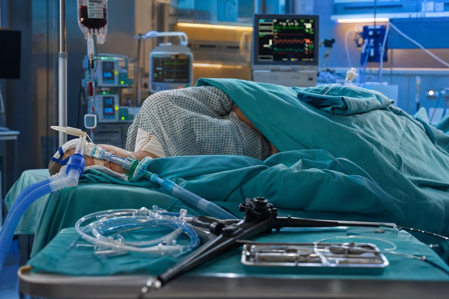
Definition
[1]There is no single universally agreed volume threshold for "massive" haemoptysis. The classical range quoted across textbooks is >100-600 mL/24 h; the most widely used operational definition is >200 mL/24 h, and many units add any bleeding that threatens the airway or causes haemodynamic instability as an equivalent trigger. The reason volume is an imperfect marker: the tracheobronchial tree of a supine, intubated patient can be flooded by a comparatively small volume, so asphyxiation risk rather than blood loss dictates urgency.[1] A practical ICU rule — any haemoptysis that contaminates the contralateral lung or impairs ventilation is massive, regardless of mL.[5]
Pathophysiology — why the bronchial circulation matters
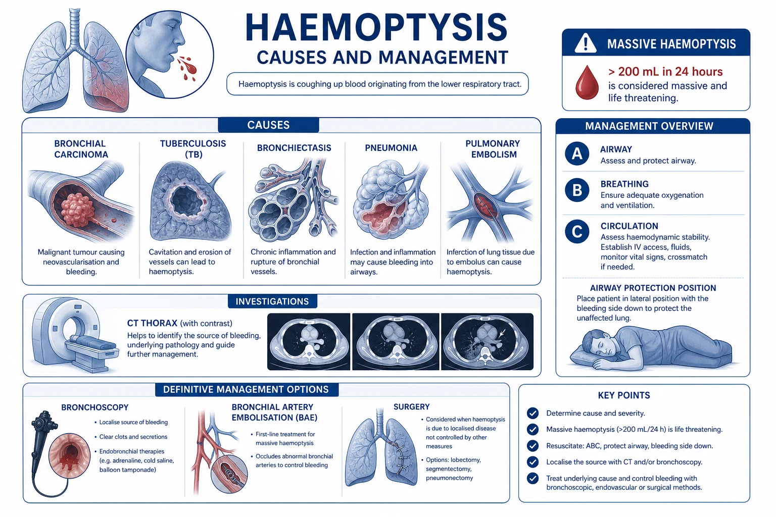
The lung has a dual blood supply: [1]
- Pulmonary circulation — low pressure (~15/8 mmHg), carries entire cardiac output. Bleeding here is usually slower and tamponades easily.
- Bronchial circulation — systemic pressure (~120/80 mmHg), arises from the descending aorta, supplies airways and is the source of ~90% of massive haemoptysis.[1]
Inflammatory, infective and neoplastic processes drive bronchial arterial hypertrophy, neovascularisation and shunting from the high-pressure systemic bronchial arteries into the low-pressure pulmonary vasculature. Because these vessels are at systemic pressure, rupture produces brisk, voluminous haemorrhage — far more dangerous than a pulmonary-artery source of similar size. This single anatomical fact explains both the urgency (asphyxiation) and the rationale for bronchial artery embolisation as first-line definitive therapy.[7]
Causes
Key causes to commit to memory for the exam — bronchiectasis (overall #1), lung cancer, tuberculosis (active + old cavity with aspergilloma), pulmonary embolism (haemorrhagic infarct), pulmonary vasculitis (GPA/Goodpasture's — diffuse alveolar haemorrhage), and pulmonary AVM (hereditary haemorrhagic telangiectasia).[6]
Diffuse alveolar haemorrhage (DAH) — a distinct subset
Not all massive haemoptysis is focal. Diffuse alveolar haemorrhage from capillaritis produces bilateral alveolar infiltrates with (often surprisingly little) haemoptysis, anaemia out of proportion to visible blood, and progressive hypoxaemia. Causes are immunological (ANCA-associated vasculitis/GPA, anti-GBM/Goodpasture's, SLE, APLA) or non-immunological (idiopathic pulmonary haemosiderosis, coagulopathy, mitral stenosis, drug toxicity). Bronchoalveolar lavage shows progressively bloodier return from successive aliquots. Management is immunosuppression (high-dose corticosteroids ± cyclophosphabmide/rituximab) ± plasma exchange (anti-GBM), not BAE — because there is no single target vessel.[1]
Management
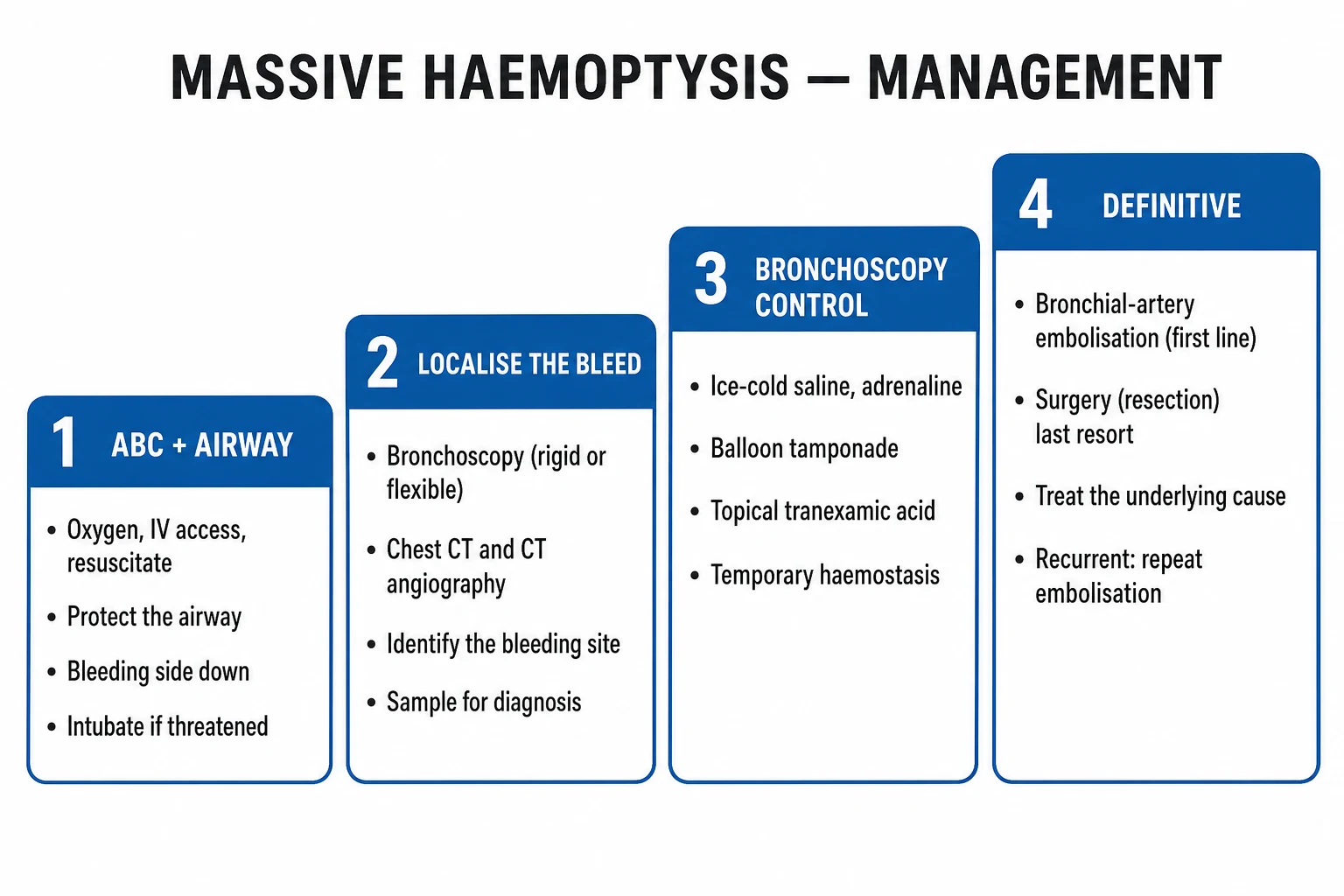
Management of massive haemoptysis
- Protect airway — ABC. Position BLEEDING SIDE DOWN (prevents blood flooding contralateral lung — main cause of death). High-flow oxygen
- Intubate if airway compromised — large single-lumen ETT (8-9mm — allows bronchoscope + suction) OR double-lumen tube (isolate bleeding lung). Consider left main bronchial intubation
- Suction and tamponade — large-bore suction. Bronchoscopic balloon tamponade (Fogarty catheter) to occlude bleeding bronchus
- Identify bleeding site — bronchoscopy (rigid preferred — better suction, allows interventions). CT chest with contrast (identifies source, vascular anatomy)
- Bronchial artery embolisation (BAE) — FIRST-LINE DEFINITIVE. Interventional radiology. Superselective catheterisation of bronchial artery + embolisation (particles, coils). 70-90% immediate success. Recurrence 10-30% (may repeat)
- Surgery — LAST RESORT (lobectomy/pneumonectomy). Indications: BAE unavailable/failed, localised surgical cause (cancer, AVM). High mortality (20-40%) in emergency setting
- Correct coagulopathy — FFP, platelets, vitamin K, reversal agents
- Treat underlying cause — antibiotics for infection, antifungal for aspergilloma, chemo/radiotherapy for cancer, immunosuppression for vasculitis
Immediate bedside actions (first 5 minutes)
- Call for help — ICU senior, anaesthetist, interventional radiology, thoracic surgery simultaneously
- Position — bleeding side DOWN (lateral decubitus); sit upright if central/unknown source to aid drainage
- Airway — clear blood with large-bore Yankauer suction; high-flow O2; prepare for intubation
- IV access × 2 large-bore — cross-match 4-6 units, FBC/coag/UEC/group+save; trend Hb every 1-2 h
- Empiric haemostasis — IV tranexamic acid 1 g (evidence modest), reverse anticoagulants, correct coagulopathy/platelets
- Localise — urgent flexible bronchoscopy if unstable; CT chest with contrast if stable enough to travel
- Definitive — activate interventional radiology suite for BAE; alert theatre for emergency thoracotomy if BAE unavailable
Airway and lung isolation
Practical pearls[8][9] — use the largest single-lumen ETT that fits (≥8.0 mm) if a bronchoscope or blocker will pass; prefer a left-sided DLT because the right main bronchus is short and the right upper lobe orifice is easily occluded; exchange a tube only if you can visualise the glottis — a blood-flooded airway converts a tube exchange into an airway disaster.[2]
Bronchoscopic haemostasis
Bronchoscopic interventions (rigid or flexible via large ETT)
- Suction and identify — clear blood; locate bleeding lobe/segment
- Iced saline lavage — 4°C normal saline in 50 mL aliquots into bleeding bronchus → vasoconstriction; repeat to total 500 mL
- Topical adrenaline (epinephrine) — 1:20,000 aliquots (dilute 1 mg in 20 mL) → local vasoconstriction
- Balloon tamponade — Fogarty/bronchial-blocker catheter inflated in bleeding bronchus; leave 24-48 h, deflate before removal
- Topical haemostatic tamponade — oxidised regenerated cellulose (Surgicel), fibrin/thrombin glue instilled via catheter
- Locoregional therapy — argon plasma coagulation (APC), Nd:YAG laser, or electrocautery for visible endobronchial lesion
- Cryotherapy — for tumour bed / visible bleeders
These are temporising measures that buy time for definitive BAE or surgery; few provide durable control in brisk bleeding.[4][2]
Bronchial artery embolisation (BAE) — first-line definitive
BAE procedural sequence (interventional radiology)
- Femoral arterial access — standard Seldinger
- Aortography / selective catheterisation — find bronchial arteries (usually arise T5-T6, often aberrant); use Cobra/Simmons catheter
- Identify bleeding vessel — hypertrophied, tortuous bronchial artery; ± contrast extravasation, AVM shunt, hypervascular tumour blush
- Catheterise superselectively — microcatheter beyond spinal/intercostal branches (avoid spinal artery supply → anterior spinal ischaemia)
- Embolise — polyvinyl alcohol (PVA) particles 150-500 µm, gelatin sponge, or microcoils
- Check completion angiogram — confirm stasis; assess non-bronchial systemic collaterals (intercostal, internal mammary, subclavian)
- Post-procedure — observe; re-bleed within 24-48 h suggests missed vessel — repeat or surgery
Surgery — lobectomy for refractory disease
Surgery is reserved for: BAE unavailable or failed, localised surgical cause (resectable cancer, AVM, mycetoma in a fit patient), or massive ongoing bleed with no IR access. Procedure is lobectomy (or segmentectomy for peripheral lesions) — pneumonectomy is avoided when possible due to high mortality. Emergency surgery carries 20-40% mortality (aspiration, haemodynamic collapse); the same operation done electively after BAE carries only 5-10% mortality. Aspergilloma with recurrent post-BAE re-bleed is the classic elective lobectomy indication.[3][6]
Differential diagnosis — true haemoptysis vs mimics
[1]Always perform an ENT examination (anterior nasal vessels, posterior epistaxis, oropharyngeal bleeding) before attributing blood to the lower airway — pseudohaemoptysis is a common pitfall.[6]
Investigation
Pharmacology adjuncts
- Tranexamic acid (TXA) — modest evidence; IV 1 g then infusion, or nebulised 250-500 mg. An integrative review supports a trial, especially while awaiting definitive therapy.[10]
- Vasopressin / terlipressin — seldom used; systemic vasoconstriction risks; superseded by selective BAE.
- Antitussives — suppress cough (codeine) to reduce mechanical disruption of clot — but avoid over-sedation that impairs airway protection.
- Coagulation correction — FFP (INR > 1.5), platelets (< 50 or antiplatelet), cryoprecipitate (fibrinogen < 1.5 g/L), vitamin K (warfarin), idarucizumab (dabigatran), andexanet alfa (apixaban/rivaroxaban), PCC.
Exam practice
SAQ — Massive haemoptysis: airway protection and bronchial artery embolisation
10 minutes · 10 marks
A 58-year-old man with longstanding bronchiectasis coughs up ~400 mL of bright red blood over 30 minutes. He is tachypnoeic (RR 32), SpO2 88% on high-flow oxygen, BP 96/58, HR 122, and is visibly distressed. The bleeding is ongoing. Bronchiectasis predominantly affects the right lower lobe.
SAQ — Diffuse alveolar haemorrhage from ANCA vasculitis
10 minutes · 10 marks
A 45-year-old man presents with progressive dyspnoea, cough with frothy blood-stained sputum, and moderate haemoptysis. Chest X-ray shows bilateral diffuse alveolar infiltrates. Hb has fallen from 132 to 88 g/L. Oxygen requirement is climbing. Urinalysis shows red-cell casts. ANCA is positive (PR3).
Clinical pearls
Red flags
Prognosis
Outcomes of massive haemoptysis (Ittrich 2017, Dtsch Arztebl Int; CIRSE BAE 2022)
Systematic review of massive haemoptysis management:
- Mortality: 7-30% (higher with delayed treatment, cancer, coagulopathy)
- BAE immediate success: 70-90% (haemostasis achieved)
- BAE recurrence (re-bleeding): 10-30% within 1 year
- Surgery mortality (emergency): 20-40%
- Surgery mortality (elective): 5-10% [1]
Causes of death: asphyxiation (80%), exsanguination (10%), complications of treatment (10%). Predictors of mortality: cancer, coagulopathy, massive amount (>1000 mL), bilateral bleeding, delayed intervention.
Bronchial artery embolisation — efficacy and recurrence (CIRSE Standards, 2022)
Pooled data across major series:
- Immediate technical success: 83-96% (superselective technique)
- Clinical success (haemostasis ≥ 30 days): 70-90%
- Recurrence: 10-30% at 1 year; up to 45% at long-term follow-up
- Highest recurrence: aspergilloma, active TB, bronchopulmonary shunt, incomplete embolisation
- Major complications: spinal ischaemia (0.5-1%), oesophageal/bronchial necrosis (rare), pulmonary infarction
- Repeat BAE: effective; multiple sessions often required in mycetoma
- BAE vs surgery (elective): comparable long-term control; BAE less invasive and preferred unless localised surgically resectable lesion [1]
Take-home: BAE is first-line; surgery reserved for BAE failure or localised resectable cause. Anticipate recurrence — follow up and offer elective definitive therapy.
Bronchoscopic tamponade for life-threatening haemoptysis (Valipour et al, Chest 2005)
Case series of bronchoscopy-guided topical haemostatic tamponade:
- Technique: cellulose mesh / fibrin glue via catheter wedged in bleeding bronchus under bronchoscopic vision
- Initial control: achieved in most patients where source could be reached
- Bridge to definitive therapy: bought time for BAE or surgery
- Limitation: requires localisable, reachable bleeding source; not effective for diffuse DAH [1]
Take-home: bronchoscopic tamponade is a temporising bridge, not definitive therapy; combine with rapid referral to interventional radiology or theatre.
Nebulised tranexamic acid for haemoptysis (Ye et al, Lung 2025 — integrative review)
Synthesis of available studies on nebulised TXA:
- Setting: mild-to-moderate haemoptysis, palliative, or where bronchoscopy/BAE delayed
- Effect: reduction in bleeding episodes and volume in observational series; limited RCT data
- Safety: generally well tolerated; cough, bronchospasm reported
- Role in massive haemoptysis: adjunct only — does NOT replace airway protection, bronchoscopy, or BAE
- Evidence base: heterogenous; large definitive RCT still needed [1]
Take-home: nebulised/IV TXA is a reasonable adjunct while arranging definitive therapy; do not delay BAE awaiting an antifibrinolytic response.
Comparison of definitive therapies
Anatomy for the exam
- Bronchial arteries — usually 2 left, 1 right; arise from descending thoracic aorta at T5-T6; may share origin with intercostal, subclavian, internal mammary, or coronary arteries.
- Non-bronchial systemic collaterals — intercostal, subclavian, internal mammary, inferior phrenic — can supply bleeding in chronic disease and must be sought if BAE fails (recurrent re-bleed).
- Artery of Adamkiewicz — major anterior spinal artery feeder; usually arises T9-L1 from an intercostal/lumbar, but aberrant origins exist — the spinal-ischaemia risk of BAE.
- Right upper lobe bronchus (RUL) — takeoff ~1 cm from carina — why right-sided DLTs are harder to position than left.
- Left main bronchus — longer, narrower, more vertical than right in some individuals (relevant to aspiration, not the often-quoted "right is more vertical" which describes the adult tracheobronchial tree — right main bronchus is wider, shorter and more vertical).[7][8]
Pitfalls and exam traps
- Treating the volume, not the airway. A "small" bleed flooding the contralateral lung is immediately fatal; a 500 mL slow ooze that the patient coughs up may not be. Triage on airway compromise.
- Positioning the GOOD side down. Catastrophic — floods the only functioning lung. Bleeding side DOWN.
- Tube exchange in a blood-flooded airway. If the single-lumen tube is working, keep it and use a blocker; lost view = lost airway.
- Attributing DAH to a focal cause and sending for BAE. Capillaritis needs immunosuppression, not embolisation.
- Forgetting non-bronchial systemic collaterals after a failed BAE — they are the usual reason for early recurrence.
- Anticoagulating a patient with recent massive haemoptysis for a "PE" — confirm the diagnosis; haemorrhagic PE infarct bleeds may be controlled by stopping, not escalating, anticoagulation, while true PE still needs treatment. Multidisciplinary decision.[1][6]
Key takeaways
- Airway first, position bleeding side down, isolate the good lung.
- 90% of bleeding is bronchial (systemic, high-pressure) — hence BAE first-line.
- Bronchoscopy localises and temporises; CT defines cause and vascular anatomy.
- BAE = 70-90% immediate success; surgery = last resort, lobectomy for refractory/localised disease.
- DAH/vasculitis is a different disease — immunosuppression, not embolisation.
- Anticipate re-bleed (10-30%) and post-bleed aspiration pneumonia — antibiotics, follow-up, elective definitive therapy. [1]
References
- [1]Ittrich H, Bockhorn M, Klose H, Simon M, et al. The Diagnosis and Treatment of Hemoptysis Dtsch Arztebl Int, 2017.PMID 28625277
- [2]Sakr L, Dutau H. Massive hemoptysis: an update on the role of bronchoscopy in diagnosis and management Respiration, 2010.PMID 20090288
- [3]Kettenbach J, Ittrich H, Gaubert JY, et al. CIRSE Standards of Practice on Bronchial Artery Embolisation Cardiovasc Intervent Radiol, 2022.PMID 35396612
- [4]Valipour A, Kreuzer A, Koller H, Koessler W, Burghuber OC. Bronchoscopy-guided topical hemostatic tamponade therapy for the management of life-threatening hemoptysis Chest, 2005.PMID 15947328
- [5]Jean-Baptiste E. Clinical assessment and management of massive hemoptysis Crit Care Med, 2000.PMID 10834728
- [6]Larici AR, Franchi P, Occhipinti M, et al. Diagnosis and management of hemoptysis Diagn Interv Radiol, 2014.PMID 24808437
- [7]Ittrich H, Klose H, Adam G. Radiologic management of haemoptysis: diagnostic and interventional bronchial arterial embolisation Rofo, 2015.PMID 25372159
- [8]Campos JH, Musselman ED, Hanada S, et al. Lung Isolation Techniques in Patients With Early-Stage or Long-Term Tracheostomy: A Case Series Report of 70 Cases and Recommendations J Cardiothorac Vasc Anesth, 2019.PMID 30190202
- [9]Collins SR, Titus BJ, Campos JH, et al. Lung Isolation in the Patient With a Difficult Airway Anesth Analg, 2018.PMID 29189274
- [10]Ye M, Chen M, Wang C, et al. Nebulized Tranexamic Acid in the Management of Hemoptysis: An Integrative Review Lung, 2025.PMID 39841268