ICU · Respiratory / airway emergencies
Acute Massive Haemoptysis — Comprehensive ICU Management
Also known as Massive haemoptysis · Life-threatening haemoptysis · Massive hemoptysis · Pulmonary haemorrhage · Bronchial artery embolisation · Rigid bronchoscopy haemostasis · Ice-cold saline lavage · Double-lumen tube lung isolation · Rasmussen aneurysm · Aspergilloma haemoptysis
Acute massive haemoptysis is a respiratory emergency in which the patient dies of ASPHYXIATION, not exsanguination — blood floods the airway, abolishes gas exchange, and causes hypoxaemic arrest long before the circulating volume is lost. Define it functionally as any volume of bleeding that threatens the airway, or classically as more than 100 mL in 24 hours (some texts use more than 600 mL/24h for 'massive'). The commonest causes are bronchiectasis, lung cancer, tuberculosis (active and old cavities), and aspergilloma (the fungal ball in an old TB cavity); around 90 per cent of brisk bleeding arises from the BRONCHIAL (systemic, high-pressure) circulation. Management is a fixed cascade: (1) protect the airway and position the patient with the BLEEDING SIDE DOWN to stop blood flooding the good lung; (2) intubate with a large (8 mm or more) single-lumen tube that admits a bronchoscope, or a double-lumen tube/bronchial blocker to isolate the bleeding lung; (3) bronchoscopy — rigid preferred for suction and tamponade — to localise bleeding and apply ice-cold saline lavage with topical adrenaline 1:20,000 and balloon tamponade; (4) BRONCHIAL ARTERY EMBOLISATION by interventional radiology (gelatin or PVA particles), the gold-standard definitive therapy with 70-90 per cent immediate haemostasis; (5) emergency surgery (lobectomy or pneumonectomy) as a last resort with high mortality; and (6) general measures — reverse coagulopathy, tranexamic acid 1 g IV (controversial; the HALT-IT trial addressed gastrointestinal bleeding, not haemoptysis), and treat the underlying cause. Mortality ranges from 7 to 30 per cent and is driven by aspiration, delay, and comorbidity.
On this page & tools
Your progress
Saved locally on this device.
Target exams
Red flags
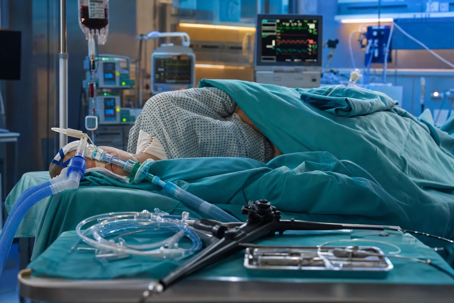
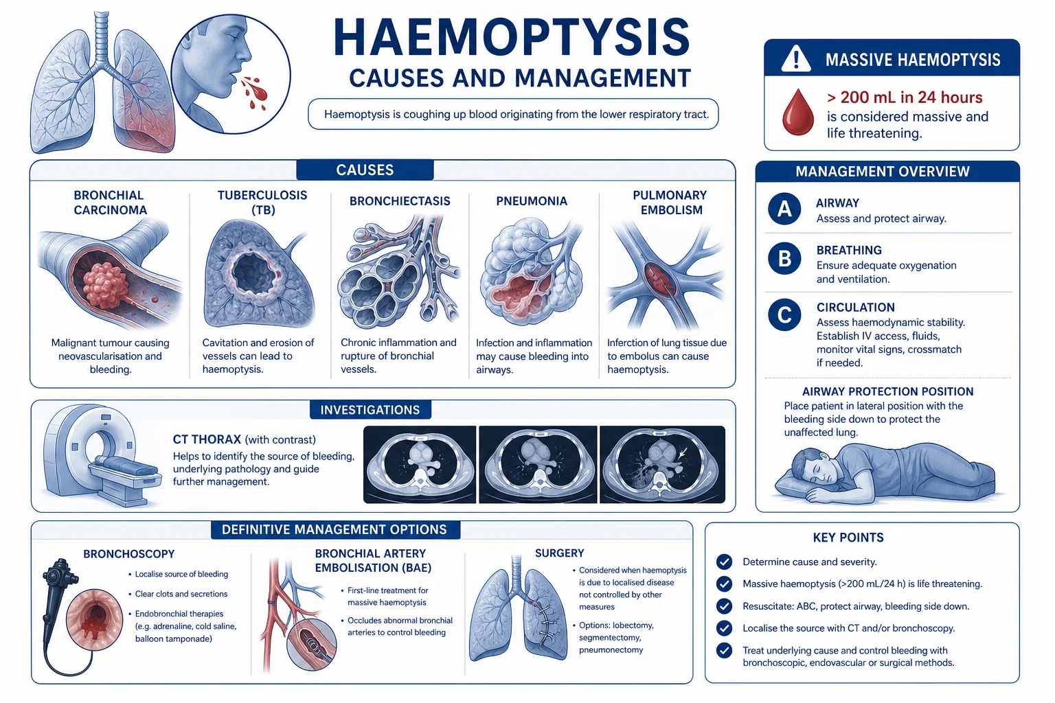
Definition — what makes haemoptysis "massive"
Haemoptysis is massive (also called life-threatening or exsanguinating) when the volume or the rate of bleeding threatens the airway and gas exchange. There is no single universally agreed volume threshold, and the examiner should know the spectrum:[1][3]
- Less than 30 mL per 24 hours — mild / submassive. Usually from bronchitis, mild infection, or airway inflammation; investigated as an outpatient or on the ward.
- 30-200 mL per 24 hours — moderate. Requires admission, investigation (bronchoscopy and CT), and observation; may be the harbinger of a larger bleed.
- More than 100 mL per 24 hours — the threshold many older texts use for massive, because this volume can acutely flood dependent airways.
- More than 600 mL per 24 hours (or more than 100 mL per hour) — the threshold others use for massive, approximating the volume at which gas exchange collapses.
- Any amount that threatens the airway — the modern functional definition championed by Larici and others: a patient who is aspirating, choking, or losing gas exchange from brisk bleeding has massive haemoptysis regardless of the millilitres, because the determinant of death is airway flooding, not blood loss.[1]
Why the patient dies — asphyxiation, not exsanguination
The lungs hold a small blood volume, and brisk haemoptysis rarely produces haemorrhagic shock. Instead, blood floods the tracheobronchial tree, displaces alveolar gas, abolishes gas exchange in dependent segments, and the patient arrests from hypoxaemia — typically within minutes to a couple of hours of onset. In autopsy and registry series the dominant cause of death is asphyxiation (around 80 per cent of deaths), with exsanguination accounting for only about 10 per cent and complications of treatment the remainder.[3][5]
Three consequences follow directly and shape every management decision: [1]
- Airway protection and lung isolation come first. Securing the airway and preventing blood from flooding the contralateral lung takes priority over finding the bleeding source, reversing coagulopathy, or transferring the patient.
- Position the bleeding side down. Gravity is a free, instant, and powerful intervention — it keeps blood in the bleeding lung and away from the good lung (see management).
- Definitive control is by tamponade or embolisation of a bronchial artery, because the bleeding source is usually the high-pressure systemic bronchial circulation. [1]
Causes — bronchiectasis, malignancy, TB, and the fungal ball
Around 90 per cent of brisk haemoptysis originates from the BRONCHIAL arteries (systemic, high-pressure, hypertrophied by chronic inflammation), and only about 5 per cent from the pulmonary artery (e.g. a Rasmussen aneurysm eroding into a TB cavity).[2][3] The cause list, in order of frequency in most modern series:[1][2][6]
Bronchiectasis (#1)
Chronic inflammation, hypertrophied bronchial arteries
- Commonest single cause in many series (up to 30-40 per cent)
- Chronic infection/inflammation drives bronchial artery hypertrophy and neovascularisation — friable, high-pressure vessels
- Cystic fibrosis, post-infective (post-TB, childhood pertussis/measles), allergic bronchopulmonary aspergillosis (ABPA), idiopathic
- Often recurrent; BAE is highly effective and may need repeating
Lung cancer
NSCLC eroding vessels; also a cause of massive bleed
- Squamous cell carcinoma especially — central, cavitating, erodes bronchial vessels
- Up to 20 per cent of massive haemoptysis; a dire prognostic sign (median survival weeks-months after a massive bleed)
- Razazi/Fartoukh 2015: in NSCLC, massive haemoptysis carries ICU mortality ~30-50 per cent and is driven by cancer stage and asphyxiation
- Iatrogenic bleeds after biopsy/resection also occur
Tuberculosis
Active and old; Rasmussen aneurysm
- Active TB — endobronchial ulceration and caseating inflammation
- Old TB cavity — the substrate for aspergilloma (below) and for a Rasmussen aneurysm
- Rasmussen aneurysm: a pulmonary artery pseudoaneurysm in a TB cavity wall that ruptures — a pulmonary-artery (5 per cent) source
Aspergilloma (fungus ball)
Mycetoma in an old cavity
- Aspergillus fumigatus colonises a pre-existing cavity (old TB, sarcoid, bulla)
- Vascular granulation tissue at the cavity wall bleeds massively and repeatedly
- CT: air-crescent sign (mobile fungal ball with crescent of air)
- BAE first-line; elective surgical excision (cavity removal) if recurrent or BAE fails
Pulmonary haemorrhage syndromes
Vasculitis and anti-GBM
- Diffuse alveolar haemorrhage (DAH): ANCA-associated vasculitis (GPA, MPA, EGPA), anti-GBM (Goodpasture), SLE
- Bleeding is diffuse and bilateral — a different beast; lung isolation is impossible
- Manage with high-dose steroids, plasmapheresis (anti-GBM), cyclophosphamide/rituximab
- Red cell casts/haematuria suggest renal-pulmonary syndrome — check anti-GBM and ANCA
Other / less common
Keep in the differential
- Chronic bronchitis (common cause of mild haemoptysis, rarely massive)
- Pneumonia / lung abscess with vascular erosion
- Pulmonary embolism / infarction; pulmonary artery rupture (rare, post-PA catheter)
- Arteriovenous malformation (AVM), hereditary haemorrhagic telangiectasia (Osler-Weber-Rendu)
- Trauma, iatrogenic (transthoracic biopsy, bronchial biopsy), coagulopathy
- Cryptogenic (~10-20 per cent) after full workup
Clinical presentation
The patient usually has a known respiratory diagnosis (bronchiectasis, old TB, known cancer) but massive haemoptysis may be the first presentation. Typical features:[1][3]
- Coughing bright-red, frothy blood — often in large aliquots; froth distinguishes true haemoptysis from haematemesis
- Choking, gurgling, dyspnoea, tachypnoea — signs of airway flooding and aspiration
- Hypoxaemia (SpO2 falling), tachycardia, anxiety; shock is a late and uncommon sign (it implies exsanguination or simultaneous other pathology)
- Diffuse crackles/ronchi on auscultation as blood spreads; localised wheeze or decreased air entry may lateralise the bleed
- Features of the underlying cause: clubbing and purulent sputum (bronchiectasis), weight loss and haemoptysis on a background of smoking (cancer), TB contact or old TB scar, haematuria (pulmonary-renal syndrome) [1]
Distinguish haemoptysis from haematemesis and pseudohaemoptysis
A rapid bedside distinction matters because it changes the service you call and the airway you prepare.[1]
| Feature | Haemoptysis | Haematemesis | Pseudohaemoptysis |
|---|---|---|---|
| Colour | Bright red, frothy | Dark red / coffee-ground | Bright red, no froth |
| Reaction | Alkaline (pH >7) | Acidic (pH <7) | Variable |
| Accompaniment | Cough, dyspnoea | Vomiting, retching | Clearing the throat / nose |
| History | Lung disease | Liver / GI / alcohol | Epistaxis, dental, ENT |
| Haematemesis test | Negative (NG aspirate bile/clear) | Positive (coffee-ground) | Negative |
Severity and amount — when to declare an emergency
Declare a massive-haemoptysis emergency and mobilise the cascade the moment any of the following are present:[1][2]
- Active brisk bleeding with choking, gurgling, or inability to clear the airway.
- Hypoxaemia (SpO2 falling on supplemental oxygen) from aspiration of blood.
- Estimated volume more than 100 mL in 24 hours, or more than 50 mL in a single episode, or bleeding that is accelerating.
- Known high-risk substrate: aspergilloma, active cavitary TB, central squamous cancer, recent transthoracic biopsy, or a coagulopathy/anticoagulant. [1]
Haemoptysis severity (click each)
>100 mL/24h or airway threat
Massive / life-threatening. Declare the emergency now: bleeding side down, high-flow oxygen, large-bore IV access, call anaesthetics/ICU + thoracic surgery + interventional radiology simultaneously, prepare for intubation and BAE.
Key numbers in massive haemoptysis
Investigations — parallel to resuscitation, never before it
Investigations must run in parallel with the airway cascade, never in sequence before it. A patient bleeding briskly is not sent to the CT scanner unsecured.[1][4]
- Arterial blood gas — the hypoxaemia and rising CO2 of aspiration; the trend guides intubation.
- Full blood count, group and crossmatch (4-6 units), coagulation (INR, aPTT, fibrinogen), U&E, lactate — detect coagulopathy and guide reversal and transfusion.
- Chest X-ray — fast and lateralising in ~half of cases (opacity in the bleeding lobe); may be normal in early or central bleeds.
- Contrast-enhanced CT chest / CT pulmonary angiography — the highest-yield localising test once the airway is secure; identifies the cause (cavity with mycetoma, tumour, bronchiectasis, AVM), bronchial artery hypertrophy (the 'shaggy' enhancing vessels), and contrast extravasation to point to the active bleeder. Perform after stabilisation or during the BAE procedure itself.[1][2]
- Bronchoscopy — both diagnostic and therapeutic; see below.
Management — the fixed cascade
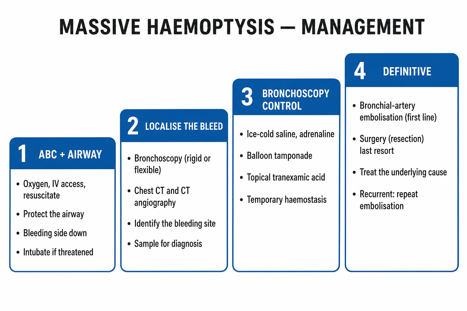
Management is a fixed cascade run in parallel by a coordinated team (ICU/anaesthetics for the airway, interventional radiology for BAE, thoracic surgery as backup). The resuscitation, airway, and referral happen simultaneously. The single overriding principle: secure the airway and protect the good lung before everything else.[1][2][5]
The massive-haemoptysis cascade — airway to embolisation
Step 1 — Protect the airway and position bleeding side DOWN
High-flow oxygen 100%. Call for help early: ICU/anaesthetics, thoracic surgery, interventional radiology simultaneously. POSITION THE PATIENT WITH THE BLEEDING SIDE DOWN (if right lung is bleeding, lie on the RIGHT side) — gravity keeps blood in the bleeding lung and prevents flooding of the good (contralateral) lung, the commonest cause of death. Sit up only if bleeding is minimal and the patient can cough effectively; in brisk bleeding, the lateral decubitus (bleeding side down) position is life-saving. Large-bore IV access x2, bloods, crossmatch 4-6 units, reverse coagulopathy in parallel.
Step 2 — Intubate: large ETT, double-lumen tube, or bronchial blocker
Intubate for airway compromise, massive ongoing bleeding, hypoxaemia, or inability to clear secretions. Use a LARGE single-lumen tube (8 mm or more — ideally 8.5-9.0 mm) so a therapeutic flexible bronchoscope passes with suction. For LUNG ISOLATION use a double-lumen tube (DLT) or a bronchial blocker placed in the bleeding main bronchus to occlude it and ventilate the good lung. Left-sided DLT is usually easier (the right main bronchus is short with an early upper-lobe takeoff). DLT/blocker requires a skilled operator and is harder in the bloody airway — have the large single-lumen tube as the fallback.
Step 3 — Bronchoscopy: localise, lavage, tamponade
Once the airway is secure, bronchoscopy localises and temporises the bleed. RIGID bronchoscopy is preferred in massive haemoptysis — large suction channel, ability to ventilate, and access for tamponade; a flexible scope can be passed through the rigid scope. If only flexible bronchoscopy is available, work through the large ETT. Therapeutic measures: ICE-COLD (4 C) saline lavage in 50 mL aliquots (vasoconstriction — the Conlan technique), TOPICAL ADRENALINE (epinephrine) 1:20,000 (5-10 mL aliquots), and BALLOON TAMPONADE (a Fogarty or dedicated bronchial-blocker balloon inflated in the bleeding bronchus for 12-24 h). All are temporising — they buy time for BAE or surgery.
Step 4 — Bronchial artery embolisation (BAE): the definitive treatment
Mobilise interventional radiology EARLY (simultaneous with intubation). BAE is the GOLD-STANDARD definitive therapy for massive haemoptysis. Angiography identifies the hypertrophied, tortuous bleeding bronchial artery (and any abnormal feeding vessels); the operator superselectively catheterises it and embolises with GELATIN particles (Gelfoam), polyvinyl alcohol (PVA) particles, or microspheres (coils for larger vessels). Immediate haemostasis 70-90 per cent. Beware a spinal artery arising from a bronchial artery — inadvertent embolisation causes cord ischaemia/paraplegia; superselective catheterisation beyond it prevents this.
Step 5 — Surgery: the last resort
Reserved for: BAE unavailable or failed, a localised surgical lesion (resectable cancer, AVM, aspergilloma in a cavity), or uncontrolled bleeding despite isolation. Lobectomy or pneumonectomy. EMERGENCY surgery carries high mortality (20-40 per cent) from aspiration and instability; ELECTIVE surgery for recurrent post-BAE bleeding is safer. Lung isolation must be in place before transfer to theatre.
Step 6 — General measures and treat the cause
REVERSE COAGULOPATHY: FFP (INR >1.5), platelets (<50 or antiplatelet), cryoprecipitate (fibrinogen <1.5 g/L), vitamin K for warfarin, idarucizumab (dabigatran), andexanet (apixaban/rivaroxaban), protamine (heparin). TRANEXAMIC ACID 1 g IV is commonly given but the evidence is weak — the HALT-IT trial (NEJM 2020) was a GI-bleed trial (no benefit, possible harm), NOT haemoptysis; a small RCT (Wand 2019) suggested reduced bleed duration in mild-moderate haemoptysis. Treat the underlying cause: antibiotics for infection, antifungal/surgery for aspergilloma, oncology/radiotherapy for cancer, steroids + plasmapheresis for vasculitis/anti-GBM.
Step 1 in detail — airway, oxygen, position, and the call
The first sixty seconds determine survival.[3][5]
- High-flow oxygen 100% via a non-rebreather mask; switch to the circuit once intubated.
- Bleeding side down (lateral decubitus, bleeding lung dependent). If you cannot lateralise, use the history and CXR/CT; if still uncertain, lie the patient on the side the bleeding seems to come from and reassess. This single manoeuvre protects the contralateral lung from drowning.
- Suction — hard Yankauer/tip suction; blood clots fast, so keep suction available and working.
- Call for help — activate the massive-haemoptysis pathway: ICU/anaesthetics (airway), interventional radiology (BAE), thoracic surgery (backup). They should be mobilised in parallel.
- IV access x2 (large bore), bloods, crossmatch 4-6 units, coagulation — and start correcting coagulopathy at once. [1]
Step 2 in detail — choosing the tube
| Tube | Pros | Cons | When |
|---|---|---|---|
| Large single-lumen (8-9 mm) | Easy to place; admits a therapeutic flexible bronchoscope; reliable ventilation | Does NOT isolate the lung — blood can still cross over | Default; most patients |
| Double-lumen tube (DLT) | True lung isolation — ventilate good lung, isolate bleeding lung | Needs a skilled operator; harder in a bloody airway; right-sided DLT risks upper-lobe occlusion | Surgeon-controlled, localised unilateral bleed |
| Bronchial blocker | Lung isolation via a single-lumen ETT; can be placed with a flexible scope | Can dislodge; less secure than a DLT | When DLT expertise unavailable; via single-lumen ETT |
Step 3 in detail — bronchoscopy: rigid versus flexible
Rigid bronchoscopy
Preferred in massive haemoptysis
- Large-bore suction channel that clears clots and blood
- Maintains ventilation through the scope and allows passage of a flexible scope through it
- Permits rigid tamponade, balloon tamponade, and ice-cold saline lavage
- Needs general anaesthesia and a bronchoscopist trained in rigid technique
- Conlan 1980: rigid bronchoscope + cold saline lavage controlled bleeding and allowed definitive management
Flexible bronchoscopy
Diagnostic; therapeutic when rigid unavailable
- Performed through a large (8 mm+) single-lumen ETT under sedation/anaesthesia
- Excellent for localising the bleeding segment/lobe and guiding BAE/surgery
- Ice-cold saline lavage, topical adrenaline 1:20,000, balloon/blocker tamponade all possible
- Smaller suction channel — struggles with heavy clot and brisk bleeding
- Hsiao 2001: flexible bronchoscopy before BAE improved localisation and outcomes
Bronchoscopic temporising measures
Step 4 in detail — bronchial artery embolisation
BAE is the definitive treatment for the common (bronchial-artery) massive haemoptysis, with immediate haemostasis in 70-90 per cent of patients and is the standard of care in centres with interventional radiology.[1][2]
- Technique — femoral arterial access, aortography then superselective catheterisation of the (usually hypertrophied) bronchial artery feeding the bleed, embolisation with gelatin (Gelfoam), PVA particles (150-700 micrometres), or tris-acryl gelatin microspheres; coils reserved for larger vessels or AVMs.
- Success — immediate haemostasis 70-90 per cent; recurrence (re-bleed) 10-30 per cent within ~1 year, higher with aspergilloma, active TB, and bronchopulmonary shunting. Re-bleed is managed by repeat BAE or elective surgery.
- Complications — the feared one is spinal cord ischaemia/paraplegia from inadvertent embolisation of a spinal (medullary) artery that arises from a bronchial artery; superselective catheterisation distal to any spinal branch prevents it. Others: chest pain, dysphagia, transient fever, non-target embolisation (stroke, visceral infarction), bronchial necrosis. [1]
Step 5 in detail — surgery
Surgery is reserved for failure or unavailability of BAE, a localised surgical lesion (resectable cancer, AVM, aspergilloma in an accessible cavity), or uncontrolled bleeding despite lung isolation. Lobectomy is preferred over pneumonectomy when feasible. Emergency surgery carries 20-40 per cent mortality from aspiration and instability; elective surgery for recurrent post-BAE bleeding is far safer. Lung isolation (DLT or blocker) must be in place before transfer to theatre.[1][3]
Step 6 in detail — general measures and the cause
- Reverse coagulopathy in parallel with the cascade: FFP for INR >1.5; platelets for count <50 or antiplatelet therapy; cryoprecipitate for fibrinogen <1.5 g/L; vitamin K for warfarin; idarucizumab for dabigatran; andexanet alfa for apixaban/rivaroxaban; protamine for heparin. Hold and reverse anticoagulants.
- Tranexamic acid 1 g IV — commonly given, but the evidence is weak and largely extrapolated. The HALT-IT trial (NEJM 2020) was a large randomised trial of tranexamic acid for gastrointestinal bleeding and showed no mortality benefit and a signal toward harm (venous thromboembolism, seizures); it was not a haemoptysis trial and should not be cited as haemoptysis evidence. A small Chinese RCT (Wand 2019) suggested a shorter bleed duration in mild-to-moderate haemoptysis. Use it, but know it is controversial and unproven in massive haemoptysis.
- Treat the cause — antibiotics for bacterial infection; antifungals and/or elective cavity excision for aspergilloma; definitive oncology therapy for cancer; high-dose corticosteroids, plasmapheresis, and cyclophosphamide/rituximab for diffuse alveolar haemorrhage from vasculitis/anti-GBM; anticoagulation review for PE-related bleeds. [1]
Diffuse alveolar haemorrhage — the exception that breaks the cascade
Diffuse alveolar haemorrhage (DAH) from ANCA-associated vasculitis, anti-GBM (Goodpasture) disease, or SLE produces bilateral, diffuse bleeding — there is no single bleeding lung to isolate, so the cascade above does not apply. Management is immunosuppression (high-dose methylprednisolone plus cyclophosphamide or rituximab) and plasma exchange (especially for anti-GBM), supportive ventilation with lung-protective settings, and treatment of any related renal failure. Suspect DAH when haemoptysis is bilateral on imaging, the patient has anaemia out of proportion, and there are red-cell casts/haematuria — send anti-GBM and ANCA urgently.[1]
Evidence and landmark references
Larici 2014 — Diagnosis and management of haemoptysis
Diagn Interv Radiol 2014
Comprehensive review on the definition, causes, and management of haemoptysis
Key finding
Recommends a functional definition (airway threat) and a structured diagnostic-therapeutic algorithm; rigid bronchoscopy for massive bleeding; BAE as first-line definitive therapy
Practice change
Codified the modern functional definition and the airway-first, BAE-centred cascade
Khalil 2015 — Severe haemoptysis: diagnosis to embolization
Diagn Interv Imaging 2015
Review of the role of imaging and bronchial artery embolisation in severe haemoptysis
Key finding
Multidetector CT angiography maps the bronchial anatomy and identifies the bleeding vessel; BAE achieves 70-90% immediate haemostasis with ~10-30% recurrence
Practice change
Established CT angiography as the planning tool and BAE as the standard of care
Razazi/Fartoukh 2015 — Severe haemoptysis in NSCLC
Eur Respir J 2015
ICU cohort of severe haemoptysis in non-small-cell lung carcinoma
Key finding
BAE achieved haemostasis in the majority; ICU mortality high (~30-50%) and driven by cancer stage and asphyxiation rather than exsanguination
Practice change
Defined outcomes and the role of BAE in cancer-related massive haemoptysis
Conlan 1980 — Rigid bronchoscopy + cold saline lavage
Thorax 1980
Case series of the rigid-bronchoscopy, cold-saline-lavage technique for massive haemoptysis
Key finding
Controlled brisk bleeding as a temporising bridge to definitive surgery, establishing the technique still taught today
Practice change
Established rigid bronchoscopy with ice-cold saline lavage as a core temporising manoeuvre
Prognosis
Outcomes in massive haemoptysis
Overall mortality ranges from 7 to 30 per cent and is driven by asphyxiation, delay to definitive control, malignancy, and coagulopathy.[1][3][6] Bronchial artery embolisation achieves immediate haemostasis in 70-90 per cent but re-bleeds in 10-30 per cent within a year (higher with aspergilloma, active TB, and bronchopulmonary shunting); recurrent bleeding is managed by repeat BAE or elective surgery. Emergency surgery carries 20-40 per cent mortality, far higher than elective resection. In non-small-cell lung cancer, a single episode of massive haemoptysis is a grim prognostic marker with ICU mortality around 30-50 per cent and short subsequent survival.[6] Predictors of a poor outcome are advanced cancer, coagulopathy or anticoagulation, a very large bleed (more than 1000 mL), bilateral aspiration, and any delay to airway protection or BAE.
Clinical pearls
Red flags
Exam practice — SAQ
SAQ — Massive haemoptysis airway and definitive control
10 minutes · 10 marks
A 58-year-old with known bronchiectasis has torrential haemoptysis, SpO2 82% on 15 L, coughing large clots. Suspected right-sided source on history.
References
- [1]Larici AR, Franchi P, Occhipinti M, Contegiacomo A, del Ciello A, Calandriello L, Storto ML, Marano R, Bonomo L. Diagnosis and management of hemoptysis Diagn Interv Radiol, 2014.PMID 24808437
- [2]Khalil A, Fedida B, Parrot A, Haddad S, Fartoukh M, Carette MF. Severe hemoptysis: From diagnosis to embolization Diagn Interv Imaging, 2015.PMID 26141487
- [3]Jean-Baptiste E. Clinical assessment and management of massive hemoptysis Crit Care Med, 2001.PMID 11378596
- [4]Hsiao EI, Kirsch CM, Kagawa FT, Wehner JH, Jensen WA, Baxter RB. Utility of fiberoptic bronchoscopy before bronchial artery embolization for massive hemoptysis AJR Am J Roentgenol, 2001.PMID 11566690
- [5]Conlan AA, Hurwitz SS. Management of massive haemoptysis with the rigid bronchoscope and cold saline lavage Thorax, 1980.PMID 7268664
- [6]Razazi K, Parrot A, Khalil A, Djibre M, Gounant V, Assouad J, Carette MF, Fartoukh M, Cadranel J. Severe haemoptysis in patients with nonsmall cell lung carcinoma Eur Respir J, 2015.PMID 25359349