ICU · Respiratory
Mechanical Ventilation
Also known as Positive-pressure ventilation · Invasive ventilation · Intermittent positive-pressure ventilation (IPPV) · Ventilator modes · Lung-protective ventilation
Mechanical ventilation applies positive pressure to the airway to support gas exchange and unload the respiratory muscles. Its overriding principle is lung-protective ventilation — tidal volume around 6 mL/kg predicted body weight, plateau pressure at or below 30 cmH2O, driving pressure at or below about 15 cmH2O, and the lowest FiO2 and PEEP compatible with adequate oxygenation — because the ventilator that saves the patient can also destroy the lung it ventilates through volutrauma, atelectrauma and biotrauma.
On this page & tools
Your progress
Saved locally on this device.
Target exams
Red flags
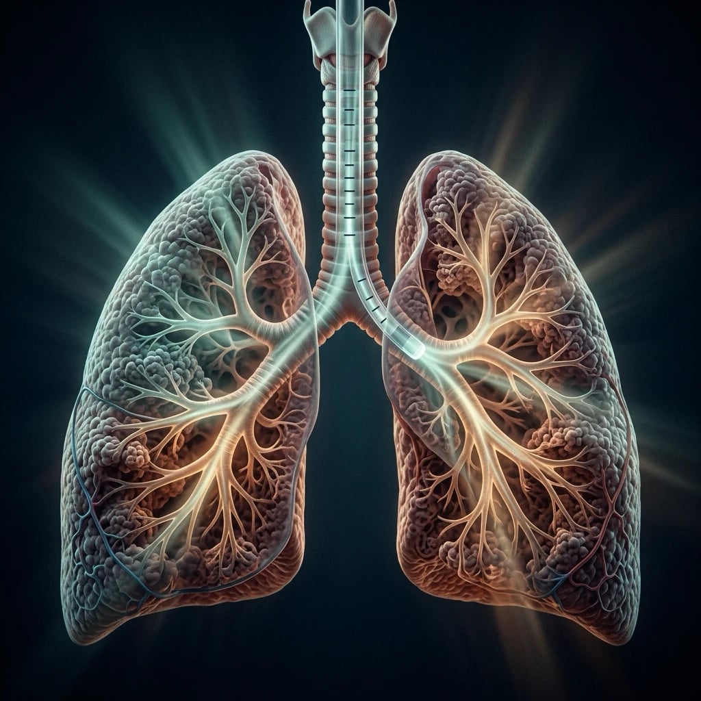
Overview — what mechanical ventilation is
Mechanical ventilation is, in its modern form, positive-pressure ventilation delivered through an endotracheal or tracheostomy tube (invasive), or through a mask or high-flow interface (non-invasive).[5] It is indicated when the patient cannot maintain one or both of the lungs' two jobs:
- Oxygenation failure — refractory hypoxaemia (a low PaO2 that does not correct with supplemental oxygen, as in ARDS or severe pneumonia).
- Ventilatory failure — an inability to move enough air to clear carbon dioxide and hold a safe pH, whether from reduced drive (central depression), neuromuscular weakness, increased load (bronchospasm, stiff lungs), or fatigue. [1]
The ventilator's job is therefore threefold: exchange gas, reduce the work of breathing, and rest the respiratory muscles — while the underlying cause is treated. The intensivist's job is to do this with the least iatrogenic harm: the lung is easily injured by over-distension and by repeated opening-and-closing of unstable units, and positive intrathoracic pressure impairs venous return and cardiac output. Every setting is a balance between supporting the patient and protecting the lung and circulation.[5]
Mechanical ventilation at a glance
Respiratory mechanics — compliance, resistance, time constants
Understanding the ventilated patient begins with three quantities from respiratory mechanics, because every mode and every setting is, in the end, an intervention on how much pressure moves how much gas over what time.[5]
- Compliance is the ease with which the lung and chest wall distend — the change in volume produced by a given change in pressure (mL/cmH2O). Two values matter at the bedside:
- Static compliance (Cstat) = VT / (Pplat − PEEPtotal). It reflects the elastic properties of the lung and chest wall with flow held to zero (the inspiratory-hold manoeuvre), and so isolates the true distensibility. A fall in Cstat means a stiffer system — ARDS, pulmonary oedema, consolidation, atelectasis, pneumothorax, or a rigid chest wall (obesity, raised intra-abdominal pressure).
- Dynamic compliance (Cdyn) = VT / (Ppeak − PEEPtotal). It is measured during flow and so includes airway resistance. Cdyn is therefore always lower than Cstat, and the gap between them reflects resistance.
- Resistance is the pressure dissipated overcoming frictional drag as gas flows through the airways and the tube — (Ppeak − Pplat) / flow. It rises with small-diameter endotracheal tubes, secretions, kinks, bronchospasm, and anything narrowing the airway.
- The time constant (τ = Resistance × Compliance) governs how quickly a lung unit fills and empties. After one time constant a unit has reached 63 percent of its volume change, after three about 95 percent, and after five essentially complete.[5] This is the single most useful concept in heterogeneous lung disease: units with long time constants (high resistance, as in COPD, or high compliance, as in emphysema) fill and empty slowly. If inspiration is too short they under-fill; if expiration is too short they fail to empty and trap gas, generating intrinsic (auto-)PEEP.
The pressure–volume curve, recruitment & PEEP
The pressure–volume (P-V) curve describes the lung's distensibility over its full range. In health it is steep and linear across most of its course. In the injured lung it develops two inflection points:[14]
- A lower inflection point, where the curve abruptly steepens as collapsed alveoli are recruited open.
- An upper inflection point, where the curve flattens as the lung reaches its elastic limit and units become over-distended. [1]
The classical (though since refined) reasoning is that PEEP should be set above the lower inflection point to hold recruited units open and prevent their cyclic collapse, and that tidal volume should stay below the upper inflection point to avoid over-distension. The modern refinement, driven by the driving-pressure analysis, is subtler: what matters most is not the absolute PEEP but the tidal change in pressure (the driving pressure), because that is the strain delivered to the aerated lung with each breath.[2]
Two features deserve emphasis. First, hysteresis — the inflation and deflation limbs differ, because it takes more pressure to open a collapsed unit than to keep an open unit inflated; this is why, once recruited, a unit can be held open at a pressure lower than that required to recruit it. Second, the "baby lung": in ARDS the lung is not uniformly stiff.[14] The disease is regionally heterogeneous — a small, normally aerated, compliant compartment coexists with consolidated dependent regions and over-distended non-dependent regions. The tidal volume is delivered preferentially to that small aerated "baby lung," so a "safe-looking" 6 mL/kg of predicted weight may still over-distend the aerated lung if too little of the lung is open. This is the physiological basis for setting tidal volume on predicted (not actual) body weight and for watching the driving pressure.[14]
Modes of mechanical ventilation
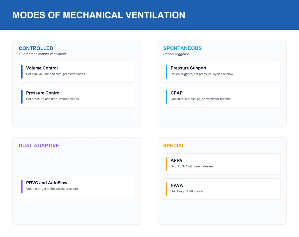
A "mode" is the control philosophy the ventilator uses to deliver breaths. Despite the bewildering array of proprietary names, every mode is defined by answering three questions: what triggers the breath (the patient or a timer), what is controlled (volume or pressure), and what cycles the breath off (a set volume, a set time, or a fall in flow).[5]
Volume control (VC)
guarantees volume
- Set tidal volume + flow + rate
- Pressure varies with compliance
- Guarantees minute ventilation
- Risk: high pressure if lung stiffens
Pressure control (PC)
pressure-limited
- Set inspiratory pressure + time + rate
- Tidal volume varies with compliance
- Pressure-limited by design (safe)
- Volume not guaranteed
Pressure support (PS)
spontaneous / weaning
- Patient triggers every breath
- Ventilator augments with set pressure
- Cycles off at % of peak flow
- Patient controls rate, depth, I-time
SIMV
hybrid (retired for weaning)
- Mandatory breaths synchronised to effort
- Spontaneous breaths allowed between
- Prolongs weaning vs PS / T-piece
- Largely retired as a primary mode
The common modes, in order of how often they appear in the exam and at the bedside: [1]
- Volume-controlled ventilation (VC / AC-VC). The clinician sets tidal volume, respiratory rate, inspiratory flow, and the inspiratory:expiratory ratio, and the ventilator delivers exactly that volume on every breath (mandatory and any patient-triggered). Pressure is the dependent variable, so airway pressure rises if compliance falls or resistance rises — hence the need for a high-pressure alarm limit. It guarantees minute ventilation but can injure a stiff lung if the volume is too large.[5]
- Pressure-controlled ventilation (PC / PCV). The clinician sets inspiratory pressure above PEEP, respiratory rate, and inspiratory time; flow is decelerating and the delivered tidal volume is the dependent variable, varying with compliance and resistance. It is pressure-limited by design, which protects against over-pressure, but it does not guarantee tidal volume — if the lung stiffens, the patient hypoventilates silently. Often preferred in ARDS for its pressure-limiting property and uniform gas distribution.[5]
- Pressure-support ventilation (PSV / PS). The patient triggers every breath; the ventilator augments the effort with a set pressure and cycles off when inspiratory flow falls to a fraction of its peak. The patient therefore controls the rate, the depth, and the inspiratory time. It is the mode of spontaneous breathing and the workhorse of weaning.[11]
- Synchronised intermittent mandatory ventilation (SIMV). The ventilator delivers a set number of mandatory breaths (volume- or pressure-controlled) synchronised to patient effort, and allows unsupported or pressure-supported spontaneous breaths in between. Once a mainstay, SIMV prolongs weaning compared with pressure-support or T-piece approaches and is now largely retired as a primary weaning mode.[11][12]
- Dual-control / adaptive modes (PRVC, AutoFlow, VC+). These set a target tidal volume but deliver it at the lowest possible pressure, adapting the pressure breath-to-breath as compliance changes. They combine volume assurance with pressure limitation — pressure-limited, volume-guaranteed.
- Airway-pressure-release ventilation (APRV). A continuous high continuous-positive-airway-pressure (CPAP) level is maintained, with brief releases to a low pressure; the patient may breathe spontaneously throughout. It is a recruitment and oxygenation tool for refractory hypoxaemia, at the cost of high mean airway pressure and the haemodynamic consequences that brings. The two set parameters are the high pressure (Phigh), the low pressure (Plow, often zero), and the time at high (Thigh) versus time at low (Tlow) — typically a long Thigh with a very short Tlow to maintain recruitment.
- Continuous positive airway pressure (CPAP) and Neurally Adjusted Ventilatory Assist (NAVA). CPAP holds a continuous pressure in a spontaneously breathing patient (no added inspiratory pressure). NAVA drives pressure off the electrical activity of the diaphragm (captured by an oesophageal catheter), delivering support proportional to neural drive. It offers the best patient–ventilator synchrony of any mode, at the cost of needing the catheter and a reliable signal.[8]
Ventilator waveforms — reading the scalars
The ventilator displays three scalar waveforms — pressure, flow and volume against time — and the disciplined clinician reads them every time they approach the bed. The shape of each curve encodes the mode, the lung mechanics, and the synchrony between patient and machine.[8]

Flow–time waveform
The flow waveform is the single most informative scalar. Inspiratory flow is shown above the baseline, expiratory flow below it. Two inspiratory flow patterns dominate: [1]
- Constant (square-wave) flow — the hallmark of classic volume control. Flow rises instantly to its set value and stays there until the set volume is delivered, then stops.
- Decelerating flow — the hallmark of pressure control (and of modern adaptive volume modes). Flow rises rapidly then falls as alveolar pressure rises toward the set inspiratory pressure; the decelerating profile gives more uniform gas distribution across units with unequal time constants.[5]
The most important expiratory finding is flow that fails to return to baseline before the next inspiration begins — the signature of incomplete expiration and intrinsic (auto-)PEEP. In a normally emptying lung, expiratory flow traces a smooth decaying curve that reaches zero. In air-trapping (COPD, asthma, or simply too high a rate), the curve is cut off short of zero, and the trapped volume accumulates.[8]
Pressure–time waveform
In volume control the pressure–time trace rises in a concave-upwards curve (because pressure = resistance × flow + volume/compliance, and volume rises throughout inspiration), reaching a peak at end-inspiration. The inspiratory pause (hold) produces the characteristic dip from peak to plateau. In pressure control the trace rises almost vertically to the set inspiratory pressure and stays there as a flat plateau throughout the inspiratory time.[5]
Volume–time waveform
The volume curve rises during inspiration to the delivered tidal volume and falls during expiration back to baseline. Failure of the expired volume to return to baseline suggests a leak in the circuit or around the cuff, or gas trapped beyond measurement. [1]
Pressure–volume and flow–volume loops
Beyond the scalar traces, the ventilator plots loops: pressure–volume (compliance) and flow–volume (resistance). A normal P-V loop is a gently sloping loop with slight hysteresis. Flattening of the inspiratory limb signals reduced compliance; a bulging expiratory limb with a "pant" appearance suggests over-distension (the loop bows to the right at high pressure). On the flow–volume loop, saw-toothed or notched expiratory flow is the signature of a leak (most often around the endotracheal tube cuff) or, in spontaneously triggered breaths, of cyclic obstruction such as secretions or kinking.[8]
Ventilator settings — the dials you turn
Whatever the mode, six primary settings define the breath: [1]
- Tidal volume (VT). Set on predicted body weight, not actual weight — actual weight over-doses the obese and under-doses the emphysematous. The lung-protective default is about 6 mL/kg predicted body weight in ARDS, often permitted up to 8 mL/kg in patients without ARDS. Predicted body weight is derived from sex and height (for a male, roughly 50 kg plus 0.91 kg for each cm over 152 cm; for a female, roughly 45.5 kg plus 0.91 kg for each cm over 152 cm).[1]
- Respiratory rate. Usually 12 to 20 breaths per minute, titrated to pH and PaCO2. Higher rates clear more carbon dioxide but shorten expiration and risk air-trapping in obstructive disease.
- Fraction of inspired oxygen (FiO2). The lowest compatible with adequate oxygenation (typically a saturation of 92 to 96 percent), to limit oxygen toxicity and absorption atelectasis.[4]
- Positive end-expiratory pressure (PEEP). Keeps alveoli recruited between breaths, improves oxygenation, and reduces atelectrauma. See the dedicated section below.
- Inspiratory time and the I:E ratio. A normal ratio is about 1:2 (longer expiration than inspiration). Inverse-ratio ventilation (inspiration longer than expiration) improves oxygenation by increasing mean airway pressure and recruitment, but it risks air-trapping and haemodynamic compromise and is poorly tolerated in obstructive disease.
- Flow and trigger. Flow is usually set or limited to a decelerating or constant pattern around 60 L/min. The trigger (pressure- or flow-based) decides how hard the patient must work to start a breath — too insensitive and efforts are missed, too sensitive and the ventilator auto-triggers off leaks and cardiac oscillation.[8]
Initial ventilator setup (a paralysed ARDS patient)
1. Calculate predicted body weight
PBW from sex + height (male 50 + 0.91×cm over 152; female 45.5 + 0.91×cm over 152). Never use actual weight.
2. Set tidal volume
Start 8 mL/kg PBW, titrate down to 6 mL/kg (4–8 range) for ARDS over hours. Plateau ≤30 cmH2O.
3. Set respiratory rate
Start 12–20, titrate to pH. Target permissive hypercapnia pH ≥7.20 in severe ARDS.
4. Set FiO2 and PEEP
FiO2 1.0 then titrate down; PEEP 5 cmH2O start, titrate up by a PEEP/FiO2 table to keep SpO2 92–96%.
5. Check the driving pressure
Inspiratory hold → ΔP = Pplat − PEEP. If ΔP >14–15 cmH2O, drop VT, re-titrate PEEP, consider proning.
6. Confirm the plateau
0.5 s inspiratory-hold manoeuvre. Pplat ≤30 cmH2O is the non-negotiable ceiling. Monitor for auto-PEEP with an expiratory hold.
PEEP physiology & intrinsic (auto-)PEEP
Extrinsic PEEP is the positive pressure maintained in the airway at end-expiration. Its benefits are reciprocal to its harms:[7]
- Benefits: recruits collapsed alveoli, increases functional residual capacity, improves ventilation–perfusion matching and oxygenation, reduces the work of breathing, and — crucially — prevents the cyclic opening and closing of unstable units that causes atelectrauma.[3]
- Harms: over-distension of already-open units (raising dead space and the risk of volutrauma), reduced venous return (lower preload and cardiac output), raised intrathoracic pressure, increased pulmonary vascular resistance (right-ventricular afterload), and the risk of barotrauma.
PEEP is titrated against FiO2 using a PEEP/FiO2 table (the ARDSNet approach), with higher PEEP reserved for the most hypoxaemic patients. Higher versus lower PEEP strategies (ART, EPVent) have shown that "more PEEP is better" is not universally true — excess PEEP over-distends the baby lung, raises the driving pressure, and can worsen outcomes; the goal is the PEEP that maximises recruitment without over-distension.[4]
Intrinsic PEEP (auto-PEEP, PEEPi) is gas trapped because expiration is incomplete — the next breath begins before the lung has emptied. It is the hallmark of obstructive disease (COPD, asthma) but also occurs with high respiratory rate, long inspiratory time, high tidal volume, or a blocked circuit.[8] Its consequences are serious: the trapped gas raises intrathoracic pressure (causing hypotension and reduced cardiac output), it must be overcome before the patient can trigger the ventilator (producing ineffective triggering and a sense of breathlessness), and it overestimates the work of breathing. You detect it by inspecting the expiratory flow waveform — if flow has not returned to baseline before the next inspiration, gas is being trapped — and you quantify it with an expiratory-hold manoeuvre, which reveals the total PEEP (extrinsic plus intrinsic).[8]
The patient–ventilator interaction — triggering, cycling, asynchrony
Every assisted breath is a negotiation between the ventilator's timing and the patient's neural drive. When the two are mismatched, the result is patient–ventilator asynchrony — common, often unrecognised, and associated with longer ventilation and worse outcomes.[8][9]
Asynchrony types (click each)
Auto-triggering
Ventilator fires breaths the patient did not request, driven by leaks, cardiac oscillation, or an over-sensitive trigger. Decrease sensitivity, fix the leak.
The common thread is that asynchrony is a symptom of a setting that does not match the patient — and the response is to adjust the trigger, the flow, the cycle-off threshold, or the sedation, and to consider a mode (such as pressure support or NAVA) that lets the patient drive the breath.[9]
Ventilator-induced lung injury (VILI)
The ventilator is a two-edged instrument: the very pressures and volumes that ventilate the lung can also injure it. Ventilator-induced lung injury (VILI) has four overlapping mechanisms, and understanding them is the rationale for the entire lung-protective strategy.[3][6]
The four mechanisms of VILI
Volutrauma — over-distension
Large tidal volumes over-stretch alveoli, tearing the alveolar–capillary membrane and flooding the lung with protein-rich oedema. The core mechanism — it is the stretch (volume), not the pressure, that injures.
Atelectrauma — cyclic opening and closing
Repeated recruitment and derecruitment of unstable units at low lung volume stresses their walls at the open–closed interface. PEEP prevents it by holding units open.
Barotrauma — alveolar rupture
Excessively high airway pressure ruptures alveoli, producing pneumothorax, pneumomediastinum and subcutaneous emphysema. The visible, dramatic end of VILI — not its dominant mechanism.
Biotrauma — inflammatory spillover
The mechanical injury releases cytokines and disrupts the alveolar–capillary barrier so mediators spill into the systemic circulation, contributing to multi-organ dysfunction.
The pivotal demonstration of the volutrauma concept was Dreyfuss and colleagues (1988), who showed that the lung injury once attributed to "high pressure" (barotrauma) was in fact caused by high volume: animals ventilated with high tidal volume were injured whether the high pressure came from a large volume or from a rigid band around the chest, while those ventilated at high pressure but low volume (chest strapped) were protected.[3]
Lung-protective ventilation — the unifying principle
The mechanisms of VILI converge on a single, evidence-based prescription: ventilate gently, with low volume, low pressure, and the lowest strain.[1]
The landmark demonstration is the ARDS Network trial of 2000, which randomised 861 patients with acute lung injury and ARDS to a low tidal volume (6 mL/kg predicted body weight, plateau pressure at or below 30 cmH2O) versus a traditional tidal volume (12 mL/kg, plateau pressure at or below 50 cmH2O). Mortality fell from 39.8 percent to 31.0 percent (P = 0.007), and the trial was stopped early for benefit.[1] It remains the most influential single trial in intensive care, and its 6 mL/kg target is the foundation of lung-protective ventilation worldwide.[1]
The refinement came from Amato and colleagues (2015), who re-analysed individual-patient data from nine randomised trials. They found that the driving pressure (ΔP = plateau pressure minus PEEP) — the tidal strain delivered to the aerated lung with each breath — was the ventilatory variable that best stratified mortality risk, and that reductions in driving pressure were most strongly associated with survival, more so than tidal volume or PEEP alone. A driving pressure above about 14 to 15 cmH2O was associated with substantially higher mortality.[2] The practical lesson: tidal volume is set on predicted weight, but the real-time check on whether that volume is safe for this lung is the driving pressure — if it cannot be brought low, the tidal volume must fall, PEEP must be re-titrated, or the lung must be recruited.[2]
ARDSNet (ARMA)
NEJM
861 patients with ALI/ARDS randomised to Vt 6 vs 12 mL/kg PBW (plateau ≤30 vs ≤50 cmH2O)
Key finding
Mortality 31.0% vs 39.8% (P = 0.007); 22% relative risk reduction
Practice change
6 mL/kg PBW + plateau ≤30 cmH2O became the standard of care
Amato — Driving Pressure
NEJM
Individual-patient data from 9 RCTs (3562 patients); mediating-variable analysis
Key finding
ΔP (Pplat − PEEP) best stratified mortality; ΔP >14–15 cmH2O associated with higher mortality
Practice change
Driving pressure is now the real-time safety check on lung-protective ventilation
PROSEVA
NEJM
466 patients with severe ARDS (PaO2/FiO2 <150); prone ≥16 h/day vs supine
Key finding
28-day mortality 16.0% vs 32.8% (P <0.001)
Practice change
Prone positioning indicated for moderate–severe ARDS
Yang & Tobin — RSBI
NEJM
Prospective study of weaning-prediction indexes in 64 + 420 patients
Key finding
Rapid shallow breathing index (f/Vt) ≤105 best predicted weaning success
Practice change
RSBI became a bedside weaning-readiness aid
Tidal volume
- ~6 mL/kg predicted body weight
- Set on height-derived PBW
- ARDSNet 2000: 31% vs 39.8% mortality
- About 22% relative risk reduction
Plateau pressure
- ≤30 cmH2O (inspiratory hold)
- Reflects alveolar pressure
- Limits volutrauma / over-distension
- A wall on the upper limit
Driving pressure
- ΔP = Pplat − PEEP
- Lowest is best, ≤~14–15 cmH2O
- Amato 2015: best mortality predictor
- The real-time safety check
The lung-protective package is completed by permissive hypercapnia — accepting a deliberately raised PaCO2 and a mildly acidotic pH to avoid injurious ventilator settings, tolerated provided the pH stays roughly above 7.20 in most patients — and by oxygen restraint (lowest FiO2 for adequate oxygenation).[4] When oxygenation remains refractory despite optimised PEEP, prone positioning (PROSEVA) and neuromuscular blockade are the next steps in moderate-to-severe ARDS.[13]
Monitoring the ventilated patient — waveforms, plateau & driving pressure
The ventilated patient generates a continuous stream of data; the disciplined clinician reads a few of them every time they approach the bed. [1]
- Peak inspiratory pressure (Ppeak) is the highest airway pressure during the breath, seen in dynamic conditions; it reflects resistance plus compliance.
- Plateau pressure (Pplat) is measured after a 0.5-second inspiratory pause (no-flow condition) and reflects compliance alone (lung + chest wall + abdomen). It is the alveolar pressure. The lung-protective ceiling is at or below 30 cmH2O.[1]
- Driving pressure (ΔP = Pplat − PEEPtotal) is the tidal strain on the aerated lung and the best single predictor of survival; keep it at or below about 14 to 15 cmH2O.[2]
- Static compliance (Cstat = VT / (Pplat − PEEPtotal)) trended over time detects a worsening lung (fall) or a recovering lung (rise).
- Auto-PEEP is revealed by an expiratory-hold manoeuvre.
Haemodynamic & systemic effects of positive-pressure ventilation
Normal spontaneous breathing generates negative intrathoracic pressure, which augments venous return. Mechanical ventilation does the opposite — it raises mean intrathoracic pressure throughout the cycle — and the circulatory consequences are immediate and often clinically decisive.[7]
- Reduced venous return (lower preload). Raised intrathoracic pressure reduces the gradient for systemic venous return, lowering right-ventricular filling and cardiac output. The effect is largest in the volume-depleted patient, who may become abruptly hypotensive at the onset of ventilation (or after a PEEP increase) — the classic reason to ensure adequate volume and avoid excessive PEEP.[7]
- Increased right-ventricular afterload. Raised alveolar pressure compresses the intra-alveolar capillaries, raising pulmonary vascular resistance and the work of the right ventricle. In a patient already on the edge of right-heart failure (massive PE, ARDS with cor pulmonale), high PEEP and high driving pressure can precipitate acute cor pulmonale — rising CVP, falling cardiac output, septal bowing on echocardiography.
- Reduced left-ventricular afterload. By raising intrathoracic pressure, positive-pressure ventilation reduces the transmural pressure the left ventricle must work against — a modest afterload reduction that can actually improve cardiac output in heart failure. This is part of why NIV relieves cardiogenic pulmonary oedema.
- Reduced renal, splanchnic and hepatic flow. Reduced cardiac output and raised venous pressures (and a fall in the atrial natriuretic peptide–renin–aldosterone axis) combine to cause sodium and water retention and a fall in urine output.[7]
Weaning, liberation & the management of withdrawal
Liberation from the ventilator is not a single event but a process — and prolonged ventilation carries its own morbidity (ventilator-associated pneumonia, ICU-acquired weakness, delirium, tracheostomy), so the goal is to liberate the patient as soon as it is safe, and not a day later.[11]
Weaning pathway
1. Screen for readiness
Cause resolving, haemodynamically stable, PaO2/FiO2 >150–200, PEEP ≤5–8, FiO2 ≤0.4, alert enough to protect airway.
2. Daily spontaneous breathing trial (SBT)
30–120 min on PS 5–7 cmH2O + PEEP 5, or T-piece. Paired with protocolised sedation interruption.
3. Interpret the trial
Success: no distress, RR <35, SpO2 >90%, HR <140, no rising BP. Failure: tachypnoea, diaphoresis, rising accessory-muscle use, desaturation.
4. Use the RSBI to support
f/Vt ≤105 predicts success; >105 predicts failure. A bedside aid — never replaces clinical judgement.
5. Extubate or treat the cause of failure
If SBT passed, extubate. If failed, return to full support and seek the reversible cause (oedema, weakness, sedation, electrolytes).
The rapid shallow breathing index (RSBI, f/VT) — the ratio of respiratory rate in breaths per minute to tidal volume in litres — was shown by Yang and Tobin (1991) to predict weaning outcome: a value at or below about 105 predicted success, above 105 predicted failure.[10] It is a simple bedside aid, though like any single index it is imperfect and is used to support, not replace, the clinical assessment.[10]
The two pivotal mode-comparison trials fixed the weaning strategy. Brochard and colleagues (1994) compared pressure-support, T-piece, and SIMV in difficult-to-wean patients and found pressure-support had the fewest failures, SIMV the most.[11] Esteban and colleagues (1995) compared four methods in 546 patients and found that a once-daily T-piece trial and pressure-support weaned patients fastest, SIMV slowest.[12] The synthesis: avoid SIMV as a weaning mode; use daily spontaneous-breathing trials with pressure-support or T-piece; and pair the trial with a protocolised, nurse- or therapist-driven pathway, which outperforms physician-directed, ad-hoc weaning.[11][12]
Prognosis, outcomes & ventilator-free days
Outcomes of mechanical ventilation are dominated less by the ventilation itself than by the underlying illness, but ventilation-specific measures matter. Ventilator-free days (the number of days alive and off the ventilator within a fixed window, commonly 28 days) is the standard composite endpoint in ICU trials precisely because it captures both survival and liberation. Prolonged ventilation predicts higher mortality, long-term cognitive impairment, ICU-acquired weakness, and the post-intensive-care syndrome — a constellation of physical, cognitive, and mental-health deficits that can persist for years. [1]
Outcomes at a glance
The ventilatory variables within the clinician's control that most influence survival are the lung-protective ones: low tidal volume, low plateau pressure, and — most of all — a low driving pressure.[1][2] The strategy that minimises VILI also minimises the inflammatory spillover that drives multi-organ failure, which is why a single ventilation trial (ARDSNet) changed intensive-care mortality across the whole unit, not just the oxygenation.[1]
Approach to the exam
A safe, repeatable structure for a ventilation SAQ, data viva, or hot-case discussion: [1]
- State the indication and the goal — oxygenation failure, ventilatory failure, or both; the ventilator supports gas exchange and rests the muscles while the cause is treated.[5]
- Choose the protective limits first — Vt ~6 mL/kg PBW, plateau ≤30 cmH2O, driving pressure ≤~15 cmH2O, lowest FiO2 and PEEP for adequate oxygenation. The mode is secondary.
- Read the waveforms and the hold — peak versus plateau (resistance vs compliance), the driving pressure, the expiratory flow for auto-PEEP.
- Solve the patient–ventilator interaction — is the patient synchronised? Adjust trigger, flow, cycle, sedation; switch to a spontaneous mode if appropriate.
- Plan liberation early — daily SBT with sedation interruption; avoid SIMV weaning; treat the cause of weaning failure, not the symptom.
Ventilation of severe ARDS — a 10-mark SAQ
15 minutes · 10 marks
A 58-year-old man (height 175 cm) is intubated for severe community-acquired pneumonia. On FiO2 1.0 and PEEP 10 cmH2O his PaO2 is 62 mmHg, PaCO2 48, pH 7.28. Chest X-ray shows bilateral diffuse infiltrates. The ventilator is set to volume control, Vt 500 mL, rate 16, I:E 1:2. Peak pressure is 38 cmH2O and, on an inspiratory hold, plateau pressure is 34 cmH2O.
Red Flags
References
- [1]The Acute Respiratory Distress Syndrome Network Ventilation with lower tidal volumes as compared with traditional tidal volumes for acute lung injury and the acute respiratory distress syndrome N Engl J Med, 2000.PMID 10793162
- [2]Amato MBP, et al. Driving pressure and survival in the acute respiratory distress syndrome N Engl J Med, 2015.PMID 25693014
- [3]Dreyfuss D, et al. High inflation pressure pulmonary edema. Respective effects of high airway pressure, high tidal volume, and positive end-expiratory pressure Am Rev Respir Dis, 1988.PMID 3057957
- [4]Fan E, et al. An Official American Thoracic Society/European Society of Intensive Care Medicine/Society of Critical Care Medicine Clinical Practice Guideline: Mechanical Ventilation in Adult Patients with Acute Respiratory Distress Syndrome Am J Respir Crit Care Med, 2017.PMID 28459336
- [5]Gallagher JJ, Adamski JL. Mechanical Ventilation: Modes, Settings, and Clinical Considerations AACN Adv Crit Care, 2025.PMID 41364850
- [6]Merola R, et al. Ventilator-Induced Lung Injury: The Unseen Challenge in Acute Respiratory Distress Syndrome Management J Clin Med, 2025.PMID 40507672
- [7]Joseph A, et al. Hemodynamic effects of positive end-expiratory pressure Curr Opin Crit Care, 2024.PMID 38085886
- [8]Holanda MA, et al. Patient-ventilator asynchrony J Bras Pneumol, 2018.PMID 30020347
- [9]Bulleri E, et al. Patient-ventilator asynchronies: types, outcomes and nursing detection skills Acta Biomed, 2018.PMID 30539927
- [10]Yang KL, Tobin MJ. A prospective study of indexes predicting the outcome of trials of weaning from mechanical ventilation N Engl J Med, 1991.PMID 2023603
- [11]Brochard L, et al. Comparison of three methods of gradual withdrawal from ventilatory support during weaning from mechanical ventilation Am J Respir Crit Care Med, 1994.PMID 7921460
- [12]Esteban A, et al. A comparison of four methods of weaning patients from mechanical ventilation. Spanish Lung Failure Collaborative Group N Engl J Med, 1995.PMID 7823995
- [13]Guérin C, et al. Prone positioning in severe acute respiratory distress syndrome N Engl J Med, 2013.PMID 23688302
- [14]Gattinoni L, et al. The baby lung became an adult Intensive Care Med, 2016.PMID 26781952
- [15]Evans L, et al. Surviving sepsis campaign: international guidelines for management of sepsis and septic shock 2021 Intensive Care Med, 2021.PMID 34599691