ICU · Oxygen & gas exchange
Oxygen and Gas Exchange
Also known as Oxygenation · Hypoxaemia · Hypoxia · Oxyhaemoglobin dissociation curve · High-flow nasal cannula · V/Q mismatch · Shunt · Dead space
Oxygenation is the carriage of oxygen from the air to the mitochondria, and its failure — the hypoxaemia and the hypoxia — is the commonest reason a patient comes to the intensive care unit. This topic builds the examiner's framework on the physiology — the oxyhaemoglobin dissociation curve and its shifters, the four mechanisms of hypoxaemia (the hypoventilation, the diffusion impairment, the ventilation-perfusion mismatch and the shunt) and the A-a gradient that separates them, the oxygen delivery and consumption and the Fick principle, and the four types of hypoxia; the oxygen therapy and the devices that deliver it, the high-flow nasal cannula (the FLORALI trial) and the oxygen-target question (the Chu 2018 review — the liberal oxygen increases the mortality), the gas-exchange failures of the V/Q mismatch, the shunt and the dead space, and the bedside monitoring of the oxygenation.
On this page & tools
Your progress
Saved locally on this device.
5 MCQs with explanations
Target exams
Overview & definition
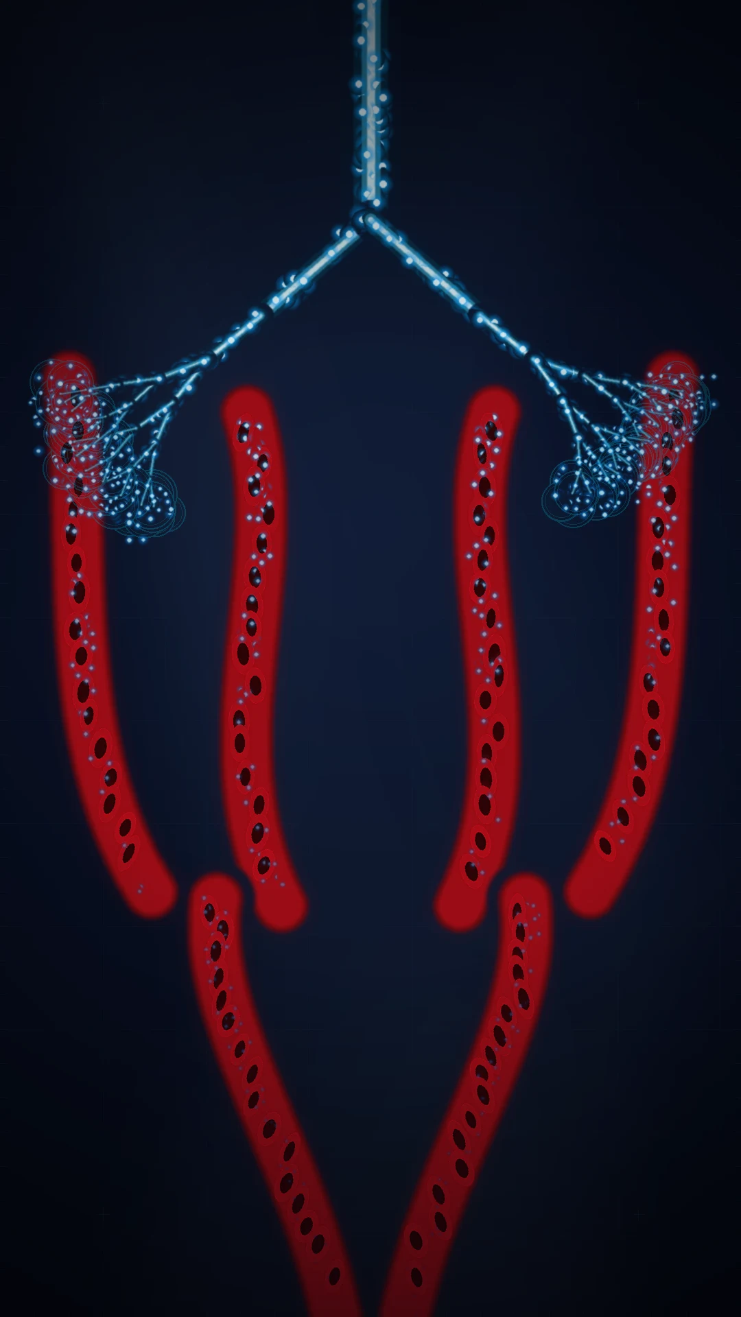
Oxygenation is the carriage of oxygen from the inspired air to the mitochondria, and its failure — the hypoxaemia (a low arterial oxygen) and the hypoxia (a low tissue oxygen) — is the commonest reason a patient comes to the intensive care unit. The intensivist's task is to identify WHERE in the carriage the failure sits, and to correct it — by the oxygen, the airway, the ventilation, the haemoglobin, the cardiac output, or the cytochrome, as the cause demands.[1][1]
The framework rests on the physiology (the oxyhaemoglobin curve, the four mechanisms of the hypoxaemia, the delivery and the consumption) and the therapy (the oxygen devices, the high-flow nasal cannula, the targeted — not the liberal — oxygenation). The over-riding principle is that more oxygen is not always better: the liberal oxygen increases the mortality (the Chu review), and the targeted, the physiological oxygenation is the goal.[2][1]
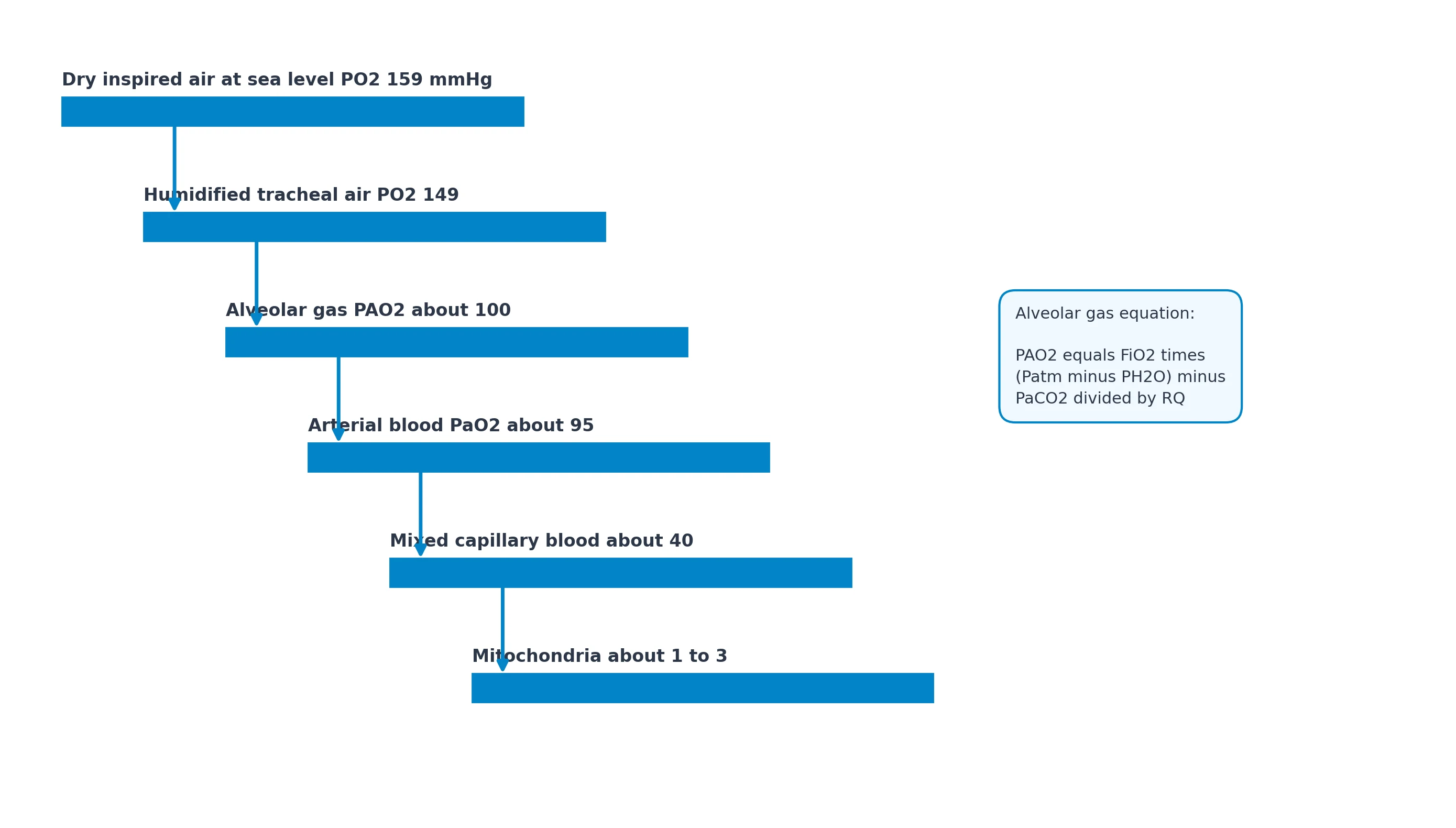
The oxygen cascade — the partial-pressure staircase
The oxygen cascade is the stepwise fall of the partial pressure of the oxygen from the dry inspired gas to the mitochondrion, and at each step work is done or pressure is spent, so the partial pressure drops. The intensivist reads the cascade as a fault-finding ladder: wherever the pressure falls more steeply than expected, the fault lies there.[1][1]
- The dry inspired gas — the PiO2. At sea level the atmospheric pressure (Patm) is 760 mmHg, the FiO2 of the room air is 0.21, and the water-vapour pressure at the body temperature (37 °C) is 47 mmHg. The PiO2 = FiO2 × (Patm − PH2O) = 0.21 × (760 − 47) = 0.21 × 713 = 150 mmHg. The dry inspired gas has lost nothing yet — but the alveolus is water-saturated and full of CO2.
- The alveolar gas — the PAO2. The alveolus is filled with the water-saturated gas into which the CO2 diffuses from the blood. The alveolar gas equation (see below) yields a PAO2 of about 100 mmHg on the room air. The drop from the PiO2 (150) to the PAO2 (100) is the cost of the CO2 — about 50 mmHg, the metabolic price of the aerobic respiration.
- The arterial blood — the PaO2. The gas crosses the alveolar-capillary membrane and is collected in the arterial blood. The PaO2 is about 90 to 95 mmHg on the room air, slightly below the PAO2 — the A-a gradient, normally under 15 mmHg in the young adult (the small anatomical shunt of the bronchial and the Thebesian veins, and the physiological shunt of the basilar atelectatic units). The drop from the PAO2 (100) to the PaO2 (90) is the cost of the membrane and the shunt.
- The capillary and the venous blood — the PvO2. The arterial blood flows into the systemic capillary, the oxygen leaves it for the tissue, and at the venous end the capillary blood has fallen to the mixed venous PO2 of about 40 mmHg (a saturation of 75 per cent). The drop from the PaO2 (90) to the PvO2 (40) is the extraction — the oxygen taken by the tissue.
- The mitochondrion. The oxygen diffuses from the interstitium to the mitochondrion, where the PO2 falls to about 1 to 3 mmHg (the critical PO2 below which the cytochrome-c oxidase cannot function and the anaerobic metabolism begins). The drop from the PvO2 (40) to the mitochondrion (1–3) is the tissue diffusion gradient. [1]
The cascade on the room air, in summary, is FiO2 21% → PiO2 150 → PAO2 100 → PaO2 90 → PvO2 40 → mitochondrion 1–3 mmHg. The two large drops — the CO2 cost (150 → 100) and the extraction (90 → 40) — are the metabolic and the tissue stations; the small A-a drop (100 → 90) is the lung station. A fault in the lung widens the A-a, a fault in the ventilation raises the PaCO2 and lowers the PAO2, and a fault in the extraction (the high-output demand or the low cardiac output) lowers the PvO2. The cascade is the diagnostic map of the hypoxaemia and the hypoxia.[1]
The alveolar gas equation — the PAO2
The alveolar gas equation relates the alveolar oxygen to the inspired oxygen and the alveolar CO2, and it is the equation that yields the A-a gradient:[1][1]
PAO2 = FiO2 × (Patm − PH2O) − [PaCO2 / RQ] [1]
where the FiO2 is the fractional inspired oxygen, the Patm is the atmospheric pressure (760 mmHg at sea level; about 630 mmHg at 1500 m; about 253 mmHg at the summit of Everest), the PH2O is the saturated water-vapour pressure at the body temperature (47 mmHg), the PaCO2 is the arterial CO2 (taken to equal the alveolar CO2, on the assumption that the CO2 exchange is complete), and the RQ is the respiratory quotient — the ratio of the CO2 produced to the O2 consumed, normally about 0.8 (0.7 on the pure fat, 0.85 on the protein, 1.0 on the pure carbohydrate). The correction by the RQ accounts for the fact that the CO2 produced is slightly less than the O2 consumed, so the CO2 occupies less alveolar volume than the O2 that was removed. [1]
Worked example on the room air: FiO2 0.21, Patm 760, PH2O 47, PaCO2 40, RQ 0.8. PAO2 = 0.21 × (760 − 47) − (40 / 0.8) = 0.21 × 713 − 50 = 150 − 50 = 100 mmHg. With a PaO2 of 90, the A-a gradient = PAO2 − PaO2 = 100 − 90 = 10 mmHg — normal (under 15 in the young, under 25 in the elderly). The age correction on the room air is A-a = 2.5 + 0.21 × age — a 70-year-old may have an A-a of 17 mmHg and still be normal. [1]
The four clinical readings of the equation: [1]
- The PaCO2 carries a negative sign. A rising CO2 (the hypoventilation) directly lowers the PAO2 — every 1 mmHg rise of the PaCO2 lowers the PAO2 by 1.25 mmHg (1/RQ). This is why the pure hypoventilation (the opiate overdose) gives a low PaO2 with a normal A-a gradient — the lung itself is normal; the ventilation is the fault, and the oxygen (or the reversal of the opiate) corrects it promptly.
- The FiO2 carries a positive sign. A high FiO2 raises the PAO2 directly — at FiO2 1.0 the PAO2 = 1.0 × 713 − 50 = 663 mmHg, the basis of the 100-per-cent-oxygen test for the shunt quantification.
- The altitude lowers the Patm. At altitude the (Patm − PH2O) term shrinks and the PAO2 falls — at the summit of Everest (Patm 253) the maximal PAO2 is barely 35 mmHg even with the maximal hyperventilation, the limit of the unacclimatised human tolerance.
- The A-a gradient widens with the FiO2. On the high FiO2 the normal A-a gradient rises to 50–100 mmHg (the nitrogen washout, the absorption atelectasis, the increased shunt fraction), and the cut-off for the "normal" must be adjusted — the a/A ratio (PaO2/PAO2, normally over 0.75) is more stable across the FiO2. [1]
The alveolar gas equation is the bedside calculator of the lung's work — it gives the PAO2 that the lung should produce, and the difference from the PaO2 it does produce (the A-a gradient) is the measure of the lung's failure. Every arterial blood gas is read against the alveolar gas equation.[1]
Pathophysiology: the oxyhaemoglobin dissociation curve
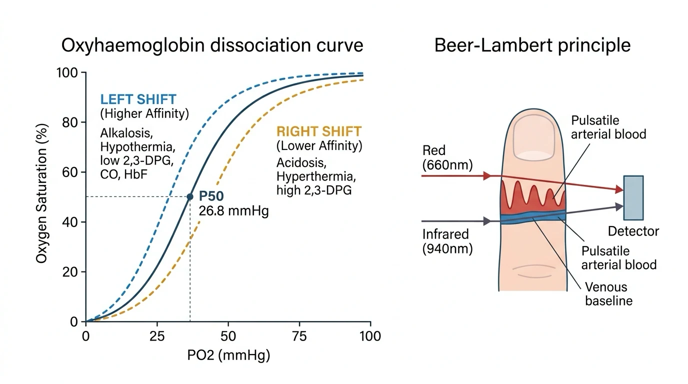
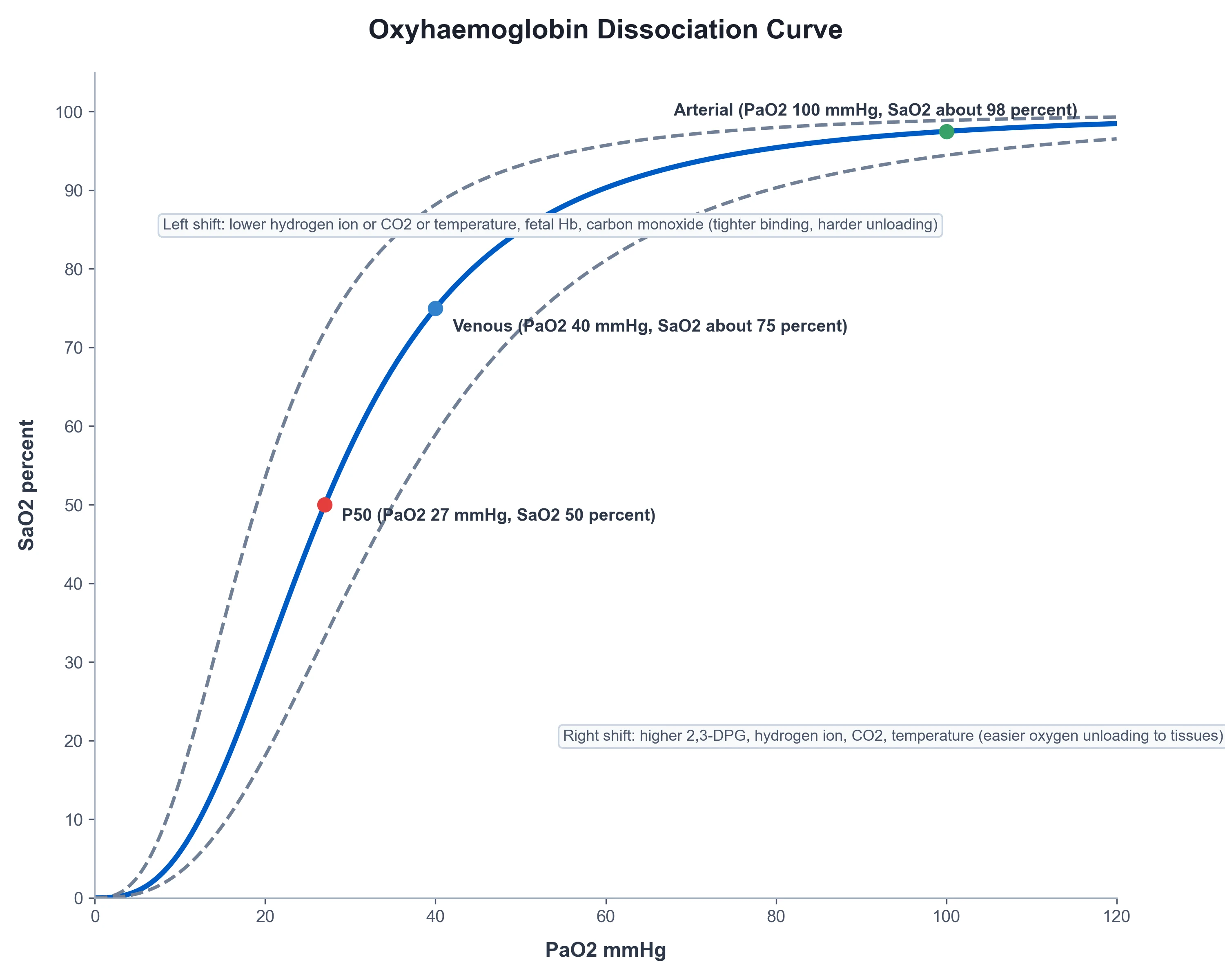
The oxyhaemoglobin dissociation curve — the sigmoid relationship between the partial pressure of the oxygen (the PaO2) and the saturation (the SaO2) — is the foundation of the oxygen carriage. Its plateau above a PaO2 of about 60 mmHg (where the saturation is over 90 per cent) is the safety margin: the saturation is held near-normal over a wide range of the PaO2, and a falling saturation signals a PaO2 that has fallen off the plateau (the steep part of the curve), a late and a dangerous sign.[1][1]
The curve is shifted by the biochemical environment. A rightward shift (the Bohr effect — the acidosis, the hypercapnia, the fever, the raised 2,3-diphosphoglycerate of the chronic hypoxia) reduces the affinity and unloads more oxygen to the tissues (a higher PaO2 for a given saturation); a leftward shift (the alkalosis, the hypocapnia, the hypothermia, the carbon-monoxide and the methaemoglobin) raises the affinity and impairs the unloading. The P50 — the PaO2 at the 50-per-cent saturation — is the measure of the affinity, normally about 26 to 27 mmHg.[1]
The four mechanisms of hypoxaemia and the A-a gradient
The hypoxaemia — a low arterial oxygen — arises from one of four mechanisms, and the A-a gradient (the alveolar-to-arterial oxygen difference) separates them.[1][1]
- The hypoventilation. A low alveolar ventilation (the opiate, the neuromuscular weakness) lowers the alveolar oxygen and raises the CO2; the PaO2 falls with a normal A-a gradient (the gas crossing the membrane is normal; the air reaching it is oxygen-poor). The oxygen corrects it readily, and the cause is the ventilation.
- The diffusion impairment. A thickened alveolar-capillary membrane (the pulmonary fibrosis, the pulmonary oedema) slows the equilibration; the A-a gradient is raised, and the oxygen (which raises the alveolar oxygen) corrects it.
- The ventilation-perfusion (V/Q) mismatch. The blood perfusing the under-ventilated alveoli leaves them under-oxygenated; the A-a gradient is raised, and the oxygen (which raises the alveolar oxygen of the under-ventilated units) largely corrects it. It is the commonest mechanism (the pneumonia, the asthma, the chronic lung disease).
- The shunt. The blood bypassing the ventilated alveoli (the anatomical shunt of the right-to-left cardiac defect, or the physiological shunt of the consolidated or the atelectatic lung) leaves the lung unoxygenated, and it does not correct with the oxygen — the oxygen cannot reach the shunted blood. The A-a gradient is markedly raised, and the shunt is the hallmark of the severe ARDS. [1]
Oxygen delivery and consumption
The global oxygen delivery (DO2) is the cardiac output × the arterial oxygen content; the consumption (VO2) is the uptake of the tissues (normally about 250 mL/min, a quarter of the delivery); and the extraction ratio (the VO2/DO2) is normally about 25 per cent. The tissues maintain the consumption across a range of the delivery by raising the extraction — until the delivery falls below a critical threshold, beyond which the consumption falls (the supply-dependence), the lactate rises, and the anaerobic metabolism begins.[1][1]
The Fick principle links them: VO2 = CO × (CaO2 − CvO2), where the CaO2 and the CvO2 are the arterial and the venous contents. The venous oxygen saturation (the ScvO2 from the central line, the SvO2 from the pulmonary artery) is the surrogate of the balance — a low ScvO2 (under about 70 per cent) signals a high extraction (the heart failing to deliver), and it was the target of the early goal-directed therapy. The consumption, the delivery and the extraction are the framework for the resuscitation of the shocked patient.[1]
The oxygen content equation — the CaO2
The arterial oxygen content (CaO2) is the volume of the oxygen carried in each decilitre of the arterial blood, and it is the sum of the oxygen bound to the haemoglobin and the oxygen dissolved in the plasma:[1][1]
CaO2 = (1.34 × Hb × SaO2) + (0.003 × PaO2) [mL O2 / dL] [1]
where the 1.34 is the Hüfner constant (the mL of O2 bound per gram of the fully saturated haemoglobin — older texts use 1.36 or 1.39, but 1.34 is the modern accepted value), the Hb is the haemoglobin in g/dL, the SaO2 is the fractional saturation (0 to 1), and the 0.003 is the solubility of the oxygen in the plasma (mL O2 per dL per mmHg of PaO2). [1]
Worked example: Hb 15 g/dL, SaO2 0.97, PaO2 90 mmHg. [1]
- Bound oxygen: 1.34 × 15 × 0.97 = 19.5 mL/dL
- Dissolved oxygen: 0.003 × 90 = 0.27 mL/dL
- Total CaO2 = 19.8 mL/dL. The dissolved fraction is barely 1.4 per cent of the total — almost all the oxygen is on the haemoglobin. [1]
Three rules of the intensive-care practice follow: [1]
- The haemoglobin dominates. The dissolved fraction (0.003 × PaO2) is negligible at any achievable PaO2; doubling the PaO2 from 90 to 180 mmHg adds only 0.27 mL/dL to the content, while raising the Hb from 7 to 10 g/dL adds 4 mL/dL. This is the physiological basis of the transfusion for the anaemic hypoxia, and the explanation of why the profound anaemia is tolerated at a normal PaO2 — until the carriage fails.
- The saturation dominates the PaO2. On the flat plateau of the curve (PaO2 above 60 mmHg) the saturation is over 90 per cent and the content is near-maximal; raising the PaO2 further adds little to the carriage. This is the physiological basis of the conservative oxygen target — the saturation of 92 per cent captures almost all the carriage, and the hyperoxia adds nothing but the harm (the oxidative stress, the absorption atelectasis, the vasoconstriction).[2]
- The dissolved fraction rises linearly with the PaO2. At a PaO2 of 600 mmHg (the FiO2 1.0) the dissolved oxygen is 1.8 mL/dL — still small but clinically meaningful in the severe anaemia, the carbon-monoxide poisoning and the hyperbaric oxygen therapy (where the PaO2 may reach 2000 mmHg and the dissolved fraction alone covers the metabolic demand, the basis of the hyperbaric therapy for the CO poisoning and the anaemic crisis in the Jehovah's Witness).
The venous content (CvO2) is the same equation with the SvO2 and the PvO2 — at Hb 15, SvO2 0.75, PvO2 40: CvO2 = 1.34 × 15 × 0.75 + 0.003 × 40 = 15.1 + 0.12 = 15.2 mL/dL. The arteriovenous O2 difference (CaO2 − CvO2) is therefore 19.8 − 15.2 = 4.6 mL/dL — the extraction, the oxygen the tissues took from each decilitre; and the heart delivers roughly 50 dL/min (a cardiac output of 5 L/min), giving a VO2 = CO × (CaO2 − CvO2) = 5000 × 0.046 = 230 mL/min — the resting metabolic demand, the Fick principle in the working numbers.[1]
The DO2, the critical delivery and the cytopathic dysoxia
The global oxygen delivery (DO2) is the cardiac output × the arterial oxygen content: [1]
DO2 = CO × CaO2 × 10 [mL O2 / min] (the ×10 converts the dL to the L). [1]
With a CO of 5 L/min and a CaO2 of 19.8 mL/dL, DO2 = 5 × 19.8 × 10 = 990 mL/min — about 1000 mL/min at rest, indexed (DO2I) to about 600 mL/min/m². The consumption (VO2) is the uptake of the tissues (normally about 250 mL/min, a quarter of the delivery); and the extraction ratio (the VO2/DO2) is normally about 25 per cent. The tissues maintain the consumption across a range of the delivery by raising the extraction — until the delivery falls below a critical threshold, beyond which the consumption falls (the supply-dependence), the lactate rises, and the anaerobic metabolism begins.[1][1]
The critical DO2 — the threshold below which the VO2 becomes supply-dependent — is about 330 mL/min/m² in the anaesthetised human (the lactate rises, the SvO2 falls below 50 per cent, the extraction ratio rises above 50 per cent). In the critical illness (and in the sepsis, in particular) the critical DO2 is shifted to the right — the impaired extraction forces the tissue into the anaerobic metabolism at a higher DO2 than the normal, the cytopathic dysoxia (the cells cannot use the oxygen even when it is delivered). The raised lactate with the normal or the high SvO2 is the hallmark of the impaired extraction — and the marker of the worse prognosis, distinguishing the macrocirculatory failure (the low DO2, the low SvO2) from the microcirculatory and the cellular failure (the normal DO2, the normal or the high SvO2, the persistent lactate). The treatment of the former is the resuscitation; the treatment of the latter is the source control, the time and (in the sepsis) the resolution of the mitochondrial injury.[1]
The venous oxygen saturations — the SvO2 and the ScvO2
The venous oxygen saturation is the integrated read-out of the delivery-consumption balance — the venous blood is what is left after the tissues have taken what they need, and a low venous saturation means the tissues took more (a higher extraction, a lower delivery).[1][1]
- The mixed venous saturation (SvO2) is sampled from the pulmonary artery (the tip of the Swan-Ganz catheter), the true mixed venous blood from all the systemic veins. The normal is 65 to 75 per cent (a PvO2 of about 40 mmHg). It is the gold standard of the delivery-consumption balance, but it requires the pulmonary-artery catheter.
- The central venous saturation (ScvO2) is sampled from the superior vena cava (the tip of the central venous catheter), the venous blood returning from the upper body (the brain, the heart, the arms). The normal is about 70 per cent — slightly higher than the SvO2, because the brain extracts less than the kidney and the splanchnic bed (which dominate the lower-body venous return). In the shock the gap widens — the splanchnic and the renal extraction rise disproportionately, the SvO2 falls below the ScvO2, and the ScvO2 overestimates the SvO2 by up to 10 per cent. The ScvO2 follows the trend reliably even if the absolute value diverges, and the central line is far more commonly placed than the pulmonary-artery catheter. [1]
The venous saturation is interpreted by the four determinants of the Fick principle — the Hb, the SaO2, the CO and the VO2 — and a low ScvO2 (under about 70 per cent in the resuscitated, under 65 per cent in the chronic) signals that one of them is failing: the anaemia (the Hb), the hypoxaemia (the SaO2), the low output (the CO) or the high demand (the VO2 — the fever, the agitation, the shivering, the sepsis). The treatment is the correction of the failing determinant — the transfusion, the oxygen, the inotrope or the sedation, respectively. The Rivers trial (NEJM 2001) made the ScvO2 ≥ 70 per cent the resuscitation end-point of the early goal-directed therapy of the severe sepsis and the septic shock, with a mortality benefit in the single-centre study.[3] The later multicentre trials (ProCESS, ARISE, ProMISe) found that the protocolised EGDT was no better than the usual care in the modern era — the improvements in the routine resuscitation had already captured its benefit — but the venous saturation remains the framework of the resuscitation, the surrogate of the Fick balance at the bedside.[4]
The trend matters more than the absolute value: a falling ScvO2 in the deteriorating patient is the early warning (before the lactate, before the hypotension), and a rising ScvO2 in the resuscitated patient is the sign of the recovery. A ScvO2 that is paradoxically high in the shocked patient (the 80s and the 90s) is the sign of the impaired extraction — the cells cannot take the oxygen — and the marker of the worse prognosis, the cytopathic dysoxia of the late sepsis and the mitochondrial failure.[1]
The shunt equation — the Qs/Qt
The shunt equation quantifies the fraction of the cardiac output that passes from the right heart to the left heart without being oxygenated — the blood that bypasses the ventilated alveoli (the true shunt, the zero-V/Q units) and the blood that perfuses the under-ventilated alveoli (the low-V/Q units, the "venous admixture"). The physiological shunt is normally 2 to 5 per cent (the bronchial and the Thebesian venous drainage); the pathological shunt rises to 30 to 50 per cent in the severe ARDS.[1][1]
Qs / Qt = (CcO2 − CaO2) / (CcO2 − CvO2) [1]
where the CcO2 is the end-capillary oxygen content (calculated from the PAO2 by the alveolar gas equation, assuming the full equilibration: CcO2 = 1.34 × Hb × 1.0 + 0.003 × PAO2), the CaO2 is the arterial content (from the arterial blood gas), and the CvO2 is the mixed venous content (from the pulmonary-artery blood gas). [1]
The 100-per-cent-oxygen test. At FiO2 1.0, the low-V/Q units are abolished (every alveolus is full of oxygen, even the under-ventilated ones), and the residual hypoxaemia is the true shunt. The rule of thumb: every 5 per cent of the shunt lowers the PaO2 by about 100 mmHg on the FiO2 1.0 — so a PaO2 of 350 mmHg on the FiO2 1.0 implies a shunt of about 30 per cent, and a PaO2 of 100 mmHg on the FiO2 1.0 implies a shunt of about 50 per cent. The shunt above 30 per cent is the oxygen-refractory hypoxaemia — the PaO2 does not rise with the FiO2 — and it is the indication for the PEEP (to recruit the collapsed alveoli), the prone ventilation (to perfuse the dorsal alveoli) and the inhaled pulmonary vasodilator (to redirect the blood from the shunted to the ventilated units). The shunt equation distinguishes the oxygen-responsive V/Q mismatch from the oxygen-refractory shunt, and it quantifies the lung's leak.[1]
The P50 and the affinity — the curve shifters revisited
The P50 — the PaO2 at the 50-per-cent saturation — is the single-number measure of the affinity of the haemoglobin for the oxygen, normally about 26 to 27 mmHg (at pH 7.40, PaCO2 40, temperature 37 °C, 2,3-DPG normal).[1]
- A raised P50 (a rightward shift) means a lower affinity — the haemoglobin releases the oxygen more readily to the tissues (a higher PaO2 for a given saturation). The rightward shifters are the acidosis, the hypercapnia (the Bohr effect), the fever, the raised 2,3-DPG (the chronic hypoxia, the chronic anaemia, the high altitude), the exercise and the pregnancy. The rightward shift is the tissue-unloading advantage, but the capillary uptake is impaired (a lower SaO2 for a given PAO2).
- A lowered P50 (a leftward shift) means a higher affinity — the haemoglobin holds the oxygen more tightly (a lower PaO2 for a given saturation). The leftward shifters are the alkalosis, the hypocapnia, the hypothermia, the lowered 2,3-DPG (the stored bank blood, the early sepsis) and the abnormal haemoglobin (the carbon-monoxide haemoglobin, the methaemoglobin and the foetal haemoglobin). The leftward shift is the pulmonary-loading advantage, but the tissue unloading is impaired (a lower PvO2, a tissue hypoxia despite a normal SaO2) — the carbon-monoxide poisoning and the methaemoglobinaemia cause a tissue hypoxia with a normal PaO2 (the saturation reads falsely high on the pulse oximeter, the curve is shifted and the carriage is impaired). [1]
The P50 is rarely measured at the bedside (it requires the tonometry and the multi-point curve), but the principle — that the affinity shifts with the biochemistry — governs the interpretation of every saturation. The pulse oximeter measures the saturation, not the content, not the delivery, not the tissue oxygen; and a normal SpO2 does not exclude a tissue hypoxia (the anaemic, the stagnant and the histotoxic hypoxia all sit at a normal SpO2). The P50 is the reason.[1]
The classification of hypoxia
The hypoxia — the low tissue oxygen — is classified by the mechanism:[1][1]
- The hypoxaemic hypoxia — a low arterial oxygen (the four mechanisms above).
- The anaemic hypoxia — a low haemoglobin (the carriage is the failure; the PaO2 is normal but the content is low).
- The stagnant (the circulatory) hypoxia — a low cardiac output (the delivery is the failure; the shock).
- The histotoxic hypoxia — the cells cannot use the oxygen (the cyanide poisoning, which blocks the cytochrome oxidase; the venous oxygen is paradoxically high). [1]
The classification directs the correction — the oxygen for the hypoxaemic, the transfusion for the anaemic, the restoration of the output for the stagnant, the antidote for the histotoxic. [1]
Oxygen therapy: the devices and the FiO2 they deliver
The oxygen devices are chosen for the FiO2 they deliver, the flow, and the precision of the control.[1]
- The low-flow devices (the nasal cannula at 1 to 6 L/min, delivering roughly 24 to 44 per cent; the simple face mask at 5 to 10 L/min, 35 to 60 per cent) deliver a variable FiO2 that depends on the patient's own peak inspiratory flow (the entrained room air dilutes the oxygen) — useful for the mild, the stable hypoxaemia, but imprecise for the severe.
- The fixed-performance devices (the Venturi mask, delivering a precise 24 to 60 per cent by the entrainment principle, the set FiO2 independent of the patient's flow) are used for the precise control — notably in the COPD patient at risk of the CO2 retention, where a target saturation of 88 to 92 per cent is the aim.
- The high-flow nasal cannula (HFNC, delivering up to 60 L/min of a humidified, warmed, oxygen-blended gas at a precise FiO2) provides the high flow, the low-level PEEP (the washout of the dead space, the reduced work of breathing), and the humidification — and it is the subject of the FLORALI trial.
- The non-invasive and the invasive ventilation deliver a precisely controlled FiO2 with the pressure support, for the respiratory failure that the oxygen alone cannot correct. [1]
High-flow nasal cannula and the FLORALI trial
The high-flow nasal cannula (the HFNC) delivers a high flow of a warmed, humidified, oxygen-blended gas through the nasal prongs, and it has three mechanisms: the low-level PEEP (a few cmH2O, splinting the alveoli), the dead-space washout (reducing the work of breathing), and the humidification (improving the secretion clearance and the tolerance).[1]
The FLORALI trial (NEJM 2015) compared the HFNC with the standard face-mask oxygen and the non-invasive ventilation in the acute hypoxaemic respiratory failure (the PaO2/FiO2 below 300). It found that the HFNC reduced the intubation rate (and a post-hoc subgroup with the PaO2/FiO2 below 200 showed a mortality benefit) — and it is now the first-line oxygen device for the moderate-to-severe hypoxaemic respiratory failure, with the non-invasive ventilation reserved for the cardiogenic pulmonary oedema and the COPD, and the intubation for the failure of the HFNC.[1]
The oxygen-target question: the liberal versus the conservative
The amount of oxygen given is itself a therapy with a harm. The liberal oxygen — the high FiO2 and the supraphysiological saturation — causes the oxidative stress, the absorption atelectasis, the coronary and the cerebral vasoconstriction, and (in the prolonged use) the lung injury; and the Chu systematic review (Lancet 2018) of the acutely ill adults found that the liberal oxygen (compared with the conservative) increased the mortality without a benefit.[2]
The synthesis is the targeted, the conservative oxygenation — a peripheral saturation of 92 to 96 per cent for most (88 to 92 per cent for the COPD at risk of the CO2 retention), the lowest FiO2 that achieves it, and the avoidance of the hyperoxia. The HOT-ICU and the ICU-ROX trials have refined the target (broadly confirming the no-benefit-to-the-higher-target), and the principle endures: the right amount of oxygen, not the most.[2][1]
V/Q mismatch, shunt and dead space — the gas-exchange failures
The two gas-exchange failures of the lung are the V/Q mismatch and the shunt (the oxygenation failures) and the dead space (the ventilation failure).[1][1]
- The V/Q mismatch — the perfusion of the under-ventilated alveoli (the low-V/Q units) is the commonest cause of the hypoxaemia, and it corrects largely with the oxygen (which raises the alveolar oxygen of the under-ventilated units).
- The shunt — the perfusion of the unventilated alveoli (the zero-V/Q units, the consolidated or the atelectatic lung) does NOT correct with the oxygen, and it is the hallmark of the severe ARDS (and the indicator for the PEEP and the prone ventilation).
- The dead space — the ventilation of the under-perfused alveoli (the high-V/Q units) is the ventilation failure, the wasted ventilation that raises the PaCO2 and the work of breathing, and it is the marker of the pulmonary vascular disease (the pulmonary embolism, the emphysema) and the severe ARDS. Bohr's dead-space equation quantifies it. [1]
Management: the targeted oxygen therapy
The oxygen therapy is the targeted, the physiological, the cause-directed.[2][1]
- Identify the mechanism of the hypoxaemia (the four mechanisms, the A-a gradient) and the type of the hypoxia.
- Give the oxygen at the lowest FiO2 that achieves the target saturation (92 to 96 per cent for most, 88 to 92 per cent for the COPD) — the conservative, the targeted, not the liberal.
- Choose the device for the severity — the low-flow for the mild, the Venturi for the precise control, the high-flow nasal cannula for the moderate-to-severe hypoxaemic respiratory failure (FLORALI), the non-invasive and the invasive ventilation for the failure.
- Treat the cause — the antibiotics for the pneumonia, the bronchodilator for the asthma, the diuretic for the oedema, the anticoagulant for the embolism, the transfusion for the anaemia, the restoration of the output for the shock.
- Avoid the hyperoxia — the supraphysiological saturation is harmful (the Chu review), and the lowest-FiO2 target is the goal.[2]
Monitoring oxygenation
Monitoring divides into the arterial, the tissue and the trend.[1][1]
- The arterial oxygen — the peripheral saturation (the continuous, the non-invasive) and the arterial blood gas (the precise, with the PaO2, the PaCO2, the pH, the base excess, the lactate). The A-a gradient is calculated from the alveolar gas equation.
- The tissue oxygen — the lactate (the marker of the anaerobic metabolism), the central or the mixed venous saturation (the surrogate of the delivery-consumption balance), the capillary refill and the urine output.
- The trend — the oxygenation is followed over time, the response to the therapy assessed, and the weaning of the FiO2 (and the device) as the cause resolves. [1]
Prognosis and the determinants of outcome
The prognosis of the hypoxaemia is the prognosis of its cause, but the duration and the depth of the hypoxia and the ** adequacy of the oxygen delivery** (the haemoglobin, the cardiac output) shape the organ injury. The avoidable harm is the liberal oxygen (the Chu review) and the delayed escalation (the failure to intubate when the HFNC or the non-invasive ventilation has failed); the principle is the targeted, the physiological oxygenation, the prompt escalation, and the treatment of the cause.[2][1]
Comparison tables
| Mechanism | Example | A-a gradient | PaCO2 | Corrects with O2? |
|---|
| Mechanism | Example | A-a gradient | PaCO2 | Corrects with O2? |
|---|---|---|---|---|
| Hypoventilation | Opiate, neuromuscular weakness, brainstem injury | Normal (< 15) | Raised | Yes — readily |
| Diffusion impairment | Pulmonary fibrosis, pulmonary oedema, emphysema | Raised | Normal (or low from hyperventilation) | Yes |
| V/Q mismatch (the commonest) | Pneumonia, asthma, COPD, atelectasis, pulmonary oedema | Raised | Normal (or low/high) | Yes — largely |
| Shunt (true, zero-V/Q) | ARDS, atelectasis, pulmonary AVM, right-to-left cardiac defect | Markedly raised | Normal (or low) | No — oxygen-refractory |
| Type | Defect | PaO2 | Hb | CO | SvO2 | Lactate | Treatment |
|---|
| Type | Defect | PaO2 | Hb | CO | SvO2 | Lactate | Treatment |
|---|---|---|---|---|---|---|---|
| Hypoxaemic | Low arterial O2 (the four mechanisms) | Low | Normal | Normal | Low | Variable | Oxygen / airway / ventilation |
| Anaemic | Low Hb carriage | Normal | Low | Normal | Low | Variable | Transfusion |
| Stagnant (circulatory) | Low CO / delivery | Normal | Normal | Low | Low | Raised | Restore output; inotrope; fluid |
| Histotoxic | Cells cannot use O2 (cyanide, CO, sepsis) | Normal | Normal | Normal | High | Markedly raised | Antidote (cyanide kit / hydroxocobalamin) |
| Device | Flow | FiO2 (approx) | Precision | Best for |
|---|
| Device | Flow | FiO2 (approx) | Precision | Best for |
|---|---|---|---|---|
| Nasal cannula | 1–6 L/min | 24–44% | Variable (patient PIF-dependent) | Mild, stable hypoxaemia |
| Simple face mask | 5–10 L/min | 35–60% | Variable | Acute moderate hypoxaemia |
| Non-rebreather mask | 10–15 L/min | 60–90% | Moderate (high, but not 100%) | Severe hypoxaemia (trauma, pre-intubation) |
| Venturi mask | Variable | 24/28/31/35/40/50/60% | Precise (set FiO2, independent of PIF) | COPD (CO2-retention risk); precise target |
| High-flow nasal cannula (HFNC) | Up to 60 L/min | 21–100% | Precise + PEEP + humidification | Moderate–severe hypoxaemic respiratory failure (FLORALI) |
| NIV / invasive ventilation | Set | 21–100% | Precise + pressure support | Failure of O2 alone; type 2 respiratory failure |
| Feature | Right shift (raised P50, low affinity) | Left shift (low P50, high affinity) |
|---|
| Feature | Right shift (raised P50, low affinity) | Left shift (low P50, high affinity) |
|---|---|---|
| P50 | > 27 mmHg | < 26 mmHg |
| Affinity | Reduced | Increased |
| Tissue unloading | Improved | Impaired |
| Pulmonary loading | Impaired | Improved |
| Causes | Acidosis, hypercapnia (Bohr), fever, raised 2,3-DPG (chronic hypoxia, anaemia, altitude), exercise, pregnancy | Alkalosis, hypocapnia, hypothermia, low 2,3-DPG (stored blood), CO-Hb, met-Hb, foetal Hb |
| Clinical | Chronic-hypoxia adaptation; better tissue O2 | CO poisoning (tissue hypoxia at normal PaO2); stored-blood transfusion |
| Feature | SvO2 (mixed venous) | ScvO2 (central venous) |
|---|
| Feature | SvO2 (mixed venous) | ScvO2 (central venous) |
|---|---|---|
| Sampling site | Pulmonary artery (Swan-Ganz tip) | Superior vena cava (central line tip) |
| What it mixes | All systemic venous return | Upper-body venous return only |
| Normal | 65–75% (PvO2 ~ 40 mmHg) | ~ 70% |
| In shock | Falls (true mixed) | Falls, but overestimates SvO2 by up to 10% |
| Catheter needed | Pulmonary-artery catheter | Central venous catheter (common) |
| Use | Gold standard; research; complex shock | Resuscitation end-point (Rivers EGDT); trend monitoring |
SAQ — The alveolar gas equation, the A-a gradient and the mechanism of hypoxaemia
10 minutes · 10 marks
A 28-year-old man is admitted to the ED after a near-drowning in fresh water. He is tachypnoeic (RR 28) and centrally cyanosed. SpO2 84 per cent on a non-rebreather mask at 15 L/min. ABG on room air before oxygen was applied: pH 7.32, PaCO2 32, PaO2 52, HCO3 18. CXR shows bilateral diffuse alveolar infiltrates.
SAQ — Oxygen delivery, the SvO2/ScvO2, and the determinants of consumption
10 minutes · 10 marks
A 62-year-old woman with septic shock from a urinary source is on noradrenaline 0.4 mcg/kg/min and vasopressin 0.03 U/min. Hb 8.2 g/dL, SpO2 96 per cent on FiO2 0.5 by face mask, blood pressure 95/50, lactate 4.2 mmol/L, urine output 0.3 mL/kg/h. A central venous line sample shows ScvO2 62 per cent. The team is debating a transfusion threshold.
Clinical pearls
Worked clinical flow steps
Approach to the hypoxaemic patient — the cascade read
- Look at the patient — the work of breathing, the accessory muscle use, the cyanosis, the consciousness. The respiratory distress with the fatigue is the indication for the immediate escalation (the HFNC, the NIV or the intubation), not for the further investigation.
- Send the arterial blood gas — the PaO2, the PaCO2, the pH, the base excess, the lactate, the CO-oximetry if the poisoning is suspected.
- Calculate the PAO2 by the alveolar gas equation (PAO2 = FiO2 × (Patm − PH2O) − PaCO2/RQ) — on room air, 0.21 × 713 − 40/0.8 = 100 mmHg.
- Calculate the A-a gradient (PAO2 − PaO2) and correct for the age (normal = 2.5 + 0.21 × age). The A-a separates the hypoventilation (normal) from the lung fault (raised).
- Read the PaCO2 — the raised with the normal A-a is the hypoventilation; the low with the raised A-a is the hyperventilating compensation for the V/Q mismatch.
- Categorise the mechanism — hypoventilation, diffusion, V/Q mismatch, shunt — by the A-a, the PaCO2 and the response to the oxygen.
- Give the targeted oxygen — the lowest FiO2 that achieves the SpO2 92–96% (88–92% in the COPD); choose the device for the severity.
- Treat the cause — the antibiotics, the bronchodilator, the diuretic, the anticoagulant, the transfusion, the inotrope.
- Monitor the trend — the SpO2, the arterial blood gas, the work of breathing; escalate to the HFNC, the NIV or the intubation at the failure.
- Avoid the hyperoxia — the lowest FiO2, the targeted SpO2, the weaning of the oxygen as the cause resolves.[2][1]
Calculating the A-a gradient — the bedside method
- Read the FiO2 the patient is on (room air = 0.21; the Venturi set; the estimated FiO2 of the nasal cannula, ~24% at 2 L/min + 4% per L/min).
- Read the PaCO2 from the arterial blood gas.
- Calculate the PAO2: PAO2 = FiO2 × (760 − 47) − (PaCO2 / 0.8).
- Read the PaO2 from the same arterial blood gas.
- Subtract: A-a = PAO2 − PaO2.
- Correct for the age: normal A-a (room air) = 2.5 + 0.21 × age; on the high FiO2 the normal rises to 50–100 mmHg — use the a/A ratio (PaO2/PAO2, normal > 0.75) instead.
- Interpret: normal A-a → hypoventilation; raised A-a → diffusion, V/Q mismatch or shunt.[1]
Calculating the oxygen delivery — the DO2
- Read the Hb, the SaO2, the PaO2 from the arterial blood gas and the full blood count.
- Calculate the CaO2: (1.34 × Hb × SaO2) + (0.003 × PaO2) — at Hb 15, SaO2 0.97, PaO2 90, CaO2 = 19.8 mL/dL.
- Measure or estimate the cardiac output (the echocardiography, the arterial-line pulse-contour, the thermodilution, or the clinical estimate).
- Multiply: DO2 = CO × CaO2 × 10 — at CO 5 L/min, DO2 = 990 mL/min (~1000 mL/min, indexed to ~600 mL/min/m²).
- Compare to the critical DO2 (~330 mL/min/m²) — below it, the lactate rises and the resuscitation is incomplete.
- Optimise the determinants: the Hb (the transfusion), the SaO2 (the oxygen, the PEEP), the CO (the fluid, the inotrope) — the lever that is failing.[1][1]
Trial cards
FLORALI — high-flow nasal cannula in acute hypoxaemic respiratory failure (NEJM 2015)
- Population: 310 adults with acute hypoxaemic respiratory failure (PaO2/FiO2 < 300), non-hypercapnic.
- Intervention: HFNC vs standard face-mask oxygen vs NIV.
- Outcome: Intubation rate (primary): HFNC 38% vs standard 47% vs NIV 50% (not significant overall).
- Subgroup (P/F < 200): HFNC reduced 90-day mortality (HR 0.32, p = 0.01) and the intubation rate.
- Take-home: HFNC is the first-line oxygen device for the moderate–severe hypoxaemic respiratory failure; NIV is reserved for the cardiogenic oedema and the COPD.[1]
Chu 2018 — liberal vs conservative oxygen (Lancet, systematic review and meta-analysis)
- Population: 25 000+ acutely ill adults (16 RCTs).
- Intervention: Liberal (higher SpO2 target, often FiO2 > 0.4) vs conservative / lower target.
- Outcome: Liberal oxygen increased mortality (RR 1.21, 95% CI 1.03–1.43) without a benefit on any other outcome.
- Harm mechanisms: Oxidative stress, absorption atelectasis, coronary and cerebral vasoconstriction, lung injury.
- Take-home: Target the lowest FiO2 that achieves the SpO2 92–96% (88–92% in the COPD); avoid the hyperoxia.[2]
Rivers EGDT — ScvO2 ≥ 70% in severe sepsis (NEJM 2001)
- Population: 263 adults with severe sepsis or septic shock, ED presentation.
- Intervention: Early goal-directed therapy — CVP 8–12, MAP ≥ 65, ScvO2 ≥ 70% (transfuse to Hct 30% if low, dobutamine if still low) within 6 hours; vs standard care.
- Outcome: In-hospital mortality 30.5% vs 46.5% (p = 0.009).
- Legacy: Made the ScvO2 the resuscitation end-point of the early sepsis care; later multicentre trials (ProCESS, ARISE, ProMISe) found no benefit of the protocolised EGDT over the modern usual care, but the Fick framework endured.[3]
ARISE — goal-directed resuscitation in early septic shock (NEJM 2014)
- Population: 1600 adults with early septic shock, 51 centres (ANZ).
- Intervention: Protocolised EGDT (Rivers-style, with the ScvO2 ≥ 70%) vs usual care.
- Outcome: 90-day mortality 18.6% vs 18.8% — no difference; no difference in organ support, length of stay or adverse events.
- Take-home: The protocolised EGDT offered no advantage over the modern usual care; the improvements in the routine resuscitation (the early antibiotics, the fluid, the lactate-guided care) had already captured the benefit. The ScvO2 remains the framework, not the mandatory target.[4]
Panwar CLOSE — conservative vs liberal oxygen in the ventilated (AJRCCM 2016, pilot)
- Population: 103 mechanically ventilated ICU adults (single-centre pilot, multicentre).
- Intervention: Conservative (SpO2 88–92%, PaO2 55–70) vs liberal (SpO2 ≥ 96%, PaO2 90–105).
- Outcome: No difference in mortality, ICU or ventilator-free days; the conservative group received less oxygen without harm.
- Take-home: The conservative target is feasible and apparently safe in the mechanically ventilated; the larger trials (ICU-ROX, HOT-ICU, the mega-registry) refined the question, broadly confirming no benefit to the higher target.[5]
Helmerhorst — hyperoxia and outcome in the critically ill (CCM 2015, meta-analysis)
- Population: Subgroups of the critically ill (the stroke, the traumatic brain injury, the cardiac arrest) from observational studies.
- Intervention (exposure): Arterial hyperoxia (PaO2 > 120) vs normoxia.
- Outcome: Hyperoxia associated with increased mortality in the cardiac arrest and the traumatic brain injury subgroups; the signal consistent with the Chu review.
- Take-home: The hyperoxia is not benign — corroborates the conservative target and the avoidance of the supraphysiological PaO2 in the resuscitated.[6]
Red flags
References
- [1]Frat JP, Thille AW, Mercat A, et al.; FLORALI Study Group. High-flow oxygen through nasal cannula in acute hypoxemic respiratory failure N Engl J Med, 2015.PMID 25981908
- [2]Chu DK, Kim LH, Young PJ, et al. Mortality and morbidity in acutely ill adults treated with liberal versus conservative oxygen therapy (IOTA): a systematic review and meta-analysis Lancet, 2018.PMID 29726345
- [3]Rivers E, Nguyen B, Havstad S, et al.; Early Goal-Directed Therapy Collaborative Group. Early goal-directed therapy in the treatment of severe sepsis and septic shock N Engl J Med, 2001.PMID 11794169
- [4]The ARISE Investigators and the ANZICS Clinical Trials Group. Goal-directed resuscitation for patients with early septic shock N Engl J Med, 2014.PMID 25272316
- [5]Panwar R, Hardie M, Bellomo R, et al.; CLOSE Study Investigators; ANZICS Clinical Trials Group. Conservative versus Liberal Oxygenation Targets for Mechanically Ventilated Patients. A Pilot Multicenter Randomized Controlled Trial Am J Respir Crit Care Med, 2016.PMID 26334785
- [6]Helmerhorst HJ, Roos-Blom MJ, van Westerloo DJ, Abu-Hanna A, de Keizer NF, de Jonge E. Association Between Arterial Hyperoxia and Outcome in Subsets of Critical Illness: A Systematic Review, Meta-Analysis, and Meta-Regression of Cohort Studies Crit Care Med, 2015.PMID 25855899