MedVellum
MCQsExamsAtlas
DashboardPricing
MBBS / Core medicine✳Dermatology✳ICU Fellowship (CICM)✳Anaesthesia✳Emergency Medicine✳Psychiatry Fellowship✳Paediatrics Fellowship✳Physician Medicine✳MCQs✳SAQs✳Vivas✳OSCE✳Evidence-first✳MBBS / Core medicine✳Dermatology✳ICU Fellowship (CICM)✳Anaesthesia✳Emergency Medicine✳Psychiatry Fellowship✳Paediatrics Fellowship✳Physician Medicine✳MCQs✳SAQs✳Vivas✳OSCE✳Evidence-first✳

MedVellum.

The folio

Exam-exhaustive medical education across every specialty — evidence-graded topics, engraved plates, and practice in every written and oral format. Educational content only — not medical advice.

llms.txt · psychiatry LLM catalog · sitemap

Atlas

  • Specialty atlas
  • MBBS / Core medicine
  • Dermatology
  • ICU Fellowship (CICM)
  • Anaesthesia
  • Emergency Medicine
  • Psychiatry Fellowship
  • Paediatrics Fellowship
  • Physician Medicine

Study & account

  • MCQ practice
  • Practice alias
  • Exam tools
  • Dashboard
  • Pricing
  • Sign in

© 2026 MedVellum. For education only — not a substitute for clinical judgement.

Folio edition · Set in Instrument Serif & Archivo

ICU Topicsrespiratory

ICU · respiratory

Oxygen Therapy and Oxygen Delivery — Comprehensive ICU Physiology

Also known as Oxygen therapy · Oxygen delivery · DO2 · VO2 · Oxygen cascade · Alveolar gas equation · Oxygen content · High-flow nasal cannula · Oxygen extraction ratio

Oxygen therapy and delivery — the physiological principles governing oxygen movement from the atmosphere to the cell, and the ICU interventions to optimise it. The oxygen cascade: atmospheric PO2 (159 mmHg at FiO2 21%) → tracheal PO2 (149, after humidification) → alveolar PO2 (PAO2 ~100, after CO2 mixing — calculated by the ALVEOLAR GAS EQUATION: PAO2 = FiO2(Patm - PH2O) - PaCO2/RQ) → arterial PO2 (PaO2 ~95, after shunt/VQ mismatch) → capillary PO2 (40, after tissue extraction) → mitochondrial PO2 (1-3, for oxidative phosphorylation). Oxygen content: CaO2 = (1.34 × Hb × SaO2) + (0.003 × PaO2) — the HAEMOGLOBIN component dominates (99% of total O2 content — dissolved O2 is negligible at normal pressures). Oxygen delivery: DO2 = CO × CaO2 × 10 — normal ~1000 mL/min. Oxygen consumption: VO2 = CO × (CaO2 - CvO2) × 10 — normal ~250 mL/min. O2 extraction ratio (ER) = VO2/DO2 — normal ~25% (the body extracts 25% of delivered O2 at rest). ICU management of hypoxaemia: increase FiO2 → increase Hb (transfuse) → increase CO (inotrope/fluid) → reduce shunt (PEEP, proning, recruit alveoli). Hyperoxia is HARMFUL (ROS, absorption atelectasis, coronary vasoconstriction) — titrate FiO2 to lowest setting maintaining SpO2 92-96%.

high3 referencesUpdated 2 July 2026
On this page & tools

Your progress

Saved locally on this device.

Target exams

CICMFFICMEDIC

Red flags

PaO2 <60 mmHg (SpO2 <90%) = SEVERE hypoxaemia — cellular ATP production is threatened — immediate intervention requiredDO2 <300 mL/min/m² = CRITICAL threshold — below this, VO2 becomes SUPPLY-DEPENDENT (anaerobic metabolism → lactate) — seen in severe shockAVOID hyperoxia (PaO2 >120 mmHg) — causes ROS generation, absorption atelectasis, coronary vasoconstriction — titrate FiO2 down to SpO2 92-96%The A-a gradient (PAO2 - PaO2) distinguishes hypoxaemia causes: NORMAL gradient (<15) = hypoventilation (opiate, neuromuscular). ELEVATED gradient (>15) = V/Q mismatch, shunt, diffusion impairment — needs OXYGEN or PEEP, not just ventilation

Your progress

Saved locally on this device.

Target exams

CICMFFICMEDIC

Red flags

PaO2 <60 mmHg (SpO2 <90%) = SEVERE hypoxaemia — cellular ATP production is threatened — immediate intervention requiredDO2 <300 mL/min/m² = CRITICAL threshold — below this, VO2 becomes SUPPLY-DEPENDENT (anaerobic metabolism → lactate) — seen in severe shockAVOID hyperoxia (PaO2 >120 mmHg) — causes ROS generation, absorption atelectasis, coronary vasoconstriction — titrate FiO2 down to SpO2 92-96%The A-a gradient (PAO2 - PaO2) distinguishes hypoxaemia causes: NORMAL gradient (<15) = hypoventilation (opiate, neuromuscular). ELEVATED gradient (>15) = V/Q mismatch, shunt, diffusion impairment — needs OXYGEN or PEEP, not just ventilation

Overview

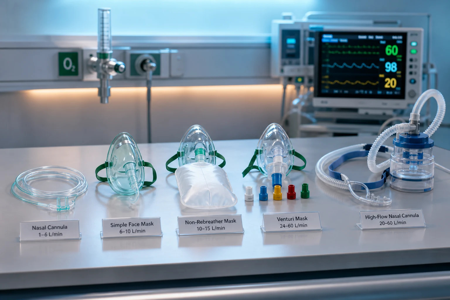
Oxygen delivery device ladder from nasal cannula to HFNC NIV invasive ventilation with SpO2 titration
FigureOxygen therapy is a ladder: match device capability (FiO2, flow, PEEP) to hypoxaemia severity and work of breathing; titrate to avoid hyperoxia.
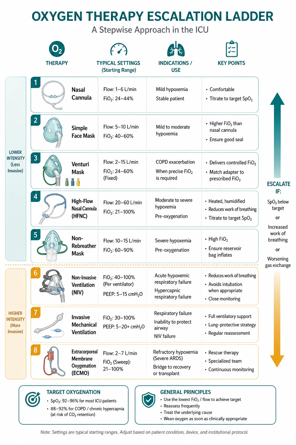
Escalation ladder NC simple mask Venturi NRB HFNC NIV MV ECMO
FigureDevice escalation: nasal cannula → Venturi/NRB → HFNC → NIV → invasive MV → ECMO when work of breathing or hypoxaemia fails the current step.

The one-paragraph exam answer

Oxygen therapy requires understanding the OXYGEN CASCADE (atmospheric → alveolar → arterial → cellular) and the OXYGEN DELIVERY equation. Alveolar gas equation: PAO2 = FiO2(Patm - PH2O) - PaCO2/RQ — the theoretical maximum PAO2 for a given FiO2. Oxygen content: CaO2 = (1.34 × Hb × SaO2) + (0.003 × PaO2) — Hb-bound O2 is >99% of total; dissolved O2 is negligible at normal pressure. Oxygen delivery: DO2 = CO × CaO2 — normal ~1000 mL/min. Oxygen consumption: VO2 = CO × (CaO2 - CvO2) — normal ~250 mL/min. Extraction ratio: VO2/DO2 ~25%. Hypoxaemia causes (5): V/Q mismatch (#1, responds to O2), shunt (does NOT respond to O2), diffusion impairment (responds to O2), hypoventilation (responds to O2, normal A-a gradient), low FiO2 (altitude). Management of hypoxaemia: increase FiO2 (oxygen therapy → HFNC → NIV → MV → ECMO) → increase Hb (transfuse if Hb <70) → increase CO (fluids/inotrope) → reduce shunt (PEEP, proning, recruit). AVOID hyperoxia (PaO2 >120 — ROS, absorption atelectasis, coronary vasoconstriction — titrate FiO2 to SpO2 92-96%). Oxygen devices: nasal cannula (24-44% FiO2), simple mask (40-60%), Venturi mask (precise 24-60%), non-rebreather (80-90%), HFNC (up to 100% with PEEP 3-5 cmH2O), NIV, mechanical ventilation, ECMO.[1][1]

The oxygen cascade — from atmosphere to mitochondrion

The oxygen cascade — each step reduces PO2

LocationPO2 (mmHg)Mechanism of reduction
Atmosphere159FiO2 21% × Patm 760 mmHg
Trachea149Humidification: subtract PH2O 47 mmHg → FiO2 × (760-47)
Alveolus (PAO2)~100CO2 mixing: subtract PaCO2/RQ (40/0.8 = 50) → the ALVEOLAR GAS EQUATION
Artery (PaO2)~95Shunt + V/Q mismatch: A-a gradient = PAO2 - PaO2 (normal <15 mmHg in young, <25 in elderly)
Capillary~40Tissue oxygen extraction — O2 diffuses from capillary to mitochondrion
Mitochondrion1-3Critical for oxidative phosphorylation — if <1 → anaerobic metabolism → lactate
Mixed venous (PvO2)~40SvO2 ~75% — reflects the balance between DO2 and VO2
[1]

The alveolar gas equation — the most important equation in respiratory physiology

PAO2 = FiO2(Patm - PH2O) - PaCO2/RQ [1]

Where:

  • FiO2 = fraction of inspired oxygen (room air = 0.21)
  • Patm = atmospheric pressure (760 mmHg at sea level)
  • PH2O = water vapour pressure (47 mmHg at 37°C)
  • PaCO2 = arterial CO2 (40 mmHg normally)
  • RQ = respiratory quotient (0.8 — ratio of CO2 produced to O2 consumed — varies with diet: carbohydrate 1.0, fat 0.7, protein 0.8) [1]

For room air: PAO2 = 0.21 × (760 - 47) - 40/0.8 = 0.21 × 713 - 50 = 149.7 - 50 = ~100 mmHg [1]

The A-a gradient (PAO2 - PaO2) is the MOST USEFUL CALCULATION for assessing the cause of hypoxaemia:

  • Normal A-a gradient (<15 young, <25 elderly) → hypoxaemia from HYPOVENTILATION alone (opiate, neuromuscular) → PaCO2 is elevated
  • Elevated A-a gradient (>15-25) → hypoxaemia from V/Q mismatch, shunt, diffusion, or low FiO2 → PaCO2 is normal or low
[1]

Worked example — the oxygen cascade step by step (room air, healthy adult at sea level)

Trace PO2 from the atmosphere to the mitochondrion for a 70 kg adult breathing room air (PaCO2 40, RQ 0.8): [1]

  1. Atmospheric PO2 = FiO2 × Patm = 0.21 × 760 = 159 mmHg.
  2. Tracheal PO2 (after humidification) = FiO2 × (Patm − PH2O) = 0.21 × (760 − 47) = 0.21 × 713 = 149 mmHg. Water vapour at 37 °C exerts 47 mmHg and displaces dry gas — this is why every respiratory gas equation subtracts PH2O.
  3. Alveolar PO2 (PAO2) — apply the ALVEOLAR GAS EQUATION: PAO2 = FiO2(Patm − PH2O) − PaCO2/RQ = 149 − 40/0.8 = 149 − 50 = ~100 mmHg. This is the single biggest step down and the one you control with FiO2.
  4. Arterial PO2 (PaO2) — subtract the A-a gradient. A young healthy adult has A-a ≈ 5-10: PaO2 ≈ 95 mmHg. An 80-year-old has A-a up to ~25: PaO2 ≈ 80 mmHg (A-a rises ~3 mmHg per decade).
  5. Systemic capillary PO2 — after tissue extraction, mixed venous PvO2 ≈ 40 mmHg (SvO2 75%).
  6. Mitochondrial PO2 — the final steep fall, to 1-3 mmHg. This is the critical pO2 for cytochrome c oxidase; below ~1 mmHg oxidative phosphorylation halts and lactate accumulates. [1]

Now repeat on 100% O2 (FiO2 1.0): PAO2 = 1.0 × 713 − 50 = 663 mmHg. Dissolved O2 = 0.003 × 663 = 2.0 mL/dL (small but no longer negligible). This is why a shunt patient's PaO2 on 100% O2 plateaus far below 600 — the shunted blood (PaO2 ~40) dilutes the well-oxygenated blood returning from ventilated alveoli. [1]

Altitude example (Denver, Patm 630 mmHg): PAO2 = 0.21 × (630 − 47) − 50 = 0.21 × 583 − 50 = 122 − 50 = 72 mmHg. PaO2 ≈ 65 — already in cyanotic territory yet tolerated by acclimatised residents. Hypobaric hypoxia is why altitude magnifies V/Q-mismatch and shunt hypoxaemia disproportionately.[1]

The oxygen-haemoglobin dissociation curve

Regions and landmarks of the oxyhaemoglobin dissociation curve

Region / landmarkPO2 (mmHg)SaturationSignificance
Plateau (top)60-10090-100%Above PaO2 60, large changes in PO2 cause tiny changes in SaO2 — a SAFETY MARGIN. This is why SpO2 is an INSENSITIVE early warning: it stays >90% down to PaO2 60, then falls off the cliff
P5027 mmHg (37 °C, pH 7.40)50%The affinity benchmark. LOWER P50 = higher affinity (curve LEFT). HIGHER P50 = lower affinity (curve RIGHT)
Steep portion20-6040-90%The WORKING range in tissue capillaries. Small drop in PO2 releases large O2 — efficient unloading. Mixed venous point (PvO2 40, SvO2 75%) lives here
Mixed venous point4075%Tissues extract ~25% of O2; the remaining 75% is the venous reserve
[1]

The Bohr effect — why CO2 and acid shift the curve RIGHT (favour unloading)

The position of the curve is governed by Hb affinity, summarised by P50. Anything that LOWERS affinity shifts the curve RIGHT (higher P50) and favours O2 UNLOADING to tissues. Anything that RAISES affinity shifts it LEFT (lower P50) and favours UPTAKE in the lung but impairs release in tissue. [1]

Right shift (↑P50, ↓affinity → better unloading):

  • ↑H+ (acidosis) — the Bohr effect (CO2 + H2O ⇌ H+ + HCO3−; CO2 also carbamylates Hb directly)
  • ↑PaCO2
  • ↑Temperature (exercising muscle, fever)
  • ↑2,3-BPG (2,3-bisphosphoglycerate) — rises in chronic hypoxia, anaemia, altitude, and chronic lung disease (an adaptive compensation)
  • Exercise (all four above combine — a massive right shift in working muscle) [1]

Left shift (↓P50, ↑affinity → better uptake, poorer unloading):

  • Alkalosis (↓H+)
  • ↓PaCO2 (hyperventilation)
  • ↓Temperature (hypothermia — bypass)
  • ↓2,3-BPG — STORED BANK BLOOD loses 2,3-BPG within days; a transfused unit releases O2 poorly for ~24 h until 2,3-BPG regenerates (one reason stored blood underperforms in the acute setting)
  • HbF (fetal haemoglobin — γ-chains bind 2,3-BPG weakly, so P50 ~19; designed to steal O2 from maternal blood across the placenta)
  • CO poisoning — carboxyhaemoglobin carries no O2 AND shifts the remaining curve LEFT (impaired release) — a DOUBLE insult. Pulse oximetry reads falsely HIGH (it cannot distinguish HbO2 from HbCO)
  • Methaemoglobinaemia — Fe³⁺ cannot bind O2 and shifts the curve LEFT; "chocolate brown" blood and a pulse-ox reading ~85% that does not move with O2 are the clues [1]

Clinical bottom line: at the same PaO2/SaO2, a right-shifted patient DELIVERS more O2 to tissues; a left-shifted patient delivers less. A right shift is usually an appropriate compensation (chronic anaemia, altitude); an inappropriate left shift (CO, metHb, old transfusion, hypothermia) causes tissue hypoxia DESPITE a normal PaO2.[1]

P50 — the single-number summary of Hb affinity

ConditionP50 (mmHg)DirectionCauseEffect
Normal (pH 7.40, 37 °C)27——Balanced uptake and release
Acidosis / hypercapnia>27RIGHTBohr effectBetter unloading
Alkalosis / hypocapnia<27LEFTBohr effectPoorer unloading
Fever / exercise>27RIGHT↑TemperatureUnloading to muscle
Stored blood (banked RBC)<27LEFT↓2,3-BPGPoor unloading for ~24 h
CO poisoning<27LEFTCarboxyhaemoglobinUptake AND release both impaired
Methaemoglobinaemia<27LEFTFe³⁺ HbCannot bind + left shift
Chronic altitude / anaemia>27RIGHT↑2,3-BPGCompensatory unloading
HbF (fetal)~19LEFTγ-chains, low 2,3-BPG bindingPlacental O2 theft
[1]

Why the sigmoid shape matters in three ICU scenarios

  1. Anaemia hides behind the plateau. Hb 50 g/L with SaO2 99% has a normal SpO2 and near-normal PaO2 — the oximeter and blood gas look fine while CaO2 is HALVED. SpO2 measures saturation, not content; it is BLIND to anaemia. Always pair SpO2 with Hb.
  2. The steep part is why shunt is catastrophic. Shunted blood (PaO2 40, sat 75%) returning to the arterial side drags the mixture down the STEEP portion — a small additional shunt fraction causes a large PaO2 drop. This is why PaO2 crumbles with modest increases in shunt (ARDS) but barely moves with pure V/Q mismatch.
  3. The plateau is why FiO2 is a blunt tool for shunt. Adding FiO2 lifts well-perfused alveolar units onto the flat plateau (no extra content gained), while shunted blood is unreachable — hence the hallmark: shunt does not correct with 100% O2.
[1]

Oxygen content, delivery, and consumption — the key equations

Oxygen transport equations — the critical calculations

ParameterEquationNormal valueClinical significance
Oxygen content (CaO2)(1.34 × Hb × SaO2) + (0.003 × PaO2)20 mL O2/dL bloodThe Hb component is >99%. Dissolved O2 (0.003 × PaO2) is negligible at normal pressure. Increasing Hb is the MOST effective way to increase CaO2
Arterial O2 delivery (DO2)CO × CaO2 × 10~1000 mL/minThe total O2 delivered to tissues per minute. Depends on BOTH cardiac output AND arterial O2 content
O2 consumption (VO2)CO × (CaO2 - CvO2) × 10~250 mL/minThe O2 actually used by tissues. Normal extraction ratio = VO2/DO2 = 25%
Extraction ratio (ER)VO2 / DO2~25%At rest, tissues extract 25% of delivered O2. In exercise/shock → ER increases (up to 70%). If DO2 falls below critical threshold → ER cannot increase further → VO2 falls → anaerobic metabolism → lactate
Mixed venous O2 (SvO2)Measured from PA catheter65-75%SvO2 <65% = inadequate DO2 (either CO low, Hb low, or SaO2 low). SvO2 <50% = severe O2 debt. SvO2 >80% = shunting (arteriovenous shunt in sepsis — blood bypasses capillaries)
[1]

Causes of hypoxaemia — the 5 mechanisms
Oxygen cascade atmosphere to mitochondrion with alveolar gas equation and A-a gradient
FigureThe oxygen cascade — atmospheric PO2 to mitochondrial PO2; the alveolar gas equation and A-a gradient anchor hypoxaemia classification.

Five causes of hypoxaemia — and which respond to oxygen

MechanismA-a gradientResponse to 100% O2ExamplesManagement
V/Q mismatch (#1 cause)ElevatedIMPROVES (well-ventilated alveoli compensate)Pneumonia, COPD, asthma, pulmonary embolism (mild), atelectasisIncrease FiO2 + treat cause (antibiotics, bronchodilators)
ShuntElevatedDOES NOT IMPROVE (blood bypasses ventilated alveoli — 100% O2 cannot reach shunted blood)ARDS, severe pneumonia, hepatopulmonary syndrome, intracardiac shunt (PFO), atelectasis (complete)PEEP (recruit collapsed alveoli → reduce shunt), proning, ECMO
Diffusion impairmentElevatedIMPROVES (increased FiO2 increases diffusion gradient)Pulmonary fibrosis, pulmonary oedema, emphysemaIncrease FiO2
HypoventilationNORMAL (<15)IMPROVES (but PaCO2 may remain elevated)Opioid overdose, neuromuscular disease (GBS, MG), CNS depressionVENTILATE (NIV or intubation) — oxygen alone is insufficient
Low FiO2 (high altitude)NORMALIMPROVESHigh altitude (low Patm → low PAO2)Increase FiO2
[1]

Oxygen therapy devices — the escalation ladder

Oxygen delivery devices — escalating FiO2

DeviceFiO2 rangeFlow rateAdvantagesDisadvantages
Nasal cannula24-44%1-6 L/minComfortable, allows eating/talking, patient can titrate. Rule of thumb: FiO2 ≈ 24% + (flow L/min × 4%)Low FiO2 only. Nasal drying/epistaxis at high flow. FiO2 varies with respiratory pattern
Simple face mask40-60%5-10 L/minHigher FiO2 than nasal cannula. Covers nose + mouthMUST have ≥5 L/min flow (to wash out CO2 from mask dead space → prevent rebreathing). Cannot eat. Claustrophobic
Venturi mask24-60% (precise)Variable (set by valve)PRECISE FiO2 (24%, 28%, 31%, 35%, 40%, 50%, 60% valves) — oxygen entrainment is fixed. Used for COPD patients who need controlled FiO2Less comfortable. FiO2 is FIXED — cannot titrate without changing valve
Non-rebreather mask80-90%10-15 L/minHIGHEST FiO2 without intubation. Reservoir bag provides O2 during inspiration. One-way valves prevent exhaled air rebreathingMask must fit tightly. Valve over reservoir must be functional. Still has some room air entrainment (not truly 100%)
High-flow nasal cannula (HFNC)Up to 100%30-60 L/minDelivers PRECISE FiO2 + FLOW (washes out dead space) + PEEP (3-5 cmH2O from high flow) + humidification + heating (improves mucociliary clearance). FLORALI: reduced intubation in hypoxaemic respiratory failureRequires specialized equipment (Optiflow/Airvo). Cannot deliver PEEP >5. Patient must be spontaneously breathing
NIV (BiPAP/CPAP)Up to 100%—Delivers POSITIVE PRESSURE (opens collapsed alveoli, reduces work of breathing, offloads LV in cardiogenic pulmonary oedema). 3CPO: CPAP improves outcome in cardiogenic pulmonary oedemaClaustrophobia, pressure ulcers, gastric insufflation. Cannot use if altered mental status (aspiration risk)
Mechanical ventilation21-100%—Full control of FiO2 + PEEP + ventilation. Can deliver ANY combinationInvasive (intubation risks: VAP, vocal cord damage, sedation)
ECMO (VV)——Bypasses the lungs entirely — oxygenates blood extracorporeally. For refractory hypoxaemia (EOLIA trial)Invasive, anticoagulation, resource-intensive
[1]

Venturi mask — entrainment physics (why FiO2 is PRECISE and FLOW is FIXED)

The Bernoulli / entrainment principle — how a Venturi valve sets FiO2

100% O2 is delivered down the supply tubing through a NARROW JET. As the jet accelerates through the constriction, the Bernoulli effect DROPS the lateral pressure and the high-velocity stream ENTRAINS (drags in) room air through calibrated SIDE PORTS. The SIZE of those ports is fixed by the colour-coded valve — that fixes the air:O2 entrainment ratio, which fixes the delivered FiO2 REGARDLESS of the patient's inspiratory pattern. This is why the Venturi mask is the only truly FiO2-ACCURATE low-flow device for the CO2-retaining COPD patient. [1]

The mixing equations:

  • Total flow = O2 flow + entrained air flow
  • Delivered FiO2 = (O2 flow + 0.21 × entrained air flow) / total flow [1]

Solving for the entrainment ratio at each valve gives the classic numbers below. Two things to notice: (a) as FiO2 RISES the entrained air FALLS, so TOTAL FLOW FALLS; (b) at the low end, total flow (~50 L/min) far exceeds peak inspiratory flow and locks FiO2, while at the high end (60%) total flow is low and a tachypnoeic patient can over-breathe the mask.

[1]

Venturi valves — entrainment ratio and total flow at each FiO2

Valve (colour)FiO2Air : O2 ratioExample total flowPractical meaning
Blue24%25 : 11 L O2 + 25 L air = 26 L/min; 2 L O2 → 52 L/minMaximal air entrainment → highest total flow, lowest FiO2. Total flow far exceeds resting peak inspiratory flow (~30 L/min) → FiO2 is LOCKED
White28%10 : 12 L O2 → 22 L/minStill above typical adult peak inspiratory flow at rest
Yellow31% / 35%6 : 1 / 5 : 13-4 L O2 → ~18-21 L/minStandard "precise moderate" range
Red40%3 : 14 L O2 → 16 L/minUpper end of the controlled-oxygen range
Green50%1.7 : 18 L O2 → ~22 L/minLess entrainment, FiO2 climbing
Pink / grey60%1 : 16-12 L O2 → 12-24 L/minMinimal entrainment → FiO2 high but TOTAL FLOW LOW. At 60% the total flow may fall BELOW peak inspiratory flow in a tachypnoeic patient → the patient entrains extra room air around the mask → true delivered FiO2 DROPS below 60%
[1]

The two iron rules of Venturi physics — and the exam traps they set

  1. As FiO2 RISES, total delivered flow FALLS. At 24% the patient gets ~50 L/min; at 60% only ~12-24 L/min. A breathless patient with high peak inspiratory flow (>30-40 L/min) will OVER-breathe a high-FiO2 Venturi and pull room air around the mask, so the TRUE FiO2 is LOWER than the valve says. Trap: never assume a "60% mask" actually delivers 60% to a tachypnoeic, high-demand patient. [1]

  2. The O2 flowmeter must be set to at least the valve's RATED flow (printed on each valve — e.g., 2, 4, 6, 8, 12 L/min). Below that flow there is not enough jet velocity to entrain the design volume of air, so FiO2 drifts UP and total flow falls. Trap: running a 24% valve at 8 L/min O2 does NOT deliver "more oxygen" — it slightly RAISES FiO2 and spoils the entrainment accuracy. The correct lever for more oxygen is to CHANGE THE VALVE, not crank the flowmeter.

[1]

High-flow nasal cannula — the four mechanisms, in physics terms

HFNC delivers up to 60 L/min of gas whose FiO2 is independently titrated (0.21-1.0), heated to 37 °C and humidified to 100% relative humidity. It is NOT "just a wet nasal cannula" — the high delivered flow fundamentally changes airway mechanics. [1]

The four mechanisms of HFNC benefit

MechanismPhysics / physiologyMeasurable effect
1. Precise, FIO2-independent FiO2Delivered flow (30-60 L/min) far exceeds peak inspiratory flow, so NO room air is entrained — the set FiO2 IS the delivered FiO2 (unlike nasal cannula / simple mask)SpO2 stabilises; FiO2 becomes a true dial, not a guess
2. Anisotropic PEEP (flow-dependent)High flow creates continuous positive pressure in the nasopharynx: ~0.5-1 cmH2O per 10 L/min with the mouth CLOSED. PEEP is ANISOTROPIC — it rises with flow and FALLS when the mouth opens (pressure vents through the oral cavity). 60 L/min + closed mouth ≈ 5-7 cmH2O; open mouth ≈ 2-3 cmH2OReduced work of breathing, splinted alveoli, reduced LV afterload (cardiogenic pulmonary oedema)
3. Dead-space washoutHigh nasal flow FLUSHES CO2-rich anatomical dead space (nasopharynx, oropharynx, large airways) between breaths. Each inspiration draws fresh gas, not the patient's own exhaled CO2. This effectively REDUCES anatomical dead space → a larger fraction of each Vt is alveolar ventilation → less minute ventilation is needed for the same CO2 clearance → lower RR, lower Vt, lower work of breathing↓RR, ↓PaCO2 (even in hypercapnic failure — the "NIV-sparing" effect in COPD), reduced WOB on oesophageal-pressure measurement
4. Heated humidified gas37 °C, 44 mg H2O/L (fully saturated). Dry cold gas paralyses mucociliary clearance, thickens secretions, and steals energy to condition the gas. Humidification PRESERVES ciliary beat frequency and mucus rheologySecretions mobilise, less atelectasis, reduced metabolic cost of gas conditioning, dramatically improved comfort and tolerance vs dry O2
[1]

The anisotropic PEEP of HFNC — flow, mouth, and leak

HFNC generates pressure that is dependent on flow AND leaky by design: [1]

  • Flow: pressure rises roughly linearly — ~1 cmH2O per 10 L/min, so 30 → ~3, 40 → ~4, 50 → ~5-6, 60 → ~6-7 cmH2O (measured in the pharynx).
  • Mouth position: with the mouth CLOSED, pressure is highest (the nasal route is the only exit). With mouth OPEN, pressure collapses to ~1-3 cmH2O regardless of flow — the oral cavity vents it. This is why you cannot RELY on a fixed PEEP number, and why HFNC pressure is called "variable" or "anisotropic" PEEP.
  • Cannula fit / leak: deliberately loose (~50% of nostril area) to allow expiration. Tighter fit → more pressure but more rebreathing; looser → less pressure.
  • Clinical implication: HFNC is NOT a PEEP-delivery device in the way CPAP is — you cannot "set 10 cmH2O." If you need reliable, titratable PEEP >5 for shunt reduction (ARDS), reach for NIV/CPAP or intubate. HFNC's pressure is a BONUS, not the primary mechanism — dead-space washout and FiO2 control are. [1]

Flow needed to wash out dead space: the nasopharyngeal + upper-airway dead space is ~50-70 mL. To flush it between breaths at RR 25 requires flow approaching peak inspiratory flow; 30-40 L/min usually achieves near-complete washout, which is why clinical benefit plateaus above ~40-50 L/min and we titrate to tolerance rather than maximising flow.[1]

Starting and titrating HFNC at the bedside — and predicting failure

  1. Set FiO2 to the patient's requirement (start 1.0 in severe hypoxaemia, wean to ≤0.4 as tolerated — target SpO2 92-96%).
  2. Set flow starting at 30 L/min and titrate UP to 50-60 L/min over 10-15 min (sudden high flow causes airway irritation, claustrophobia, and aerophagia). Higher flow = more dead-space washout + more PEEP.
  3. Confirm heat and humidity — circuit temperature 37 °C, chamber water level adequate. A dry circuit negates the mucociliary benefit and dries secretions.
  4. Apply the ROX index to predict success or failure: ROX = (SpO2/FiO2) / RR. ROX ≥4.88 at ≥2 h predicts HFNC success; ROX <3.85 predicts failure and should trigger intubation planning. Trend it serially — a falling ROX is an early warning.[2]
  5. Monitor for failure (any): persistent RR >30-35, SpO2 <90% on maximal settings, increasing accessory-muscle use / paradoxical breathing, agitation or altered mental status, rising PaCO2, acidosis (pH <7.3). Do NOT delay intubation for a "HFNC trial" that is failing — the delay-to-intubation itself worsens outcome.
  6. Wean by stepping FiO2 down first (to ≤0.4), THEN reducing flow to 30 L/min, then stepping to standard O2. A rising ROX with a comfortable patient supports continuing; a static or falling ROX does not.

Hyperoxia injury — the reactive oxygen species chemistry

Why hyperoxia is not benign — the ROS cascade at the mitochondrion and beyond

At FiO2 1.0, the partial pressure of O2 in mitochondria rises 100-600× above its Km for cytochrome c oxidase. The normal 1-2% electron "leak" from the electron transport chain (complexes I and III) now produces a FLOOD of the one-electron reduction product superoxide (O2•−). Superoxide itself is a modest oxidant, but it is the STARTING MATERIAL for a chain of progressively more damaging species. This is the molecular basis of the IOTA finding that liberal oxygen KILLS.[1]

Step 1 — Superoxide (O2•−): O2 + e− → O2•−. Produced by mitochondrial complexes I and III, xanthine oxidase, NADPH oxidase, and uncoupled nitric oxide synthase. Disarmed rapidly by superoxide dismutase (SOD): 2 O2•− + 2 H+ → H2O2 + O2. BUT superoxide also INACTIVATES protective signalling via reaction with nitric oxide: O2•− + NO → peroxynitrite (ONOO−), a potent cytotoxic oxidant that nitrates tyrosine residues and damages proteins and DNA. [1]

Step 2 — Hydrogen peroxide (H2O2): the product of SOD. Relatively STABLE and membrane-permeable (crosses membranes readily), so it propagates damage away from its site of production. Cleared by catalase (2 H2O2 → 2 H2O + O2) and the glutathione peroxidase / glutathione (GSH) system. Not a radical itself, but a ready substrate for Step 3. [1]

Step 3 — The Fenton reaction → hydroxyl radical (•OH): in the presence of free iron (Fe2+) — released from ferritin during inflammation, ischaemia-reperfusion, or transfusion of stored blood — Fe2+ + H2O2 → Fe3+ + •OH + OH−. The hydroxyl radical is the most reactive species in biology: it reacts at diffusion-limited rates with the FIRST molecule it contacts (half-life nanoseconds, radius a few Ångström). There is NO enzymatic defence against •OH — the only protection is to prevent its formation (chelate iron; scavenge H2O2 before it meets iron). This is why iron overload and massive transfusion amplify hyperoxia injury. [1]

Step 4 — Downstream tissue damage:

  • Lipid peroxidation: •OH abstracts a hydrogen atom from a polyunsaturated fatty-acid side chain → lipid radical → a propagating chain reaction (lipid radical + O2 → lipid peroxyl radical → attacks the next neighbour) → membrane destruction, loss of barrier integrity, surfactant inactivation. Measurable end-products: malondialdehyde (MDA), 4-hydroxynonenal (4-HNE). The lung, with its enormous surface area and lipid-rich surfactant, is the prime target.
  • Protein damage: oxidation of sulphydryl (-SH) groups, carbonylation, and nitration (via ONOO−) → enzyme inactivation, protein aggregation, loss of function. Mitochondrial Fe-S cluster enzymes (complex I, aconitase) are directly stripped by O2•− and •OH → energetic failure.
  • DNA damage: •OH attacks guanine → 8-oxo-guanine (mis-pairs with adenine → GC→TA transversions), single- and double-strand breaks, and activation of PARP → NAD+ depletion → further ATP collapse. In the lung this drives apoptosis of alveolar epithelial and endothelial cells.
[1]

The hyperoxia injury cascade — ROS species, sources, and defences

SpeciesFormulaSourceReactivity / half-lifeEnzymatic defenceTissue marker
SuperoxideO2•−Mitochondrial ETC (I, III), xanthine oxidase, NADPH oxidase, uncoupled NOSModerate, short (µs)Superoxide dismutase (SOD)Reacts with NO → peroxynitrite
Hydrogen peroxideH2O2SODStable, membrane-permeableCatalase, glutathione peroxidase (GSH)Substrate for Fenton
Hydroxyl radical•OHFenton: Fe2+ + H2O2EXTREME, ns (diffusion-limited)NONE — prevent formation8-oxoG (DNA), MDA / 4-HNE (lipid)
PeroxynitriteONOO−O2•− + NOStrong, msNone effectiveNitrotyrosine
[1]

Beyond ROS — three additional mechanisms of hyperoxia harm

  1. Absorption atelectasis. Nitrogen (79% of room air) is poorly soluble and "splints" alveoli open. Breathing 100% O2 washes out nitrogen → the gas in poorly ventilated alveoli is absorbed into blood faster than it is replaced → alveolar collapse → shunt. Visible within minutes during preoxygenation for intubation; one reason a pre-oxygenated patient desaturates fast if laryngoscopy is prolonged.
  2. Coronary and cerebral vasoconstriction. O2 is a vasoconstrictor (the mirror image of hypoxic vasodilation). Hyperoxia raises coronary and cerebral vascular resistance → reduced blood flow; in the post-cardiac-arrest and STEMI settings this can WORSEN tissue O2 delivery despite a higher arterial content — the physiological rationale for avoiding hyperoxia after ROSC.
  3. Atelectrauma and VILI amplification. Hyperoxia primes inflammatory pathways (NF-κB, cytokine release) and sensitises the alveolus to ventilator-induced lung injury; in animal models oxygen toxicity combined with even modest tidal volumes produces ARDS-like histology. [1]

Antioxidant defences (the system hyperoxia overwhelms): SOD, catalase, glutathione peroxidase, the glutathione (GSH/GSSG) redox couple, vitamins C and E, and urate. Premature infants have immature defences — the basis of retinopathy of prematurity (ROP) and bronchopulmonary dysplasia (BPD), the classic oxygen-toxicity syndromes, and the reason neonatal FiO2 is titrated to SpO2 88-95%. The same logic, less dramatically, applies to adults: titrate FiO2 to the LOWEST effective level.[1]

Clinical pearls

Clinical pearl

  1. The A-a gradient is the MOST USEFUL calculation for assessing hypoxaemia. PAO2 - PaO2. Normal <15 (young), <25 (elderly — increases 3 mmHg per decade). Normal A-a gradient → hypoxaemia from HYPOVENTILATION (PaCO2 elevated — opiate, neuromuscular). Elevated A-a gradient → V/Q mismatch, shunt, or diffusion impairment.[1]

  2. Increasing Hb is the MOST effective way to increase CaO2. CaO2 = (1.34 × Hb × SaO2) + (0.003 × PaO2). The Hb-bound component is >99%. Increasing PaO2 from 80 to 400 mmHg (on 100% O2) only adds 0.96 mL O2/dL (0.003 × 320) — negligible. Increasing Hb from 70 to 100 g/L adds 4.0 mL O2/dL (1.34 × 0.30 × 0.97) — SIGNIFICANT. In severe anaemia, OXYGEN THERAPY is less effective than TRANSFUSION.[1]

  3. AVOID hyperoxia (PaO2 >120 mmHg). Hyperoxia causes: (a) Reactive oxygen species (ROS) — damage to lipids, proteins, DNA. (b) Absorption atelectasis — nitrogen washout from alveoli → alveolar collapse. (c) Coronary vasoconstriction — reduced coronary blood flow. IOTA meta-analysis: liberal oxygen (SpO2 >96%) increased mortality vs conservative. LOCO2: conservative oxygen target (PaO2 60-90) was SAFE. Titrate FiO2 to SpO2 92-96%.[1][1]

  4. Shunt does NOT respond to 100% oxygen. In shunt, blood bypasses ventilated alveoli (e.g., ARDS, consolidation, atelectasis). Giving 100% O2 only oxygenates the blood that ALREADY passes through ventilated alveoli — it cannot reach the shunted blood. This is why shunt requires PEEP (recruit collapsed alveoli → reduce shunt fraction), not just more FiO2.[1]

  5. V/Q mismatch RESPONDS to oxygen. V/Q mismatch (pneumonia, COPD, asthma, PE) — some alveoli are underventilated but still perfused. Giving O2 increases PAO2 in ALL alveoli → even the underventilated alveoli achieve adequate PAO2 → improves oxygenation. This is why V/Q mismatch patients improve with O2 therapy but shunt patients don't.[1]

  6. HFNC reduces intubation in hypoxaemic respiratory failure (FLORALI trial). HFNC delivers: (a) Precise FiO2, (b) High flow (washes out dead space → reduces work of breathing), (c) PEEP 3-5 cmH2O (splints open alveoli), (d) Heated humidified gas (improves mucociliary clearance + patient comfort). FLORALI: HFNC reduced intubation rate vs standard O2 or NIV in hypoxaemic respiratory failure (PaO2/FiO2 <300).[1]

  7. The FLORALI trial — HFNC vs NIV vs standard O2. FLORALI (Frat 2015, NEJM): HFNC vs NIV (BiPAP) vs standard face mask O2 in patients with hypoxaemic respiratory failure (PaO2/FiO2 <300, non-hypercapnic). Result: HFNC had LOWEST intubation rate (38% vs 50% NIV vs 47% standard — not individually significant). In the subgroup with PaO2/FiO2 <200: HFNC SIGNIFICANTLY reduced intubation (35% vs 53% NIV — significant). HFNC is PREFERRED for moderate hypoxaemic respiratory failure.[1]

  8. SvO2 (mixed venous saturation) reflects the balance between DO2 and VO2. Normal SvO2 = 65-75% (tissues extract 25-35% of delivered O2). SvO2 <65% = DO2 is inadequate (either CO low, Hb low, or SaO2 low). SvO2 <50% = severe O2 debt (anaerobic metabolism). SvO2 >80% = O2 extraction is impaired (shunting, sepsis, cyanide toxicity — blood passes through capillaries without releasing O2 to tissues). Measured from PA catheter (true mixed venous) or ScvO2 from central line (central venous — less accurate but simpler).[1]

  9. The critical DO2 threshold — below this, VO2 becomes supply-dependent. Normal DO2 ~1000 mL/min → VO2 ~250 mL/min (VO2 is supply-INDEPENDENT — tissues regulate their O2 extraction to maintain constant VO2 regardless of DO2). When DO2 falls below the CRITICAL THRESHOLD (~300 mL/min/m²) → VO2 becomes SUPPLY-DEPENDENT → VO2 falls in parallel with DO2 → anaerobic metabolism → lactate rises. This is the basis of 'shock' — inadequate DO2 for tissue needs.[1]

  10. IOTA meta-analysis — liberal oxygen INCREASES mortality. IOTA (Chu 2018, Lancet): meta-analysis of 25 RCTs (16,037 patients). Liberal oxygen therapy (SpO2 >96%) vs conservative oxygen. Result: liberal oxygen INCREASED mortality (RR 1.21) without improving any other outcome. Interpretation: hyperoxia is HARMFUL — titrate FiO2 to the LOWEST setting maintaining SpO2 92-96%.[1]

  11. Venturi mask for COPD — precise FiO2 is critical. COPD patients who are CO2 retainers need LOW FiO2 (24-28% via Venturi mask) — excessive O2 can worsen hypercapnia via: (a) V/Q mismatch worsening (O2 relaxes hypoxic pulmonary vasoconstriction → blood flow to poorly ventilated alveoli → increased dead space), (b) Haldane effect (O2 displaces CO2 from haemoglobin → CO2 released into blood → increased PaCO2). But NEVER WITHHOLD OXYGEN from a hypoxic COPD patient — hypoxia kills faster than hypercapnia.[1]

  12. HFNC provides PEEP 3-5 cmH2O — useful for cardiogenic pulmonary oedema. The positive pressure from HFNC: (a) reduces preload (decreases venous return → reduces pulmonary congestion), (b) reduces afterload (decreases LV transmural pressure → improves LV output), (c) splints open alveoli (reduces work of breathing). This is why HFNC is effective for cardiogenic pulmonary oedema (similar mechanism to CPAP but less invasive).[1]

  13. ICU-ROX and LOCO2 — conservative oxygen targets are SAFE. ICU-ROX (Stewart 2019, NEJM): conservative (SpO2 88-92%) vs liberal (SpO2 >96%) in mechanically ventilated ICU patients — no difference in outcome. LOCO2 (Barrot 2020, JAMA): conservative (PaO2 60-90) vs liberal (PaO2 150-180) in ARDS — conservative was SAFE (no excess mortality). These trials support TITRATING FiO2 to the LOWEST effective level (SpO2 92-96%) rather than routinely giving high FiO2.[1][1]

  14. The RQ (respiratory quotient) matters for the alveolar gas equation. RQ = CO2 produced / O2 consumed. RQ varies with diet: carbohydrate = 1.0, fat = 0.7, protein = 0.8. Mixed diet = 0.8 (standard value used in calculations). On a PURE carbohydrate diet → RQ = 1.0 → PaCO2/RQ = 40/1.0 = 40 (instead of 50) → PAO2 increases by 10 mmHg. On a PURE fat diet → RQ = 0.7 → PaCO2/RQ = 40/0.7 = 57 → PAO2 decreases by 7 mmHg. This is why high-carbohydrate nutrition can improve oxygenation (and why high-fat nutrition can worsen it — but the effect is small).[1]

  15. The oxyhaemoglobin dissociation curve's plateau is why SpO2 is BLIND to early hypoxaemia AND to anaemia. Above PaO2 60 mmHg the curve is flat — large falls in PaO2 (e.g., 100 → 65) barely move SpO2 (99% → 91%), so SpO2 is an INSENSITIVE early-warning monitor and lags PaO2. Worse, SpO2 measures SATURATION not CONTENT — a Hb of 50 g/L with SaO2 99% looks "normal" on the oximeter while CaO2 is halved. Always pair SpO2 with Hb.[1]

  16. Venturi: as FiO2 rises, total flow FALLS — a 60% valve can be a lie in a breathless patient. At 24% (25:1) total flow ≈ 50 L/min (FiO2 locked); at 60% (1:1) only ≈ 12-24 L/min. A tachypnoeic patient (peak inspiratory flow >40 L/min) over-breathes a 60% Venturi and entrains room air at the mask edge → true delivered FiO2 far below 60%. For a controlled trial of precise FiO2 use the LOW valves (24-28%); for high FiO2 in a distressed patient abandon Venturi for HFNC or a non-rebreather. [1]

  17. HFNC's PEEP is anisotropic — it depends on flow AND mouth position. Closed mouth, 60 L/min ≈ 5-7 cmH2O; open mouth collapses it to ~2-3. You cannot "dial PEEP" with HFNC, and you cannot reliably deliver >5 cmH2O. If a patient needs titratable PEEP >5 for shunt (ARDS), HFNC is the wrong tool — use NIV/CPAP or intubate.[1]

  18. HFNC washes out dead space — this is its NIV-sparing trick in hypercapnia. By flushing CO2 from the nasopharynx between breaths, HFNC reduces anatomical dead space so each tidal volume delivers more alveolar ventilation → lower RR and lower PaCO2 without positive pressure. This is why moderate COPD/pneumonia hypercapnia can improve on HFNC and avoid intubation, whereas shunt-driven hypoxaemia (ARDS) cannot. [1]

  19. Stored blood is LEFT-shifted — it releases O2 poorly for ~24 h. 2,3-BPG degrades during storage; transfused red cells hold onto O2 (low P50) until 2,3-BPG regenerates. A massively transfused patient may have a normal Hb and PaO2 yet suffer tissue hypoxia from impaired unloading — one more reason to transfuse only what is needed and to correct the cause, not the number.[1]

  20. CO poisoning is a DOUBLE insult — absent carriage PLUS a left shift, and the oximeter lies. Carboxyhaemoglobin carries no O2 and shifts the remaining curve LEFT (impaired release); pulse oximetry cannot distinguish HbO2 from HbCO and reads falsely HIGH (often ~95-100%). Diagnose with CO-oximetry (a multi-wavelength ABG), treat with 100% O2 (halves CO half-life from ~320 to ~80 min), and consider hyperbaric oxygen for pregnancy, neuro signs, or high COHb.[1]

  21. Hyperoxia's ROS damage is iron-amplified — the Fenton reaction is why transfusion and reperfusion magnify oxygen toxicity. Fe2+ + H2O2 → •OH + OH−; the hydroxyl radical has NO enzymatic defence (unlike superoxide and H2O2). Free iron from ischaemia-reperfusion, haemolysis, or stored-blood transfusion therefore converts a manageable H2O2 load into the most damaging species in biology — restrict unnecessary iron load AND unnecessary FiO2 together.[1]

  22. The ROX index tells you when HFNC is failing — measure it at 2, 6, and 12 h. ROX = (SpO2/FiO2)/RR. ROX ≥4.88 at 2 h predicts success; a falling ROX or ROX <3.85 predicts failure and should trigger early intubation. A "HFNC trial" that is failing is more dangerous than timely intubation — delayed intubation after HFNC failure carries excess mortality.[2]

Red flags

AVOID hyperoxia — titrate FiO2 to SpO2 92-96%

Hyperoxia (PaO2 >120) causes ROS generation, absorption atelectasis, coronary vasoconstriction. IOTA meta-analysis: liberal oxygen increases mortality. Titrate FiO2 to the LOWEST setting maintaining SpO2 92-96%.[1]

Shunt does NOT respond to 100% O2 — needs PEEP

If hypoxaemia persists despite 100% FiO2 → the cause is SHUNT (ARDS, consolidation). Increasing FiO2 further is useless. Need PEEP (recruit collapsed alveoli → reduce shunt fraction), proning, or ECMO.[1]

SpO2 is BLIND to anaemia — a normal oximeter does not exclude tissue hypoxia

SpO2 measures saturation, not content. A patient with Hb 50 g/L and SaO2 99% has a normal oximeter and near-normal PaO2 but HALF the oxygen content (CaO2 ≈ 10 mL/dL). In haemorrhagic or haemolytic shock the oximeter may read 97% while tissues are dying. Always pair SpO2 with Hb; transfuse to the target that restores delivery, not the saturation.[1]

A failing HFNC trial kills by delay — apply the ROX index and intubate early

HFNC failure recognised late carries excess mortality (the delay-to-intubation itself worsens outcome). Track ROX = (SpO2/FiO2)/RR at 2, 6, and 12 h: ROX <3.85, a falling trend, or any of RR >35, SpO2 <90% on maximal settings, rising PaCO2, pH <7.3, or increased work of breathing = failure → intubate now.[2]

Pre-oxygenation causes absorption atelectasis — intubate promptly after a good pre-ox

Breathing 100% O2 washes the nitrogen splint out of alveoli; dependent alveoli collapse within minutes, creating shunt that blunts the very oxygenation you just built up. A perfectly pre-oxygenated patient desaturates FAST if laryngoscopy is prolonged. Use PEEP / recruitment after intubation to re-open what the pre-ox collapsed.

[1]

A 60% Venturi is not reliable in a breathless patient — total flow is too low

At 60% (air:O2 1:1) total flow is only ~12-24 L/min. A tachypnoeic patient over-breathes the mask, entrains room air, and receives far less than 60%. If a high FiO2 is genuinely needed in a high-work-of-breathing patient, use HFNC, a non-rebreather, or intubate — do not trust a high-FiO2 Venturi number.

[1]

Prognosis

Oxygen therapy outcomes — the evidence

StrategyOutcomeEvidence
HFNC vs standard O2HFNC reduces intubation in PaO2/FiO2 <200FLORALI trial
Conservative vs liberal O2Conservative is SAFE (no excess mortality)ICU-ROX, LOCO2
Liberal O2INCREASES mortality (RR 1.21)IOTA meta-analysis
CPAP for cardiogenic pulmonary oedemaReduces intubation + mortality3CPO trial
NIV for COPD exacerbationReduces intubation + mortalityPlant trial
[1]

Key trials and evidence

FLORALI trial — HFNC vs NIV vs standard O2 (PMID 22642477)

Study design

Randomised — 310 patients with hypoxaemic respiratory failure

Population

Adults with PaO2/FiO2 <300, non-hypercapnic

Intervention

HFNC vs NIV (BiPAP) vs standard face mask O2

Primary outcome

Intubation rate at 28 days: 38% (HFNC) vs 47% (standard) vs 50% (NIV) — overall not significant

Subgroup (PaO2/FiO2 <200)

HFNC SIGNIFICANTLY reduced intubation: 35% vs 53% (NIV) — p=0.009

Clinical bottom line

HFNC is PREFERRED for moderate hypoxaemic respiratory failure — especially PaO2/FiO2 <200

[1]

IOTA meta-analysis — Liberal vs conservative oxygen (PMID 27568755)

Study design

Meta-analysis — 25 RCTs, 16,037 patients

Population

Acutely ill adults (sepsis, trauma, cardiac, stroke, critical illness)

Intervention

Liberal oxygen (SpO2 >96%) vs conservative (room air or titrated O2)

Primary outcome

Mortality: liberal oxygen INCREASED mortality (RR 1.21, 95% CI 1.03-1.43)

Key finding

Hyperoxia is HARMFUL — every unnecessary unit of O2 increases risk without benefit

Clinical bottom line

Titrate FiO2 to SpO2 92-96% — do NOT give 100% O2 routinely — conservative oxygen is the new standard

[1]

ROX index — predicting HFNC success or failure (PMID 30576221)

Study design

Multicentre derivation and validation — adults with acute hypoxaemic respiratory failure on HFNC

Index

ROX = (SpO2 / FiO2) / respiratory rate

Thresholds

ROX ≥4.88 at ≥2 h predicts HFNC success (low intubation risk); ROX <3.85 predicts failure (high intubation risk)

Use

Measure serially at 2, 6, and 12 h. A falling ROX is an early warning of failure — pair with bedside work-of-breathing assessment

Clinical bottom line

ROX is a simple, bedside gauge of whether HFNC is working — but it supplements, never replaces, clinical judgement; never delay intubation for a borderline number

[1]

HOT-ICU — lower vs higher oxygenation targets in ICU (PMID 33471452)

Study design

Multicentre RCT — 2928 adults with acute hypoxaemic respiratory failure

Intervention

Lower target PaO2 60 mmHg (8 kPa) vs higher target PaO2 90 mmHg (12 kPa)

Primary outcome

90-day mortality: NO DIFFERENCE between groups

Key finding

A lower oxygenation target was SAFE but did not reduce mortality — alongside ICU-ROX and LOCO2, conservative targets are non-inferior, supporting titration to SpO2 92-96% and avoidance of hyperoxia

Clinical bottom line

There is no benefit to deliberately supranormal PaO2; target the lowest effective FiO2 (SpO2 92-96%, or 88-92% in CO2-retaining COPD)

[1]

Oxygen-target trials at a glance — conservative is safe, liberal is harmful

TrialPopulationComparisonResultTake-home
IOTA (Chu 2018)Acutely ill adults (25 RCTs)Liberal vs conservative O2Liberal ↑ mortality RR 1.21Hyperoxia is HARMFUL — titrate down
ICU-ROX (Stewart 2019)Mechanically ventilated ICUConservative (SpO2 88-92%) vs liberalNo outcome differenceConservative is SAFE
LOCO2 (Barrot 2020)ARDSConservative (PaO2 60-90) vs liberal (150-180)Conservative SAFE (no excess mortality)Conservative is SAFE in ARDS
HOT-ICU (Schjørring 2021)Acute hypoxaemic RFPaO2 60 vs PaO2 90No difference in 90-day mortalityNo benefit to higher PaO2
FLORALI (Frat 2015)Hypoxaemic RF (PaO2/FiO2 <300)HFNC vs NIV vs standard O2HFNC ↓ intubation in PaO2/FiO2 <200HFNC preferred for moderate hypoxaemia
[1]

SAQ — Oxygen delivery ladder and conservative targets

10 minutes · 10 marks

A 62-year-old man with community-acquired pneumonia has SpO2 88% on room air, RR 32, and is starting to tire. You are asked to escalate oxygen therapy and set targets.

References

  1. [1]Pittman RN, et al. Commentary on providing guidance to patients: physicians' views about the relative responsibilities of doctors and religious communities South Med J, 2013.PMID 23820320
  2. [2]Roca O, et al. An Index Combining Respiratory Rate and Oxygenation to Predict Outcome of Nasal High-Flow Therapy Am J Respir Crit Care Med, 2019.PMID 30576221
  3. [3]Schjørring OL, et al. Lower or Higher Oxygenation Targets for Acute Hypoxemic Respiratory Failure N Engl J Med, 2021.PMID 33471452