ICU · Respiratory / ventilation
PEEP — Physiology, Setting & Optimisation
Also known as Positive end-expiratory pressure · PEEP · Extrinsic PEEP · Intrinsic PEEP · Auto-PEEP · Best PEEP · Alveolar recruitment · Recruitment manoeuvre · Atelectrauma · Driving pressure
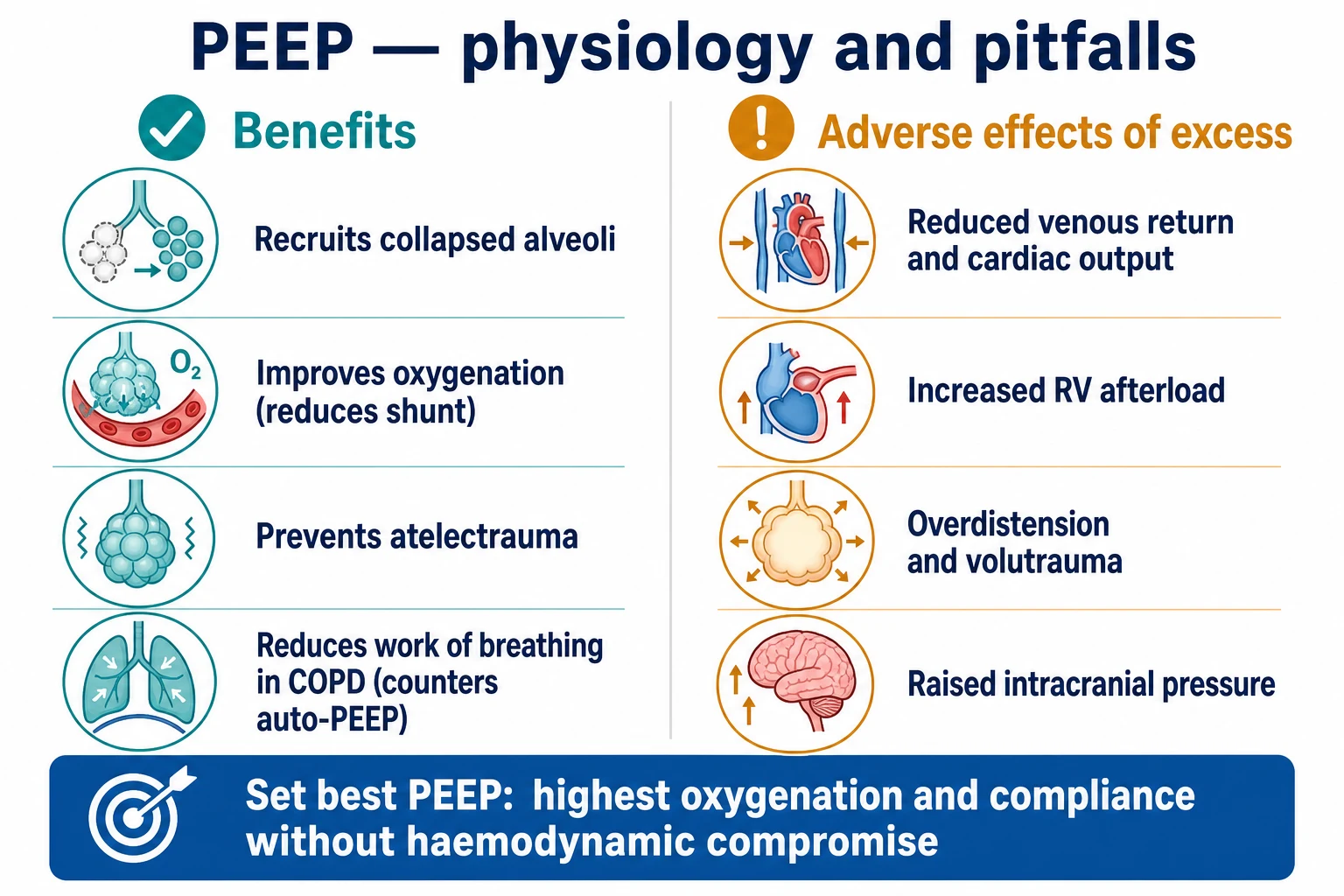
Positive end-expiratory pressure (PEEP) is positive pressure maintained in the airways above atmospheric at end-expiration. It recruits collapsed alveoli, raises functional residual capacity, improves oxygenation by reducing shunt, and prevents the cyclic collapse-reopening of atelectrauma, which is central to lung-protective ventilation (the ARDSnet PEEP/FiO2 tables). Excessive PEEP reduces venous return and cardiac output, raises RV afterload, overdistends alveoli (volutrauma), and raises intracranial pressure. 'Best PEEP' maximises oxygenation and compliance without haemodynamic compromise. Intrinsic (auto-) PEEP from incomplete expiration in COPD and asthma causes hyperinflation; applying external PEEP at about 75-80 per cent of auto-PEEP counteracts the inspiratory threshold load.
On this page & tools
Your progress
Saved locally on this device.
Target exams
Overview & definition
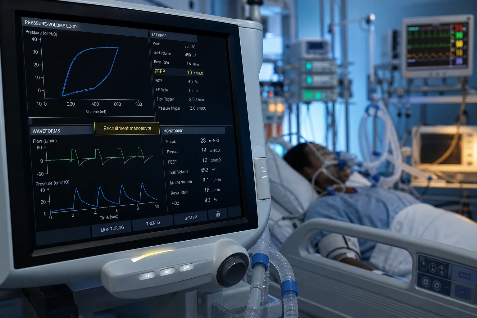
Positive end-expiratory pressure (PEEP) is positive pressure maintained in the airways at end-expiration, above atmospheric. It is set on the ventilator (applied, or extrinsic, PEEP) via the expiratory valve. Intrinsic (auto-) PEEP is positive pressure that builds dynamically within the lungs at end-expiration when expiration is incomplete (air-trapping). PEEP's central role is to recruit and hold open alveoli, improving oxygenation and reducing ventilator-induced lung injury.[1][1]
Physiology — what PEEP does
PEEP exerts its effects through the alveoli:[1]
- Recruits collapsed alveoli and increases the functional residual capacity (FRC), holding alveoli open throughout the respiratory cycle.
- Improves oxygenation by raising mean airway pressure, improving ventilation-perfusion matching, and reducing the shunt fraction (blood passing through non-ventilated, collapsed alveoli).
- Prevents atelectrauma — the repetitive opening and closing (shear injury) of unstable alveoli at low lung volumes. Combined with a low tidal volume, PEEP is central to lung-protective ventilation.[1]
- Reduces the work of breathing in obstructive disease by counteracting the inspiratory threshold load of intrinsic PEEP (below).[1]
The mechanism of alveolar recruitment — Laplace, surfactant, and the time constant
PEEP does not "pop open" alveoli in an instant. Recruitment is the gradual reopening of collapsed alveolar units as a transpulmonary pressure (the airway pressure minus the pleural pressure, Pₗ) exceeds their opening pressure. Once open, alveoli are held open at a lower closing pressure — the lung exhibits hysteresis on its pressure-volume curve, so the pressure needed to keep alveoli patent is less than the pressure needed to reopen them.[1][10]
Two biophysical principles govern this: [1]
- The law of Laplace ($P = 2T/r$). A small alveolus (low $r$) requires a higher pressure to keep open than a large one. In the surfactant-deficient injured lung, surface tension ($T$) is high and the radius small, so collapsed alveoli need substantial transpulmonary pressure to reopen. PEEP holds recruited alveoli above their closing volume, preventing the radius from shrinking to the point where Laplace pressure overwhelms the re-expansion force.[1]
- Surfactant function. PEEP preserves surfactant by reducing the cyclic surface-area change (tidal deflation-reinflation) that squeezes and inactivates surfactant molecules. Atelectatic alveoli, once collapsed, also show surfactant dysfunction — so PEEP that prevents closure also preserves the surfactant film.[1]
- Regional time constants. Lung units empty and fill at different rates ($\tau = R \times C$). In injured lung, fast units fill first and overdistend while slow units (high resistance, low compliance) fill late — a major cause of injurious stress at high PEEP. This heterogeneity is why "one PEEP for all units" cannot be optimal, and why regional tools (EIT, transpulmonary pressure) are attractive.[10]
The recruitable lung volume is finite. Gattinoni's landmark CT study showed that recruitability varies enormously between patients — from near-zero in focal/non-dependent disease to over 50 per cent in diffuse ARDS. A lung that recruits little cannot benefit from high PEEP; it can only be overdistended by it. This is the conceptual basis for individualised PEEP rather than a blanket high-PEEP strategy.[10]
The four mechanisms by which PEEP improves oxygenation
Alveolar recruitment → reduced shunt
Collapsed, fluid-filled, or atelectatic alveoli in dependent regions are reopened and held open through the respiratory cycle. Blood that previously shunted past these non-ventilated units now makes contact with gas, so the shunt fraction falls and PaO2 rises. This is the dominant oxygenation mechanism in ARDS and lobar pneumonia. Recruitment is greatest in the first 30-60 minutes after a PEEP change and plateaus thereafter — a PEEP trial that is judged too early will under-estimate its effect.<Cite id="10" />
Increased functional residual capacity (FRC)
PEEP lifts end-expiratory lung volume above the closing volume of small airways, keeping them patent throughout the cycle. A higher FRC places the lung on a stiffer, more linear portion of the P/V curve (higher compliance) and reduces the work spent reopening collapsed units each breath. This also increases the alveolar oxygen reservoir, buffering against transient desaturation.
Improved V/Q matching
By redistributing ventilation from overdistended non-dependent units toward recruited dependent units (where blood flow is greatest), PEEP narrows the spread of ventilation-perfusion ratios. Low-V/Q units (perfused, poorly ventilated) are improved; high-V/Q dead-space units (ventilated, poorly perfused) are reduced. The net effect is a fall in both shunt and dead space, and a more efficient gas exchange surface.<Cite id="1" />
Reduced work of breathing (in obstruction)
In COPD/asthma with intrinsic PEEP, applying external PEEP at ~80 per cent of auto-PEEP counterbalances the trapped alveolar pressure upstream of a flow-limited choke point. The inspiratory threshold load (the negative pressure the patient must first generate to "climb" the auto-PEEP) falls, ineffective triggering resolves, and the work of breathing drops. This mechanism is specific to flow-limited obstruction — it does not apply to fixed upper-airway obstruction.<Cite id="1" />
| Lung unit state | Response to PEEP | Clinical correlate | The risk |
|---|
| Lung unit state | Response to PEEP | Clinical correlate | The risk |
|---|---|---|---|
| Collapsed / atelectatic (recruitable) | Reopens at a threshold pressure; oxygenation and compliance rise | Dependent ARDS, lobar pneumonia, post-operative atelectasis | If PEEP is too LOW: stays closed → atelectrauma and shunt |
| Open and compliant (normal) | Minimal further recruitment; small compliance change | Non-dependent healthy lung adjacent to injury | If PEEP is too HIGH: overdistension, volutrauma, dead space rises |
| Open but stiff (fibrotic / late ARDS) | Little recruitment; high opening pressure | Late/organising ARDS, fibrosis | High PEEP adds stress without benefit; RV afterload rises |
| Already overdistended | Further pressure worsens stress; capillaries compressed | Tension pneumothorax, breath-stacking in asthma | Barotrauma, RV failure, reduced cardiac output |
| Flow-limited obstructed (COPD) | External PEEP ≈ 80% of auto-PEEP relieves threshold load | COPD with dynamic hyperinflation | External PEEP > auto-PEEP adds to hyperinflation |
Prevention of cyclic atelectrauma — the VILI pillar PEEP addresses
Ventilator-induced lung injury (VILI) has four recognised mechanisms: volutrauma (overstress from excessive transpulmonary pressure/tidal volume), atelectrauma (shear injury from cyclic opening-closing), barotrauma (extra-alveolar air), and biotrauma (inflammatory mediator release). PEEP is the specific counter to atelectrauma. When unstable alveoli collapse at end-expiration and are wrenched open with the next tidal breath, the shear stress at the alveolar-capillary interface and at the junction between open and closed units can exceed 100 cmH2O locally — far above the airway pressure reading — because of the geometry of the boundary. Setting PEEP above the lower inflection point of the P/V curve (or, more practically, to the level that minimises driving pressure) keeps these units open throughout the cycle and removes the injurious stress.[1][1]
Critically, atelectrauma prevention works synergistically with low tidal volume: the ARDSNet bundle is not two independent interventions but a coherent protective strategy. PEEP without low Vt overdistends; low Vt without adequate PEEP permits derecruitment and atelectrauma. The combination — low Vt, adequate PEEP, plateau <30, driving pressure minimised — is the only ventilator intervention proven to reduce mortality in ARDS.[1]
Adverse effects of excessive PEEP
Too much PEEP is harmful:[1]
- Reduced venous return and cardiac output — raised intrathoracic pressure reduces the venous return gradient, causing hypotension (worse in hypovolaemia).
- Increased pulmonary vascular resistance and RV afterload — overdistended alveoli compress the alveolar capillaries, raising RV afterload and precipitating acute cor pulmonale (RV dilatation and septal shift on echo).
- Overdistension (volutrauma) — PEEP that overdistends open alveoli causes injurious lung stress.
- Barotrauma — pneumothorax (less common with modern low-tidal-volume strategies).
- Reduced organ perfusion — renal and splanchnic flow fall with a reduced cardiac output.
- Raised intracranial pressure — reduced cerebral venous drainage; caution in TBI.[1]
The haemodynamic cascade — why a PEEP change can drop the blood pressure in seconds
The cardiovascular impact of PEEP is the single most important practical hazard. Raised intrathoracic pressure reduces venous return (the gradient from the systemic venous reservoir to the right atrium), raises right ventricular afterload (overdistended alveoli compress intra-alveolar capillaries, creating "zone 1/zone 2" physiology), and may reduce biventricular contractility through neurohumoral and mechanical effects. The net effect is a fall in stroke volume and cardiac output, most pronounced in the hypovolaemic or vasodilated patient and in those with pre-existing RV dysfunction.[1]
The bedside test: if raising PEEP drops the blood pressure, the lungs and circulation are competing. Reduce the PEEP, give fluid if hypovolaemic, reassess the right heart on echo (RV dilatation, septal shift, TR velocity — the markers of acute cor pulmonale), and treat the cause. Never assume "it's just sedation". A trial of reducing PEEP by 3-5 cmH2O with haemodynamic observation is a quick, reversible, and often diagnostic manoeuvre.[1]
The haemodynamic cascade of excessive PEEP — from intrathoracic pressure to shock
Raised intrathoracic pressure
PEEP transmits directly to the pleural space (less so in the stiff chest wall). Mean intrathoracic pressure rises by approximately the change in mean airway pressure, modified by chest-wall compliance. This is the proximate insult.
Reduced venous return
The pressure gradient from the extrathoracic venous system (systemic venous reservoir) to the right atrium falls. Right atrial pressure measured by CVP rises, but this is a false "full" — it reflects downstream pressure, not preload. True preload (the gradient) drops. Stroke volume falls. Worst in hypovolaemia, vasodilation (sepsis), and high PEEP with low chest-wall compliance.
Increased RV afterload
Overdistended alveoli stretch and compress the intra-alveolar capillaries ("West zone 1" creation), sharply raising pulmonary vascular resistance (PVR). The thin-walled RV is afterload-intolerant: it dilates, the interventricular septum shifts leftward (paradoxical/bowtie septum on echo), LV filling is impaired, and acute cor pulmonale develops. Tricuspid regurgitation worsens.
Reduced LV preload + biventricular interdependence
Septal shift impairs LV diastolic filling; the pericardial constraint amplifies this. Cardiac output falls further. The patient now has RV failure, LV underfilling, and falling systemic pressure — easily mistaken for LV failure or sepsis, but the cause is the ventilator.
Reduced organ perfusion — renal, splanchnic, hepatic, cerebral
Falling cardiac output and rising intrathoracic/CVP reduce the perfusion gradient to every organ. Renal venous congestion and reduced renal arterial flow → oliguria and creatinine rise (a PEEP-induced AKI that mimics ATN). Splanchnic perfusion falls, risking ischaemic hepatitis and gut barrier dysfunction. Cerebral venous drainage is impeded → raised ICP.
| Organ system | Mechanism of harm | Bedside sign | What to do |
|---|
| Organ system | Mechanism of harm | Bedside sign | What to do |
|---|---|---|---|
| Heart (LV preload) | Reduced venous return gradient; raised intrathoracic pressure | Hypotension, low pulse pressure, cool peripheries after a PEEP increase | Reduce PEEP; give fluid bolus if hypovolaemic; reassess |
| Heart (RV) | Raised PVR from alveolar overdistension; acute cor pulmonale | Rising CVP out of proportion to wedge pressure; RV dilatation, septal shift, TR on echo | Reduce PEEP; correct hypoxaemia/acidosis (raise PVR); consider inotropes/pulmonary vasodilators |
| Kidney | Reduced renal blood flow + renal venous congestion; raised intra-abdominal pressure | Oliguria, rising creatinine, sodium retention (RAAS activation) | Optimise haemodynamics; do NOT chase a urine output with more fluid if venous congested |
| Liver / splanchnic | Reduced portal and hepatic arterial flow; hepatic venous congestion | Rising transaminases, bilirubin, lactate; ischaemic hepatitis | Treat the haemodynamic cause; reduce PEEP; check for acalculous cholecystitis |
| Brain | Reduced cerebral venous drainage → raised ICP; reduced CPP if BP falls | Rising ICP (if monitored); worsening neurological exam | Avoid high PEEP in TBI; elevate head of bed; maintain MAP/CPP |
| Lung (injured) | Volutrauma — overdistension of open units; raised dead space | Rising plateau and driving pressure for the same Vt; falling compliance | Reduce PEEP; use best-compliance/decremental titration; check for pneumothorax |
Fluid overload and the high-PEEP trap
A subtle but important interaction: patients managed with a high-PEEP strategy tend to be in positive fluid balance, both because the PEEP-induced reduction in cardiac output prompts fluid resuscitation (the "chase the BP with saline" reflex) and because high intrathoracic pressure promotes sodium and water retention (RAAS, ADH, reduced atrial natriuretic peptide). The FACTT trial of conservative vs liberal fluid strategies in ARDS showed that a conservative fluid approach improved lung function and reduced ventilator days without increasing renal failure. Excess fluid worsens pulmonary oedema, increases the dead space and shunt, and raises intra-abdominal pressure — which in turn pushes the diaphragm up, raises pleural pressure, and necessitates even higher airway pressures to achieve the same transpulmonary pressure. Breaking this vicious cycle requires diuresis/ultrafiltration and a lower-rather-than-higher PEEP philosophy in the volume-overloaded patient.[1]
The hepatorenal dimension
Persistently high intrathoracic pressure has been linked to a "hepatorenal-like" syndrome in prolonged critical illness: hepatic congestion from reduced venous return and raised CVP impairs synthetic and metabolic function, while splanchnic and renal venous engorgement drive sodium retention and oliguria. In the patient on high PEEP who develops unexplained rising creatinine and bilirubin, consider that the ventilator settings may be contributing — not every organ failure in ICU is from the primary disease.[1]
Setting PEEP — best PEEP
There is no single best method; clinical practice combines several:[1][1]
- The ARDSnet PEEP/FiO2 tables — a pragmatic, empirical titration of PEEP to the FiO2 needed for oxygenation (lower and higher-PEEP tables), paired with low tidal-volume ventilation. The original ARDSnet trial (NEJM 2000) established lung-protective ventilation; the higher-PEEP trials (ALVEOLI 2004; LOVS and ExPress 2008) showed a modest benefit of higher PEEP in moderate-severe ARDS but no benefit in mild disease.[1]
- Best oxygenation — the PEEP that maximises the PaO2 (or SpO2) without haemodynamic compromise.
- Best compliance — the PEEP at which the static respiratory-system compliance is highest (and the driving pressure, ΔP, lowest) for a given tidal volume; minimising driving pressure is itself associated with survival.[1]
- Pressure-volume (P/V) curve — historically, PEEP was set just above the lower inflection point (the alveolar recruitment threshold); less used now.
- Recruitment manoeuvre then a decremental PEEP trial — recruit the lung, then step PEEP down to find the point at which it derecruits (and set PEEP just above it). The ART trial (JAMA 2017) cautioned that aggressive recruitment plus very high PEEP may harm.[1]
- Oesophageal-pressure (transpulmonary-pressure) guided — set PEEP so the end-expiratory transpulmonary pressure is around zero or positive, avoiding lung collapse.[1]
- Electrical impedance tomography (EIT) — regional PEEP optimisation.[1]
In practice: start at about 5 cmH2O in the non-ARDS lung, titrate by the ARDSnet table or best compliance in ARDS, and reassess the haemodynamics and the right heart at each change.[1]
Optimal PEEP determination — the methods compared
The quest for "best PEEP" predates modern ARDS care. Suter's 1975 study defined best PEEP as the level that maximised oxygen delivery (the product of arterial oxygen content and cardiac output) without a fall in compliance — a "total oxygen delivery" endpoint that anticipated by 50 years the modern preoccupation with both lung and circulation.[11] Today the candidate endpoints cluster into four families, each measuring a slightly different thing, and each with a limitation:
| Endpoint family | What it optimises | The target | The blind spot |
|---|
| Endpoint family | What it optimises | The target | The blind spot |
|---|---|---|---|
| Best oxygenation | PaO2, PaO2/FiO2, SpO2 | The PEEP that maximises PaO2 (or allows the lowest FiO2) | Chasing PaO2 ignores compliance, dead space, and the circulation; can push the lung into overdistension for a marginal O2 gain |
| Best compliance / lowest driving pressure | Static respiratory-system compliance (Crs); ΔP = Pplat − PEEP | The PEEP that maximises Crs (minimises ΔP) for the set Vt | Crs includes the chest wall; in obesity/abdominal hypertension it under-estimates lung stress. The best-validated mortality-surrogate endpoint.[7] |
| Lowest dead space | Vd/Vt (Bohr; or ventilator-derived volumetric capnography) | The PEEP that minimises wasted ventilation | Less routinely available; complements compliance rather than replacing it |
| Best gas exchange + circulation | Oxygen delivery (DO2), mixed venous saturation, lactate | The PEEP that maximises DO2 (CaO2 × CO) | Requires a PA catheter or serial cardiac-output monitoring; the most physiological and least used in practice |
Best compliance and driving pressure — the mortality-surrogate endpoint
For a given tidal volume, the driving pressure (ΔP = plateau pressure − PEEP) is the ratio of the tidal volume to the static respiratory-system compliance (ΔP = Vt / Crs). It is the variable that most directly reflects the dynamic strain imposed on the lung per breath. In Amato's landmark re-analysis of nine RCTs, ΔP was the ventilator variable most tightly associated with survival — more than Vt, plateau pressure, or PEEP individually. The PEEP setting that minimises ΔP (i.e. maximises compliance for the given Vt) is the practical, bedside, repeatable best-PEEP target: it identifies the point where recruitment (good) tips into overdistension (bad).[7]
How to do it at the bedside: hold Vt constant at 6 mL/kg predicted body weight. Step PEEP up in 2 cmH2O increments from 5 to a safe ceiling (plateau <30). At each step, perform a 0.5-second inspiratory hold to read the plateau pressure and calculate ΔP. The best PEEP is the level at which ΔP is lowest. If ΔP rises as PEEP is added, the lung is being overdistended — go back down. This takes 5 minutes and a single hold at each level.[7]
Decremental PEEP after a recruitment manoeuvre — recruit, then find the closing pressure
This strategy (associated with the work of Borges, Amato and the ART investigators) proceeds in two phases. First, recruit the lung with a sustained inflation (e.g. 40 cmH2O for 40 seconds) or a stepwise ramp, opening all recruitable units. Second, step PEEP down from a high level (e.g. 20-25 cmH2O) in 2-3 cmH2O decrements, watching compliance, oxygenation, and (if available) EIT. The point at which compliance/oxygenation abruptly falls is the closing pressure — the lung is derecruiting. Set PEEP 2 cmH2O above that point.[8][10]
The logic exploits hysteresis: once recruited, alveoli stay open at a lower pressure than the opening pressure, so the optimal "keep-open" PEEP lies below the recruitment pressure. The method is conceptually elegant and works in the recruitable lung. The ART trial tempered enthusiasm by showing that an aggressive, prolonged recruitment plus very high PEEP strategy increased 28-day mortality in moderate-severe ARDS — likely from haemodynamic compromise, RV failure, and overdistension in patients with low recruitability. The lesson: recruit gently, titrate PEEP down to physiology, and abandon the strategy if the circulation deteriorates.[8]
The decremental PEEP trial — step by step
Confirm the patient is recruitable and stable enough
Best in moderate-severe ARDS (PaO2/FiO2 <200) with confirmed recruitable lung (CT, EIT, or a rise in compliance/oxygenation with a test recruitment). NOT in shock, severe acidosis, RV failure, or a clearly non-recruitable (focal) pattern. Ensure adequate sedation/paralysis to allow the holds.
Perform a gentle recruitment manoeuvre
A stepwise ramp (e.g. PCV with driving pressure 15, PEEP rising in 5 cmH2O steps to 25-30 over 2 min) or a modest sustained inflation (30-40 cmH2O for 30-40 s). Stop immediately for haemodynamic compromise (MAP drop, desaturation, arrhythmia). Avoid the aggressive 50 cmH2O/2 min manoeuvre tested in ART.<Cite id="8" />
Set PEEP high (20-25 cmH2O) and step down in 2-3 cmH2O decrements
At each PEEP level, allow 3-5 min equilibration, then measure: oxygenation (PaO2/SpO2), static compliance and ΔP (inspiratory hold), and (if available) EIT regional compliance or end-expiratory lung volume. Hold the ventilator settings constant between steps except for PEEP.
Identify the derecruitment (closing) pressure
The closing pressure is the PEEP level at which compliance or oxygenation abruptly falls (typically a >10% drop in Crs or a >10% fall in PaO2), or where EIT shows dependent derecruitment. Above this, the lung is recruited; below it, alveoli are closing.
Set PEEP 2 cmH2O above the closing pressure
This is the "best PEEP" — high enough to keep the lung open through the cycle, low enough to avoid overdistension and haemodynamic compromise. Re-check the haemodynamics, the right heart (echo), and the driving pressure. Reassess daily as the lung disease evolves.
Abort criteria
Stop and revert if: MAP drops >20% and does not recover with fluid/vasopressor; new RV failure or septal shift on echo; desaturation despite recruitment; pneumothorax or air leak. The strategy is a tool, not a religion — the patient comes first.<Cite id="8" />
Oesophageal pressure (transpulmonary pressure) guidance
The airway pressure is only half the story; the transpulmonary pressure (Pₗ, airway minus pleural pressure) is the true distending stress on the lung. Pleural pressure is estimated with an oesophageal balloon catheter, which sits in the mid-lower oesophagus and reads the intra-pleural pressure (with cardiac pulsations and small offsets in the supine patient that must be acknowledged). The strategy, tested in the EPVent trial, sets PEEP so that the end-expiratory transpulmonary pressure is zero or slightly positive — neither collapsing (negative Pₗ) nor grossly overdistending (very positive Pₗ). This is the most physiologically direct method and is particularly valuable in the obese patient (high chest-wall pressure compresses the lung), in abdominal hypertension, and in asymmetric ARDS.[6]
EPVent (Talmor, NEJM 2008) found that oesophageal-pressure-guided PEEP setting improved oxygenation and trended toward lower mortality (though it was underpowered and the larger EPVent-2 trial did not confirm a mortality benefit). It remains a powerful physiological tool for the difficult case — the patient in whom empirical PEEP titration gives contradictory oxygenation and compliance signals, or in whom chest-wall mechanics confound the airway pressures.[6]
Electrical impedance tomography (EIT) — regional optimisation
EIT is a non-invasive, radiation-free, real-time bedside imaging modality that measures regional changes in lung air content from a belt of electrodes around the chest. It resolves the fundamental limitation of global endpoints (the lung is heterogeneous): EIT shows, breath by breath, which regions are recruited, which are overdistended, and at what PEEP each transition occurs. A PEEP trial guided by EIT identifies the level that maximises regional recruitment while minimising overdistension — a precision tool.[1]
EIT is most valuable in moderate-severe ARDS, in the obese patient, and in any case where the global signal (compliance, oxygenation) is ambiguous. Its adoption is limited by equipment cost and training. As a research and specialist tool it has reframed how PEEP is understood: "best PEEP" is a regional as much as a global concept, and no single airway pressure can be optimal for every lung unit simultaneously.[1]
The P/V curve and the lower inflection point — historical context
Historically, PEEP was set just above the lower inflection point (Pflex) of the static pressure-volume curve — the pressure at which compliance abruptly improves, interpreted as the opening pressure of collapsed alveoli. While conceptually important (it introduced the recruitment idea to clinical practice), the method is rarely used now: the curve is laborious to obtain, the inflection point is often indistinct, and the static curve does not capture the regional heterogeneity that EIT and transpulmonary pressure reveal. The principle survives in the modern best-compliance/decremental methods, which are effectively dynamic, bedside versions of the same idea.[1]
The ARDSNet PEEP/FiO2 tables — the pragmatic default
When physiology-guided methods are unavailable or impractical, the ARDSNet PEEP/FiO2 tables provide a pragmatic, protocolised titration of PEEP to the FiO2 the patient needs. The logic is simple: a lung that needs more oxygen (higher FiO2) is sicker and deserves more PEEP. The tables come in two versions — a lower-PEEP table (the original ARDSNet approach) and a higher-PEEP table (the strategy refined by the higher-PEEP trials). Both are paired with low tidal-volume ventilation (6 mL/kg predicted body weight) and plateau pressure <30 cmH2O.[1]
Lower-PEEP table (ARDSNet 2000) — pairs low PEEP with rising FiO2: [1]
| FiO2 | 0.30 | 0.40 | 0.50 | 0.60 | 0.70 | 0.80 | 0.90 | 1.00 |
|---|---|---|---|---|---|---|---|---|
| PEEP (cmH2O) | 5 | 5-8 | 8 | 10 | 10 | 10-12 | 12-14 | 14-16 |
Higher-PEEP table (refined by ALVEOLI, LOVS, ExPress) — pairs higher PEEP with the same FiO2 ladder, targeting an "open lung" approach: [1]
| FiO2 | 0.30 | 0.40 | 0.50 | 0.60 | 0.70 | 0.80 | 0.90 | 1.00 |
|---|---|---|---|---|---|---|---|---|
| PEEP (cmH2O) | 5-8 | 8-10 | 10 | 10-12 | 14 | 14 | 14-16 | 18-20-24 |
Higher vs lower PEEP strategy — what the trials showed
The question of whether a systematically higher PEEP strategy improves outcomes is one of the most studied in critical care. The individual trials (ALVEOLI, LOVS, ExPress) were individually underpowered or neutral, but the consistent direction of effect prompted Briel's 2010 individual-patient-data meta-analysis, which found that higher PEEP was associated with lower mortality in the subgroup with PaO2/FiO2 <200 (moderate-severe ARDS), with no benefit and possible harm in milder disease (PaO2/FiO2 200-300).[2][3][4][5]
The synthesis that emerged: use the higher-PEEP table (or a physiology-guided higher PEEP) in moderate-severe ARDS where the lung is recruitable; use the lower-PEEP table (or PEEP 5) in mild ARDS and in the non-ARDS lung. The ART trial then added an important caveat: a blanket strategy of aggressive recruitment plus very high PEEP is harmful — individualisation to recruitability, compliance, and the circulation is essential.[8]
| Feature | Lower-PEEP strategy | Higher-PEEP strategy |
|---|
| Feature | Lower-PEEP strategy | Higher-PEEP strategy |
|---|---|---|
| Typical PEEP | 5-10 cmH2O | 12-16 cmH2O (up to 20-24 in severe) |
| Best for | Mild ARDS (PaO2/FiO2 200-300), non-ARDS lung, post-op, normally compliant | Moderate-severe ARDS (PaO2/FiO2 <200), recruitable diffuse disease |
| Oxygenation | Adequate for mild disease | Better PaO2/FiO2, lower FiO2 requirement |
| Mortality signal | No benefit of higher PEEP in this group; trial-neutral | Mortality benefit in PaO2/FiO2 <200 (Briel meta)[5] |
| Main risk | Under-recruitment, atelectrauma if too low in sick lung | Overdistension, RV failure, hypotension, fluid retention if applied to non-recruitable lung |
| Caution | Don't under-treat severe disease | Don't apply to mild disease or non-recruitable lung (ART harm)[8] |
The driving-pressure principle — why ΔP matters more than PEEP alone
A re-analysis by Amato (NEJM 2015) reframed how the PEEP setting is judged. Across nine RCTs, the variable most tightly associated with survival was not Vt, PEEP, or plateau pressure alone, but the driving pressure (ΔP = plateau − PEEP = Vt/Crs). This is profound for PEEP setting: adding PEEP is beneficial when it lowers ΔP (recruitment increases compliance) and harmful when it raises ΔP (overdistension decreases compliance). The best PEEP is therefore the one that minimises ΔP — a single bedside number that integrates recruitment and overdistension. The implication: do not chase oxygenation or a table in isolation; check the driving pressure at every PEEP change, and prefer the level with the lowest ΔP.[7]
Intrinsic (auto-) PEEP
Intrinsic PEEP (PEEPi, auto-PEEP) is the positive alveolar pressure remaining at end-expiration when expiration is incomplete — alveoli do not empty fully before the next breath. It arises from:[1]
- Airflow obstruction — COPD and asthma (the narrowed airways slow emptying).
- A short expiratory time — a high respiratory rate, a large tidal volume, or an inverted (high) I:E ratio.
Detection. On the flow-time trace, the expiratory flow does not return to baseline before the next breath; or, with an end-expiratory hold (occluding the expiratory port), the ventilator reads the trapped alveolar pressure.[1]
Adverse effects. Hyperinflation raises the work of breathing (the patient must generate enough negative pressure to overcome the PEEPi before they can trigger a breath — the inspiratory threshold load); it impairs venous return and the circulation; and it risks barotrauma.[1]
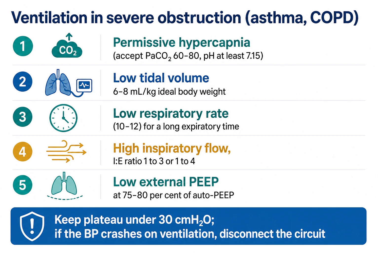
Management. Treat the cause — bronchodilation, a larger endotracheal tube, and ventilator adjustments (a lower respiratory rate, a shorter inspiratory time, a lower tidal volume, a longer expiratory time). Apply external PEEP at about 75-80 per cent of the PEEPi — this counteracts the inspiratory threshold load (the patient triggers more easily) without worsening hyperinflation. (Setting external PEEP higher than the PEEPi would add to the hyperinflation.)[1]
PEEP in COPD and asthma — the 80% rule and the Starling resistor
In obstructive disease, intrinsic (auto-) PEEP is the central mechanical problem, and external PEEP is a targeted therapy — not a recruitment tool. The physiology is distinct from ARDS and the setting rule is different. [1]
The mechanism of auto-PEEP in obstruction. In COPD, the small airways are flow-limited: they collapse during expiration at a choke point (a Starling resistor), so that alveolar pressure exceeds airway-opening pressure throughout expiration. Gas is trapped; end-expiratory alveolar pressure is positive even though the ventilator reads zero at the airway. In asthma, bronchospasm and mucus raise airway resistance uniformly, lengthening the expiratory time constant so the lung cannot empty before the next breath.[1]
The inspiratory threshold load. To trigger a breath, the patient must first generate enough negative intrathoracic pressure to offset the auto-PEEP and then create flow — so much effort is "wasted" before any ventilator assistance arrives. This manifests as ineffective triggering (the patient makes a respiratory effort but the ventilator does not deliver a breath), tachypnoea, and accessory-muscle use. In the spontaneously breathing COPD patient on a ventilator, this load is a major driver of failure-to-wean.[1]
The 80% rule (external PEEP ≈ 80% of auto-PEEP). Applying external PEEP at approximately 75-80 per cent of the measured auto-PEEP counterbalances the trapped alveolar pressure upstream of the flow-limited choke point. Because the choke point acts as a Starling resistor, raising downstream (airway) pressure does not increase flow or lung volume until it exceeds the choke-point pressure — so external PEEP below the auto-PEEP reduces the gradient the patient must overcome (the threshold load falls, triggering becomes effective, work of breathing drops) without adding to hyperinflation. Once external PEEP exceeds the auto-PEEP, the choke point is overcome and lung volume rises — harmful. Hence the ceiling at ~80%.[1]
How to measure auto-PEEP. In a paralysed/sedated patient, an end-expiratory hold (occluding the expiratory port for 0.5-2 s) lets the alveolar pressure equilibrate with the airway, and the ventilator displays the auto-PEEP. In a spontaneously breathing patient the hold under-reads (only the lowest units equilibrate); a more sensitive method is oesophageal manometry or watching the flow-time trace — expiratory flow that does not return to baseline before the next breath signals air-trapping.[1]
| Obstruction type | Mechanism | Effect of external PEEP | Setting |
|---|
| Obstruction type | Mechanism | Effect of external PEEP | Setting |
|---|---|---|---|
| COPD — flow-limited (dynamic airway collapse) | Starling resistor at the small airways; a choke point downstream | Reduces the threshold load WITHOUT raising lung volume, up to the choke-point pressure | External PEEP ≈ 80% of measured auto-PEEP |
| Asthma — fixed high resistance | Diffuse bronchospasm/mucus; uniformly raised resistance, long time constant | Less predictable — reduces the work of triggering but may add volume in some patients; titrate to triggering and haemodynamics | External PEEP ≈ 50-80% of auto-PEEP; start low and observe |
| Fixed upper-airway obstruction (tight ETT, stridor) | No choke point downstream | ADDS to hyperinflation — harmful | Do not apply; treat the cause (change ETT, racemic adrenaline) |
Managing auto-PEEP at the bedside — beyond external PEEP
External PEEP addresses the threshold load but does not treat the underlying air-trapping. The primary manoeuvres are ventilator adjustments to lengthen expiratory time and reduce the volume to be exhaled: [1]
Managing auto-PEEP — the bedside sequence
Recognise — and quantify — the air-trapping
Signs: hypotension (especially after intubation or a rate increase), high plateau pressure, a rising CVP with a narrow pulse pressure, ineffective triggering on the ventilator (retriggered breaths that do not deliver). Confirm with an end-expiratory hold (auto-PEEP) and the flow-time trace (expiratory flow not reaching baseline). In a crashing intubated COPD/asthma patient, suspect a tension pneumothorax AND breath-stacking — they coexist.
Lengthen expiration
Reduce the respiratory rate (e.g. to 8-12/min); shorten the inspiratory time (high inspiratory flow 60-90 L/min; I:E 1:3 or 1:4); accept permissive hypercapnia (pH >7.15-7.20). The single most effective change is usually lowering the rate — it gives each breath more time to exhale.
Reduce the volume to exhale
Lower the tidal volume (4-6 mL/kg predicted body weight). Less volume per breath means less to empty before the next cycle. Watch the minute ventilation and PaCO2 — permissive hypercapnia is the price.
Treat the obstruction
Bronchodilators (nebulised salbutamol/ipratropium; IV salbutamol/magnesium in severe asthma), steroids, treat the COPD/asthma exacerbation. Clear secretions. Consider a larger endotracheal tube if the tube itself is contributing (a #8 is fine for most adults; a #6 adds substantial resistance).
Apply external PEEP at 80% of auto-PEEP
Once air-trapping is quantified and the ventilator is optimised, set external PEEP to ~75-80% of the measured auto-PEEP. This relieves the inspiratory threshold load and improves triggering without worsening hyperinflation. Re-measure auto-PEEP after each change — it shifts as the obstruction and ventilation change.
If haemodynamically crashing — disconnect the circuit
The single most useful manoeuvre in the arresting intubated COPD/asthma patient: disconnect the circuit at the Y-piece and let the trapped gas escape over 15-30 s. The lung deflates, intrathoracic pressure falls, venous return returns, and the blood pressure usually recovers in seconds. Then ventilate gently and exclude a tension pneumothorax.
The intubation hazard — auto-PEEP arrest
A feared scenario: the COPD or asthma patient who arrests or crashes immediately after intubation. The freshly intubated, obstructed patient ventilated at a "normal" rate and tidal volume traps air with every breath — dynamic hyperinflation, rising intrathoracic pressure, falling venous return, and pulseless electrical activity within minutes. The differential is tension pneumothorax vs breath-stacking, and the first move is the same: disconnect the circuit and watch the BP. If it recovers, it was breath-stacking; reduce the rate and tidal volume, allow permissive hypercapnia, and treat the obstruction. If it does not recover, exclude pneumothorax with ultrasound and a chest drain. Intubating the severe COPD/asthma patient is high-risk: start with a low rate (8-10), a low Vt (6 mL/kg), a long expiratory time, and minimal PEEP, and watch the haemodynamics like a hawk.[1]
[1]Red flags
Landmark trials — the PEEP evidence base
The PEEP literature is one of the richest in critical care. The trials below define when higher PEEP helps, when it harms, and how to individualise the setting. Read together, they tell a coherent story: low tidal volume is the foundation; higher PEEP helps the moderate-severe recruitable lung; aggressive recruitment plus very high PEEP harms the non-recruitable lung; and physiology-guided (transpulmonary pressure, driving pressure, EIT) titration is the frontier.[1]
ARDSNet (ARMA)
N Engl J Med 2000
Multicentre RCT, 861 patients with ALI/ARDS — Vt 6 mL/kg PBW (plateau ≤30) vs Vt 12 mL/kg (plateau ≤50)
Key finding
Low Vt REDUCED mortality (31% vs 40%, NNT 11). Established low-tidal-volume lung-protective ventilation paired with the lower-PEEP/FiO2 table as the foundation of ARDS care.
Practice change
Low tidal volume + protocolised PEEP/FiO2 became the universal standard for ARDS — the single best-evidenced ventilator intervention.
ALVEOLI (Brower)
N Engl J Med 2004
Multicentre RCT, 549 patients with ALI/ARDS — higher-PEEP/FiO2 (recruitment + PEEP up to 34) vs lower-PEEP/FiO2 table
Key finding
No difference in mortality or ventilator-free days. Higher PEEP improved oxygenation but not outcomes — the first signal that "more PEEP is always better" is false.
Practice change
Did not establish higher PEEP as routine; prompted the larger LOVS/ExPress trials.
LOVS (Meade)
JAMA 2008
Multicentre RCT, 983 patients with ALI/ARDS — high PEEP (open-lung: recruitment + PEEP up to ~20-24) vs control table
Key finding
No overall mortality difference, but reduced use of rescue therapies and a trend to benefit. Higher PEEP was safe; benefit appeared in the sicker (PaO2/FiO2 <200) patients.
Practice change
Supported higher PEEP in moderate-severe ARDS; contributed to the Briel meta-analysis conclusion.
ExPress (Mercat)
JAMA 2008
Multicentre RCT, 767 patients with ALI/ARDS — PEEP titrated to plateau ~28-30 (high, ~15) vs minimal PEEP (low, ~6)
Key finding
No overall mortality difference. Higher PEEP improved oxygenation and lung compliance and reduced ventilator-free days, with no excess barotrauma. Benefit concentrated in moderate-severe ARDS.
Practice change
Reinforced that higher PEEP is safe and beneficial in moderate-severe ARDS; pooled with LOVS in meta-analysis.
Briel meta-analysis
JAMA 2010
Individual-patient-data meta-analysis of 3 trials (ALVEOLI, LOVS, ExPress), 2299 patients with ALI/ARDS — higher vs lower PEEP
Key finding
Higher PEEP NOT beneficial overall, but significantly LOWER mortality in the PaO2/FiO2 <200 subgroup (moderate-severe ARDS). No benefit and possible harm in mild ARDS (PaO2/FiO2 200-300).
Practice change
Established that higher PEEP should be targeted to moderate-severe ARDS, not applied universally.
EPVent (Talmor)
N Engl J Med 2008
Single-centre RCT, 61 patients with ALI/ARDS — PEEP titrated by oesophageal/transpulmonary pressure (end-expiratory PL ≥0) vs ARDSNet table
Key finding
Transpulmonary-pressure-guided PEEP set higher PEEP and significantly improved oxygenation and compliance; trended toward lower mortality (underpowered). EPVent-2 did not confirm a mortality benefit.
Practice change
Established transpulmonary-pressure-guided PEEP as the most physiological method; a specialist tool for difficult cases.
ART (Cavalcanti)
JAMA 2017
Multicentre RCT, 1010 patients with moderate-severe ARDS — aggressive recruitment (incremental PEEP to 45 + 40 cmH2O 2 min) + decremental high PEEP vs standard low-Vt + lower-PEEP table
Key finding
INCREASED 28-day mortality (16% vs 10% relative increase) and more pneumothorax, barotrauma, and vasopressor use. Trial stopped for harm.
Practice change
CAUTIONED against aggressive recruitment + very high PEEP as a blanket strategy; individualise to recruitability and haemodynamics.
Amato (driving pressure)
N Engl J Med 2015
Re-analysis of 9 RCTs (3562 patients) — association of ventilator variables (Vt, PEEP, plateau, ΔP) with survival
Key finding
DRIVING PRESSURE (ΔP = Pplat − PEEP = Vt/Crs) was the ventilator variable most strongly associated with survival. Each 1 cmH2O rise in ΔP predicted higher mortality, independent of Vt and PEEP.
Practice change
Reframed PEEP setting: the best PEEP is the one that MINIMISES driving pressure — integrates recruitment and overdistension in one bedside number.
SAQ — Setting PEEP in moderate-severe ARDS
10 minutes · 10 marks
A 45-year-old woman with severe pneumonia is intubated for severe ARDS. Ventilator settings: Vt 6 mL/kg PBW, RR 28, PEEP 5, FiO2 1.0. ABG: pH 7.30, PaCO2 50, PaO2 56, HCO3 24. P/F 56. Plateau pressure 24 cmH2O, driving pressure 19 cmH2O. The team asks whether to escalate PEEP and how to do it.
SAQ — Intrinsic (auto-)PEEP in status asthmaticus
10 minutes · 10 marks
A 32-year-old woman with acute severe asthma is intubated for exhaustion. Settings: Vt 500 mL, RR 14, I:E 1:3, PEEP 0, FiO2 0.6. Five minutes after starting ventilation she becomes hypotensive (BP 78/40) and tachycardic (HR 128), SpO2 90 per cent. The peak inspiratory pressure is 38 cmH2O. The ventilator waveform shows the expiratory flow fails to return to baseline before the next breath.
Clinical pearls
Additional red flags
Key facts
References
- [1]The Acute Respiratory Distress Syndrome Network Ventilation with lower tidal volumes as compared with traditional tidal volumes for acute lung injury and the acute respiratory distress syndrome N Engl J Med, 2000.PMID 10793162
- [2]Brower RG, Lanken PN, MacIntyre N, et al. Higher versus lower positive end-expiratory pressures in patients with the acute respiratory distress syndrome N Engl J Med, 2004.PMID 15269312
- [3]Meade MO, Cook DJ, Guyatt GH, et al. Ventilation strategy using low tidal volumes, recruitment maneuvers, and high positive end-expiratory pressure for acute lung injury and acute respiratory distress syndrome: a randomized controlled trial JAMA, 2008.PMID 18270352
- [4]Mercat A, Richard JC, Vielle B, et al. Positive end-expiratory pressure setting in adults with acute lung injury and acute respiratory distress syndrome: a randomized controlled trial JAMA, 2008.PMID 18270353
- [5]Briel M, Meade M, Mercat A, et al. Higher vs lower positive end-expiratory pressure in patients with acute lung injury and acute respiratory distress syndrome: systematic review and meta-analysis JAMA, 2010.PMID 20197533
- [6]Talmor D, Sarge T, Malhotra A, et al. Mechanical ventilation guided by esophageal pressure in acute lung injury N Engl J Med, 2008.PMID 19001507
- [7]Amato MBP, Meade MO, Slutsky AS, et al. Driving pressure and survival in the acute respiratory distress syndrome N Engl J Med, 2015.PMID 25693014
- [8]Cavalcanti AB, Suzumura ÉA, Laranjeira LN, et al. Effect of Lung Recruitment and Titrated Positive End-Expiratory Pressure (PEEP) vs Low PEEP on Mortality in Patients With Acute Respiratory Distress Syndrome: A Randomized Clinical Trial JAMA, 2017.PMID 28973363
- [9]Guérin C, Reignier J, Richard JC, et al. Prone positioning in severe acute respiratory distress syndrome N Engl J Med, 2013.PMID 23688302
- [10]Gattinoni L, Caironi P, Cressoni M, et al. Lung recruitment in patients with the acute respiratory distress syndrome N Engl J Med, 2006.PMID 16641394
- [11]Suter PM, Fairley B, Isenberg MD Optimum end-expiratory airway pressure in patients with acute pulmonary failure N Engl J Med, 1975.PMID 234174