ICU · Respiratory / ventilation
Pleural Disease — Effusion, Pneumothorax & Chest Drains
Also known as Pleural effusion · Pneumothorax · Chest drain · Tube thoracostomy · Light's criteria · Underwater seal · Tension pneumothorax · Safe triangle · Empyema · Re-expansion pulmonary oedema
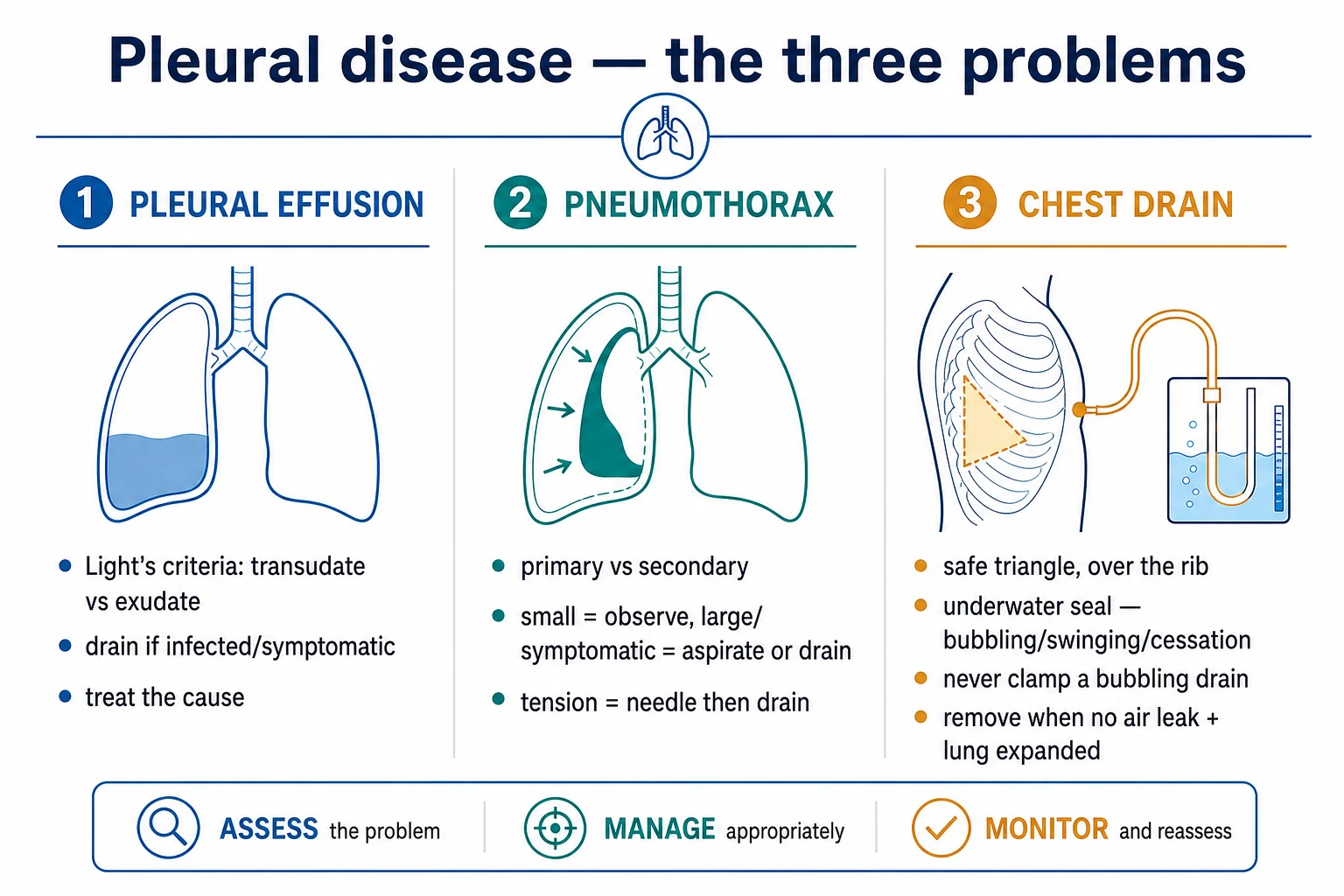
Pleural disease in the ICU covers three problems: pleural effusion (a transudate or an exudate, distinguished by Light's criteria, drained if infected or symptomatic), pneumothorax (primary or secondary; managed by observation, aspiration, or a chest drain; tension pneumothorax is a clinical diagnosis treated by immediate needle decompression then a chest drain), and the chest drain itself (inserted in the safe triangle, over the rib, connected to an underwater seal; bubbling indicates an air leak, swinging indicates patency, and cessation indicates resolution; never clamp a bubbling drain). The chest drain is removed when the air leak has resolved and the lung is expanded on CXR. Re-expansion pulmonary oedema is a risk if a large effusion or pneumothorax is drained too rapidly.
On this page & tools
Your progress
Saved locally on this device.
Target exams
Overview & definition
Pleural disease in the ICU covers three problems: the pleural effusion (fluid in the pleural space), the pneumothorax (air in the pleural space), and the chest drain (the tube thoracostomy that treats both). The BTS guidelines are the standard reference for their management.[1][1]
Pleural effusion
Fluid in the pleural space is classified by Light's criteria:[1]
- Transudate — protein effusion-to-serum ratio under 0.5, LDH ratio under 0.6, and LDH under two-thirds of the upper limit of normal serum LDH. Causes: heart failure, cirrhosis, nephrotic syndrome, hypoalbuminaemia, atelectasis.
- Exudate — fails one or more of the Light's criteria. Causes: infection (parapneumonic effusion, empyema), malignancy (mesothelioma, metastatic), pulmonary embolism, autoimmune (rheumatoid, SLE), pancreatitis.
Management — drain if the effusion is infected (empyema — a chest tube plus antibiotics; consider intrapleural fibrinolytics for loculation), large and symptomatic (dyspnoea), or persistent. A diagnostic tap (thoracentesis) for the unexplained effusion. Treat the underlying cause. Pleurodesis for the recurrent malignant effusion.[1][1]
Pneumothorax
Air in the pleural space, classified by the underlying lung:[1]
- Primary spontaneous — no underlying lung disease; typically tall thin young males; apical blebs.
- Secondary spontaneous — underlying disease (COPD, cystic fibrosis, PCP, LAM).
- Tension pneumothorax — air under pressure, compressing the mediastinum and the great vessels — a clinical diagnosis (hypoxaemia, hypotension, tracheal deviation, absent breath sounds, hyperresonance). Do not wait for the CXR — decompress immediately with a needle (a large-bore cannula in the 2nd ICS mid-clavicular or the 5th ICS mid-axillary), then place a formal chest drain.[1]
Management:[1]
- Small (under 2 cm rim, asymptomatic) — observe, give high-flow oxygen (nitrogen washout).
- Moderate or symptomatic — aspirate (a 16G cannula in the 2nd ICS or the safe triangle) or insert a chest drain.
- Ventilated patient — always drain (the positive pressure converts a small pneumothorax into a tension).[1][1]
The chest drain (tube thoracostomy)
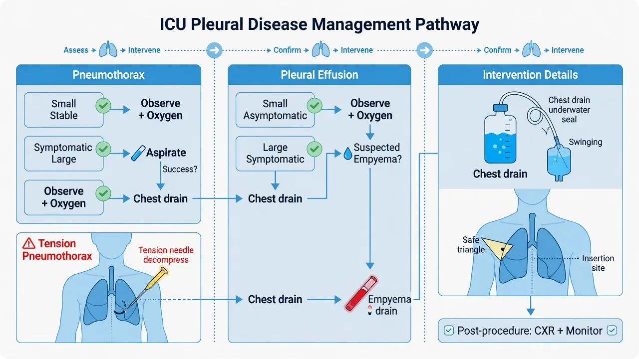
Indications — a pneumothorax (tension after decompression, large symptomatic, or any in a ventilated patient), a haemothorax, a pleural effusion (empyema, large symptomatic), and post-thoracic surgery.[1]
- Site: the safe triangle — anterior to the mid-axillary line, posterior to the lateral border of the pectoralis major, inferior to the axilla, and above the 5th intercostal space (to avoid the diaphragm, the liver, and the spleen).
- Approach: blunt dissection for a large-bore drain (for blood or empyema) or a Seldinger technique for a small-bore drain (for air or a simple effusion).
- The drain passes over the upper border of the rib (the neurovascular bundle runs along the lower border — avoid it).
- Confirm the position with a CXR.
The underwater seal system:[1]
- Bubbling = an air leak (from a bronchopleural fistula or a system leak).
- Swinging (the fluid level moves with respiration) = the drain is patent and in continuity with the pleural space.
- No bubbling = the air leak has resolved (good), or the drain is blocked, kinked, or malpositioned (bad — assess).
- Suction (at minus 10 to minus 20 cmH2O) — for a persistent air leak that does not resolve on the underwater seal alone.
- Clamping — never clamp a bubbling drain (the trapped air causes a tension pneumothorax). Clamp only with a plan (for example, a trial of clamping before removal, or during transport).[1]
Removal — when the air leak has resolved and the lung is expanded on the CXR (for a pneumothorax), or the drainage has reduced to a minimal volume (for an effusion). Remove during a Valsalva manoeuvre (a forced expiration against a closed glottis) to prevent air re-entry.[1]
Complications — pain, infection (cellulitis, empyema), blockage, malposition, re-expansion pulmonary oedema (if a large effusion or pneumothorax is drained too rapidly — the re-expanding alveoli leak fluid), injury to the intercostal vessels, and subcutaneous emphysema.[1]
[1]SAQ — Tension pneumothorax in the ventilated patient
10 minutes · 10 marks
A 28-year-old man with severe ARDS from H1N1 pneumonia is intubated and ventilated (Vt 6 mL/kg PBW, RR 28, PEEP 14, FiO2 1.0). Over 5 minutes he becomes acutely hypotensive (BP 70/40 from 110/70), tachycardic (HR 132), SpO2 falls to 84 per cent, peak airway pressure rises to 48 cmH2O, and there is tracheal deviation to the right with absent breath sounds and hyperresonance over the left hemithorax. He is on vasoactive support and the team is preparing to take him to CT.
SAQ — Pleural effusion in the ICU: transudate vs exudate and parapneumonic effusion
10 minutes · 10 marks
A 65-year-old man with severe community-acquired pneumonia has a large right pleural effusion on CXR and bedside ultrasound showing anechoic fluid with internal septations and a 3 cm rim of consolidated lung. Diagnostic thoracentesis returns thick, turbid, foul-smelling yellow fluid with pH 7.10, glucose 1.2 mmol/L, LDH 1200 U/L and a pleural fluid protein of 4.2 g/dL (serum protein 6.8 g/dL).
Red flags
Pleural effusion in the ICU — expanded
The pleural space normally holds 5–15 mL of lubricating fluid, with a net movement of roughly 1 L per day governed by Starling forces across the parietal and visceral pleura. Any disturbance of hydrostatic pressure, oncotic pressure, capillary permeability, lymphatic drainage, or pleural anatomy produces an effusion. In the ICU, the prevalence is very high — around 60 per cent of mechanically ventilated patients have a clinically detectable effusion on bedside ultrasound within a week, and roughly 8 per cent develop a large effusion (>25 per cent of hemithorax volume). Most ICU effusions are hydrostatic (cardiogenic, hypo-oncotic, atelectatic), but a small fraction are infected, bloody, chylous, or biliary — and these drive the drainage decisions.[1][3]
Light's criteria — the cornerstone classification
Light's 1972 paper remains the most-cited diagnostic study in pleural medicine and the foundation of every exam answer.[1] An effusion is an exudate if any one of three criteria is met; it is a transudate only if all three fail:
- Pleural fluid / serum protein ratio > 0.5
- Pleural fluid / serum LDH ratio > 0.6
- Pleural fluid LDH > two-thirds of the upper limit of normal serum LDH (≈ > 200 IU/L in most labs) [1]
Light's criteria are highly sensitive (~99 per cent) for exudates — they almost never miss an exudative process — but only moderately specific (~78 per cent), so they misclassify some cardiac transudates as exudates (especially after diuresis, when the protein and LDH concentrate). This is the well-known diuretic-treated heart failure trap. Two rescue tests were derived precisely for this situation: [1]
- Serum–pleural fluid albumin gradient — subtract pleural albumin from serum albumin. A gradient > 12 g/L (1.2 g/dL) reclassifies a "pseudoexudate" as a transudate of cardiac or hepatic origin. This is the single most useful rescue test and is the BTS-endorsed method.[3]
- NT-proBNP in pleural fluid or serum — a pleural NT-proBNP > 1500 pg/mL strongly supports a cardiac effusion. This is the BTP (B-type natriuretic peptide / brain natriuretic peptide test) that anchors the modern pleural workup.[3]
The pleural fluid "panel" — what to send and why
Every diagnostic thoracentesis should send the core biochemical, microbiological, and cytological panel. The ICU senior should be able to interpret each vial:[3]
| Test | What it tells you | Cut-off / pattern |
|---|---|---|
| Protein + LDH (fluid & serum) | Light's criteria | See above |
| Glucose | Low in infection, rheumatoid, malignancy, TB | < 3.3 mmol/L — complicated parapneumonic, empyema, RA, TB |
| pH | Low pH = neutrophilic, infected, or malignant | < 7.20 — drain; < 7.30 — re-sample. Measure by blood-gas analyser, not litmus |
| Gram stain + culture | Empirical antibiotic guidance | Aerobes, anaerobes, mycobacteria |
| Cytology | Malignancy | Repeated samples increase yield (~65 % after 3 taps) |
| Amylase | Pancreatic or oesophageal source | High in pancreatic pleural effusion, Boerhaave syndrome |
| Triglycerides | Chylothorax vs pseudo-chylothorax | > 1.1 mmol/L (with chylomicrons) = chylothorax |
| Cholesterol | Pseudo-chylothorax (chronic) | > 5.2 mmol/L, cholesterol crystals |
| ADA (adenosine deaminase) | Tuberculosis (lymphocytic effusion) | > 40 U/L sensitive for TB in endemic areas |
| Differential cell count | Lymphocytic vs neutrophilic vs eosinophilic | Neutrophils acute; lymphocytes chronic; eosinophilia benign/drug |
| Haematocrit (fluid/serum) | Haemothorax | Fluid Hct > 50 % of serum Hct = haemothorax |
Always use a blood-gas analyser for pleural pH and glucose — a bedside pH meter or litmus paper is inaccurate, and air or local anaesthetic in the sample falsely alters the reading. Send pH and glucose in a heparinised syringe, and the rest in the standard containers.[3]
ICU-specific causes — beyond the textbook
In the ICU, the effusion differential narrows to a handful of high-frequency causes, each with a distinctive fluid signature and management implication. [1]
Cardiogenic (heart failure) — the commonest
Right-sided, bilateral, or shifting; a clear transudate (unless diuresed). Confirm with serum–pleural albumin gradient or NT-proBNP. Management is diurese, not drain — a diagnostic tap is reserved for an atypical or unilateral effusion, a febrile patient, or failure to respond after 3 days of aggressive diuresis. Therapeutic taps in heart failure are typically reserved for the symptomatic, diuretic-resistant effusion, and the BTS recommends against routine drainage.[3]
Parapneumonic effusion and empyema — the drainage trigger
Parapneumonic effusions evolve through three classic stages, each with progressively worse biochemistry and progressively stronger indication for a chest drain:[1][4]
- Uncomplicated (exudative) stage — sterile sympathetic fluid, pH > 7.20, glucose > 3.3 mmol/L, LDH < 1000, free-flowing. Treat the pneumonia with antibiotics; the effusion resolves.
- Complicated (fibrinopurulent) stage — bacterial invasion, neutrophil influx, fibrin deposition and loculation; pH < 7.20, glucose < 2.2 mmol/L, LDH > 1000, positive Gram stain. Drain now — a chest tube plus antibiotics. Consider intrapleural tPA/DNase for loculation (see evidence below).
- Empyema (organising) stage — frank pus or positive culture, fibroblast ingrowth with a peal limiting drainage. Requires drainage, often surgical; intrapleural tPA/DNase if unsuitable for surgery. [1]
The BTS pleural infection risk score (RAPS / RAPID score: Renal, Age, Purulence/biochemistry, Infection source, Diabetes) stratifies mortality at 3 months and helps identify the patient who needs the surgical team early.[4]
Haemothorax
Blood in the pleural space. Defined as pleural fluid haematocrit > 50 per cent of the serum haematocrit (a haemothorax has roughly equal Hct to peripheral blood; a haemorrhagic effusion from malignancy or PE is usually 5–40 per cent of serum Hct). Causes in ICU: blunt or penetrating chest trauma, iatrogenic (post-CVC, post-thoracotomy, post-CPR, post-chest drain insertion), anticoagulation, aortic dissection, ruptured pulmonary artery catheter, and spontaneous (rare, on anticoagulants). Drain immediately with a large-bore (28–36 Fr) chest tube — small-bore drains clot and block. A retained haemothorax (incomplete evacuation within days) becomes an empyema or a fibrothorax and mandates VATS. Massive or ongoing haemothorax (> 1500 mL initial drainage or > 200 mL/h for 2–4 h) requires thoracic surgery.[1][1]
Chylothorax
Chyle in the pleural space from thoracic duct disruption. Pleural triglycerides > 1.1 mmol/L (with chylomicrons on electrophoresis) confirm it. The fluid is milky-white, but post-prandial patients look milky; fasted/nil-by-mouth ICU patients may have a clear chylothorax, so send triglycerides whenever the cause is in doubt. Causes in ICU: post-oesophagectomy or post-thoracic surgery (the commonest ICU cause — the duct crosses the chest at T5–T7), central venous thrombosis (occluding lymphatic return), mediastinal tumour (lymphoma), trauma, and congenital. Management: drain (often an IPC for nutrition losses), medium-chain triglyceride diet or NPO with TPN (MCTs are absorbed directly into the portal vein and bypass the duct), and octreotide to reduce lymph flow. Surgical thoracic duct ligation or embolisation is reserved for high-output (> 1 L/day) chylothorax failing 1–2 weeks of conservative care. Watch and replace the nutritional and immunological losses — chylothorax drains T-cells, fat-soluble vitamins, and protein; a chronic drain becomes a profound immunodeficiency and malnutrition syndrome.[1]
Pseudo-chylothorax (cholesterol effusion)
A chronic effusion (present for years) rich in cholesterol, producing a milky appearance but with triglycerides < 1.1 mmol/L and cholesterol crystals on microscopy. Classic causes: chronic rheumatoid pleurisy, tuberculosis, old empyema. Do not confuse it with chylothorax — MCT diet and octreotide are pointless; treat the underlying cause. [1]
Hepatic hydrothorax
Transudative effusion from cirrhosis and portal hypertension, occurring in 5–10 per cent of cirrhotics, usually right-sided (the diaphragmatic defects are more common on the right). May dominate the clinical picture and produce respiratory failure even with minimal ascites. Salt restriction, spironolactone, and albumin are first line; a TIPSS is effective for the refractory case. Avoid a chest drain — the protein and electrolyte losses, the high mortality, and the infection risk make a chronic drain a poor choice; an IPC is occasionally used as a bridge to transplant.[1][3]
Other ICU effusions worth naming
- Atelectatic — commonest ICU effusion of all; a clear transudate from negative pleural pressure pulling fluid in as the lung collapses. Treat the atelectasis (recruitment, physiotherapy), not the effusion.
- Uraemic — serositis with a neutrophilic exudate; resolves with dialysis.
- Pancreatic — left-sided, exudative, amylase > 200 U/L, in pancreatitis or a pancreatic pseudocyst.
- Dressler / post-cardiac injury — a pericardial/pleural reaction days to weeks after MI, surgery, or ablation; NSAIDs or colchicine.
- Pulmonary embolism — virtually any effusion; small, exudative, often haemorrhagic.
- Drug-induced — amiodarone, nitrofurantoin, methotrexate, bromocriptine.
- Post-CABG / post-cardiotomy — common; usually left-sided; differentiate early (< 30 days, bloody, eosinophilic) from late (> 30 days, clear, cardiac transudate).[1]
Drainage options — choosing the right device
| Device | Bore / technique | Best use | Pitfall |
|---|---|---|---|
| Therapeutic thoracentesis | Needle / small catheter | Diagnostic + symptomatic tap, single drainage | Re-accumulation; re-expansion oedema if > 1.5 L drained fast |
| Small-bore (pigtail) chest tube | 8–14 Fr, Seldinger | Air, simple effusion, malignant effusion, IPC | Clots/blocks in blood or pus; requires frequent flush |
| Large-bore chest tube | 24–36 Fr, blunt dissection | Haemothorax, empyema, large air leak (BPF) | More painful, larger scar; neurovascular bundle risk |
| Indwelling pleural catheter (IPC) | 15–16 Fr tunneled (e.g. PleurX) | Recurrent malignant effusion, hepatic hydrothorax bridge | Long-term external device; infection risk lower than expected |
| Pigtail + IPC hybrid | Same as IPC | Same indications | — |
| Surgical (VATS) | General anaesthesia | Loculated empyema, recurrent pneumothorax, BPF | Requires single-lung ventilation and a fit patient |
The modern evidence has reversed the older dogma that "pus and blood need a big tube". Both the BTS guidelines and the Australasian PICO data show small-bore (pigtail) catheters are as effective as large-bore tubes for most pleural infections, with less pain, when used alongside intrapleural tPA/DNase to maintain patency.[4][1] Large-bore tubes remain first-line only for frank pus with debris that blocks a pigtail, and for haemothorax.[1]
Transudate vs exudate — the full Light's-criteria answer side by side
| Feature | Transudate | Exudate |
|---|---|---|
| Light's criteria | Fails ALL three | Meets ANY one |
| Protein fluid/serum | < 0.5 | > 0.5 |
| LDH fluid/serum | < 0.6 | > 0.6 |
| Fluid LDH | < two-thirds ULN | > two-thirds ULN (~200 IU/L) |
| Macroscopic | Clear, straw-coloured | Cloudy, bloody, purulent, milky |
| Common ICU causes | CHF, cirrhosis, nephrotic, atelectasis, hypoalbuminaemia, hepatic hydrothorax, uraemia, myxoedema, peritoneal dialysis | Pneumonia/empyema, malignancy, PE, pancreatitis, rheumatoid/SLE, TB, chylothorax, Dressler, uraemia (occasionally), drugs |
| Albumin gradient (serum–fluid) | > 12 g/L (1.2 g/dL) | < 12 g/L |
| NT-proBNP (BTP) | > 1500 pg/mL (cardiac) | < 1500 pg/mL |
| Glucose | Normal (> 3.3 mmol/L) | Variable; low in infection, RA, TB, malignancy |
| pH | > 7.40 | Variable; < 7.20 = drain |
| Management | Treat the cause; do NOT routinely drain | Drain if infected, large, symptomatic, or persistent; treat cause |
Parapneumonic effusion stages — the drainage decision
| Stage | Pathology | pH | Glucose (mmol/L) | LDH (IU/L) | Gram stain | Drain? |
|---|---|---|---|---|---|---|
| 1. Uncomplicated (exudative) | Sterile sympathetic fluid | > 7.20 | > 3.3 | < 1000 | Negative | No — antibiotics only |
| 2. Complicated (fibrinopurulent) | Bacterial invasion, fibrin, loculation | < 7.20 | < 2.2 | > 1000 | May be positive | Yes — chest tube + antibiotics ± tPA/DNase |
| 3. Empyema (organising) | Frank pus, peal formation | < 7.20 | < 2.2 | > 1000 | Positive (often) | Yes — drain (often surgical) |
Drainage device choice by problem — the ICU bedside logic
| Problem | First-line device | Why | Exception |
|---|---|---|---|
| Cardiogenic effusion | None — diurese | Treats the cause | Diagnostic tap if unilateral/atypical |
| Simple parapneumonic | Small-bore pigtail | Equivalent efficacy, less pain | Switch to large-bore if blocks |
| Frank empyema | Large-bore 24–28 Fr | Debris blocks small tubes | Add tPA/DNase for loculation |
| Haemothorax | Large-bore 28–36 Fr | Small tubes clot | VATS if retained |
| Chylothorax | Small-bore or IPC | Nutrition loss management | Surgical ligation if high-output |
| Malignant (recurrent) | IPC (PleurX) ± talc | Outpatient drainage, auto-pleurodesis | Talc pleurodesis if patient ambulant |
| Pneumothorax (ventilated) | Small-bore pigtail | Effective for air | Large-bore for large BPF air leak |
Pneumothorax in the ICU — expanded
Classification by mechanism
| Type | Mechanism | ICU relevance |
|---|---|---|
| Primary spontaneous (PSP) | Apical subpleural bleb rupture in a tall, thin, young, non-smoking male | Post-PSP ICU admission for observation or drainage |
| Secondary spontaneous (SSP) | Underlying lung disease: COPD (commonest), asthma, CF, PCP, LAM, alpha-1-antitrypsin, idiopathic pulmonary fibrosis, sarcoidosis, TB | Higher mortality, higher recurrence, usually drained |
| Traumatic | Blunt (rib fracture lacerating lung) or penetrating | Often associated haemothorax; needs drain, sometimes surgery |
| Iatrogenic | CVC insertion, thoracentesis, transthoracic biopsy, barotrauma (high PEEP, ARDS), CPR, mechanical ventilation | The commonest pneumothorax in ICU — anticipate after every line / drain |
| Tension | Any pneumothorax + a one-way valve → mediastinal shift, obstructive shock | Clinical diagnosis — immediate decompression |
| Catamenial | Endometriosis-related, recurrent, right-sided, in women 30–40 y | Recurrence prevention with hormonal suppression |
Iatrogenic pneumothorax — the ICU hazard
Approximately one in four ICU pneumothoraces are iatrogenic. The big four causes are internal jugular or subclavian central venous catheter insertion (mechanical puncture of the apex — risk falls steeply with ultrasound guidance), barotrauma from positive-pressure ventilation (high plateau pressures, high PEEP, ARDS, status asthmaticus — the classical "volutrauma" of the 1990s), thoracentesis and transthoracic needle biopsy (risk reduced by ultrasound), and CPR (rib fractures). The intensivist's mitigation bundle: ultrasound-guided CVC, lung-protective ventilation (Vt 6 mL/kg, plateau < 30 cmH2O), ultrasound-guided pleural procedures, and post-procedure CXR for any supradiaphragmatic line or drain.[1]
Traumatic pneumothorax
In major trauma, a pneumothorax may be occult (seen only on CT, not CXR), open (a sucking chest wound — needs a three-sided occlusive dressing then a formal drain), or tension (immediate decompression). A traumatic pneumothorax in a ventilated patient always needs a chest drain — positive pressure will convert a small occult pneumothorax into a tension within minutes. The traumatic pneumothorax often coexists with a haemothorax (haemopneumothorax) — use a large-bore tube.[1]
Tension pneumothorax — the updated decompression site
The classical teaching is 2nd ICS mid-clavicular line (MCL) with a 14–16 G cannula. Modern evidence and the BTS / ITLS guidance have shifted: in adults the 5th ICS, anterior axillary line (the same site as a chest drain) is preferred, because the classical 2nd ICS site misses the pleural space in up to half of patients (thick subcutaneous tissue and pectoralis muscle, especially in women and the muscular or obese patient) and the cannula is often too short. Either site is acceptable in the exam answer, but acknowledge the change. After needle decompression, always place a formal chest drain — the needle is a bridge, not a treatment.[1][2]
Management by size and type — the BTS approach
The BTS 2010 pneumothorax guideline (and 2023 update) stratifies management by size (small < 2 cm rim vs large ≥ 2 cm rim, measured at the hilum on a PA CXR) and symptom status, with separate algorithms for PSP and SSP.[2]
Primary spontaneous:
- Small, asymptomatic — observe, high-flow oxygen (nitrogen washout — only in non-COPD patients).
- Small, symptomatic or large — aspiration first (BTS preference) with a 16 G cannula in the 2nd ICS MCL; if it fails or recurs, a small-bore chest drain.
- Large symptomatic — small-bore (pigtail) chest drain; large-bore rarely needed. [1]
Secondary spontaneous:
- Small, asymptomatic — observe in hospital (not outpatient); high-flow oxygen only if not CO2-retaining.
- Symptomatic or large — small-bore chest drain (aspiration has a higher failure rate in SSP and is not preferred). [1]
Tension:
- Immediate needle decompression (5th ICS AAL preferred) → formal chest drain → CXR. [1]
Ventilated patient (any pneumothorax):
- Always drain — positive pressure will convert any pneumothorax to a tension. [1]
Pigtail catheters — the modern default
Small-bore (8–14 Fr) Seldinger pigtail catheters have largely replaced large-bore tubes for air. They are equivalent in efficacy, less painful, smaller scar, and easier to insert, with comparable resolution and recurrence rates. The BTS endorses them as first-line for PSP and SSP drainage and for most iatrogenic pneumothoraces. Their main limitation is in frank pus or blood, where they clot and block.[2][1] Flush the pigtail with 20 mL saline 6–8-hourly to maintain patency, and connect to an underwater seal as for any chest drain.
Bronchopleural fistula (BPF)
A persistent communication between the bronchial tree and the pleural space — the diagnosis whenever a chest drain bubbles continuously for more than 48 hours on the underwater seal. In ICU the commonest causes are post-pneumonectomy or post-lobectomy stump leak (presents days to weeks post-op with fever, cough, and a falling pleural fluid level on CXR), necrotising pneumonia (especially with cavitation — MRSA, Klebsiella, anaerobes), lung abscess rupture, trauma (persistent alveolar–pleural communication), and barotrauma in severe ARDS or status asthmaticus.[1]
Why a BPF is dangerous in a ventilated patient
A BPF in a ventilated patient is one of the most challenging problems in critical care. Each positive-pressure breath escapes through the fistula rather than ventilating the lung — producing a tidal-volume thief, persistent hypoxaemia and hypercapnia, failure to wean, and a risk that the pleural infection seeds proximally. The bigger the fistula and the higher the airway pressures, the larger the leak. [1]
Management — a staged approach
- Drain the pleural space — a functioning chest drain is mandatory; never clamp a BPF drain.
- Reduce airway pressures — the most effective immediate manoeuvre. Lower tidal volume, lower PEEP, allow permissive hypercapnia, switch to pressure-control, or use independent lung ventilation via a double-lumen tube.
- High-frequency jet ventilation (HFJV) or oscillation can reduce mean airway pressure and leak flow when conventional ventilation fails.
- Position the affected side dependent (good lung up) to protect the healthy lung from contamination and to improve V/Q matching.
- Definitive closure — bronchoscopic glue, silver-nitrate, one-way endobronchial valves, or surgical closure (muscle flap, omental patch, completion pneumonectomy). The decision is multidisciplinary with thoracic surgery.[1]
The chest drain (tube thoracostomy) — detailed
Three-bottle system logic
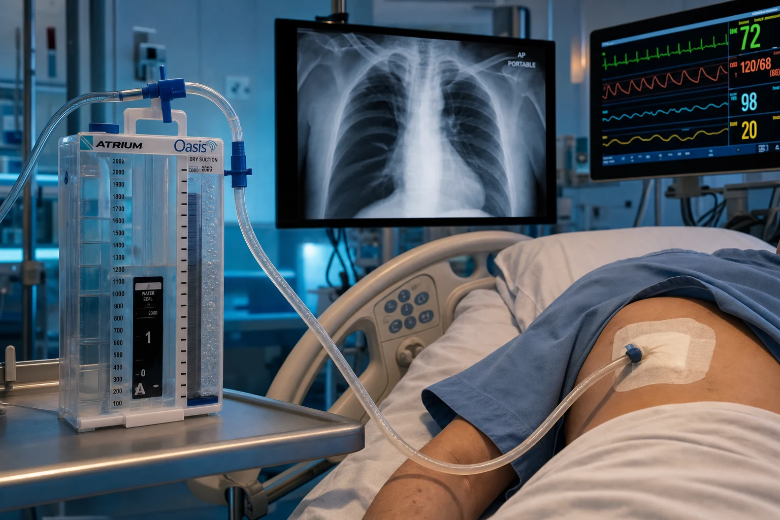
The traditional "bottle" system is conceptualised as three chambers in series, now integrated into a single commercial unit (e.g. Atrium, Pleur-evac):[1]
- Collection bottle — receives pleural fluid and air; sterile.
- Water-seal bottle — a 2 cm column of water through which air bubbles out on expiration (one-way valve). The water level swings with respiration if the system is patent.
- Suction-control bottle — a third column of water set to the desired suction (typically −10 to −20 cmH2O), so the wall suction can be turned up high without over-applying pressure (the water simply bubbles, capping the transmitted vacuum). [1]
Reading the underwater seal
- Bubbling = an air leak. Grade it: grade 1 = forced expiration only (cough); grade 2 = normal expiration; grade 3 = inspiration (the worst — a significant BPF). A new or worsening leak warrants CXR and reassessment.
- Swinging = patent. The fluid meniscus moves 2–6 cm with each breath; if it stops swinging, the tube is blocked, kinked, malpositioned, or the lung is fully expanded against the tube.
- No bubbling, lung expanded = resolved — ready for trial of clamp and removal.
- No bubbling, lung not expanded = blocked or malpositioned — flush, re-image, replace. [1]
Suction
Apply low-pressure suction at −10 to −20 cmH2O for a persistent air leak that does not resolve within 24–48 hours. There is no proven benefit to high suction (it does not heal a fistula faster) and it can worsen a BPF by increasing flow across the defect. BTS guidance: suction is optional, not routine.[2]
Trial of clamping and removal
Before removal, clamp the drain for 4–12 hours (with close observation) and confirm with a CXR that the lung remains expanded. This is the only acceptable reason to clamp a chest drain in ICU — never clamp a bubbling drain. Remove during a Valsalva (or at end-inspiration in a ventilated patient) with a sterile, occlusive dressing.[1][1]
Prophylactic antibiotics for chest drains
BTS guidance: prophylactic antibiotics are NOT routinely required for chest drain insertion in non-trauma patients, but are recommended for trauma (where the infection risk is higher) until 24 hours after drain removal. In ICU, weigh the risk — many patients are already on therapeutic antibiotics.[1]
Complications — full list
- Immediate (insertion): pain, vasovagal, intercostal vessel injury (arterial bleeding — the "intercostal artery pseudoaneurysm" is a delayed hazard), damage to lung (haemopneumothorax), damage to diaphragm/liver/spleen (if site too low), intrathoracic placement of the trocar (large-bore), hypoxaemia, bronchospasm.
- Early (hours-days): blockage, kinking, malposition (intraparenchymal, subcutaneous, intrafissural), subcutaneous emphysema, infection (cellulitis, empyema), re-expansion pulmonary oedema, persistent air leak (BPF).
- Late (weeks): retained haemothorax, empyema, fibrothorax, drain-site hernia, drain-site malignancy seeding (mesothelioma), intercostal artery pseudoaneurysm.[1][1]
Re-expansion pulmonary oedema (REPO) — the avoidable killer
REPO occurs when a large effusion or pneumothorax is drained too rapidly — the rapidly re-expanding alveoli leak proteinaceous fluid because the chronic collapse has remodelled the capillaries and the lymphatics. It is ipsilateral (can become bilateral), develops within minutes to 24 hours, and carries a mortality up to 20 per cent. Prevention: limit a single therapeutic tap to 1.5 L (or stop if the patient develops chest tightness or cough), use a controlled flow rate on a chest drain (rather than free flow), and re-image early. Treatment is supportive — oxygen, diuretics, NIV or intubation for severe cases.[1][1]
Indwelling pleural catheter (IPC, e.g. PleurX)
A tunneled 15–16 Fr catheter placed for outpatient management of recurrent malignant effusion (and occasionally hepatic hydrothorax). The patient or carer drains 600–1500 mL two to three times a week via a vacuum bottle. Auto-pleurodesis occurs in around 40–50 per cent within 60 days, allowing catheter removal. Compared with talc pleurodesis, IPC gives equivalent symptom control and fewer admissions, with a small risk of catheter-tract metastasis (especially mesothelioma) and infection (~5 per cent, usually cellulitis).[1][1][1]
Pleurodesis
Mechanical or chemical obliteration of the pleural space to prevent recurrent effusion or pneumothorax. Options: talc slurry (via chest drain, the most effective agent, ~75 per cent success in malignancy, ~90 per cent in pneumothorax), talc poudrage (insufflated at VATS, the most effective of all), doxycycline or bleomycin (less effective, alternatives when talc contraindicated), and autologous blood patch (for persistent air leak). Talc must be graded (particle size > 15 µm to avoid systemic spread; non-graded talc was linked to ARDS). Contraindications: lung trapped by malignancy (no apposition possible — IPC instead), active pleural infection, severe coagulopathy.[1][1]
Bedside ultrasound (POCUS) — the ICU standard
Ultrasound is the first-line imaging for the ICU pleural space — it outperforms CXR for detecting and quantifying effusion, distinguishes anechoic from septated fluid (predicting drainage success), locates the diaphragm and the safe site for a tap, and rules out pneumothorax in the post-procedural patient (lung sliding + lung pulse exclude pneumothorax with high sensitivity). The BTS mandates ultrasound guidance for every pleural intervention; the era of blind taps is over. The classic POCUS signs: an anechoic space above the diaphragm; atelectatic lung waving within the effusion (jellyfish sign); septa suggest empyema or haemorrhagic fluid; the sinusoid sign on M-mode confirms fluid. For pneumothorax: absent lung sliding, absent B-lines, lung point (the specific sign where sliding meets non-sliding, ~66 per cent sensitive but near 100 per cent specific).[3][1]
ICU pleural effusion — the diagnostic and management algorithm
Detect (POCUS + CXR)
Bedside ultrasound for every unexplained respiratory deterioration; quantify (small, moderate, large); identify anechoic vs septated; check the diaphragm and the heart.
Decide: transudate pattern or not?
Bilateral, symmetric, clear effusion in a cardiac/failure/cirrhotic/low-albumin patient = transudate pattern → treat the cause, do NOT drain. Atypical (unilateral, septated, febrile, no obvious cause) → diagnostic tap.
Diagnostic thoracentesis (ultrasound-guided)
Send the full panel: protein, LDH (fluid + serum), glucose, pH by blood-gas analyser, Gram + culture, cytology, amylase, triglycerides, cell count. Add ADA if TB suspected, NT-proBNP (BTP) if cardiac.
Apply Light's criteria + albumin gradient + BTP
Exudate if ANY Light criterion met; rescue with serum–pleural albumin gradient > 12 g/L (or NT-proBNP > 1500 pg/mL) for the diuretic-treated cardiac case.
Decision: drain or not?
Drain if pH < 7.20, glucose < 3.3, frank pus, loculated, large/symptomatic, or to relieve respiratory failure. Do not drain a transudate or an uncomplicated parapneumonic effusion.
Choose device
Pigtail for air/simple effusion/malignant; large-bore for pus-with-debris or haemothorax; IPC for recurrent malignant. Insert in the safe triangle, ultrasound-guided, over the upper rib border.
Manage the drain
Underwater seal; grade the air leak; suction −10 to −20 cmH2O only for persistent leak; flush pigtail 6–8-hourly; daily CXR; for empyema add tPA/DNase for loculation.
Remove
When the air leak resolves and the lung is expanded (pneumothorax), or drainage < 200 mL/day (effusion). Trial of clamp 4–12 h, CXR, remove on Valsalva with an occlusive dressing.
Tension pneumothorax — immediate management in the ICU
Recognise the clinical diagnosis
Hypoxaemia, hypotension, tachycardia, absent breath sounds, hyperresonance, tracheal deviation (late), increased airway pressures (in the ventilated patient). DO NOT wait for the CXR.
Call for help and 100% oxygen
Increase FiO2 to 1.0; call the senior and the thoracic team; prepare a chest drain trolley.
Immediate needle decompression
Large-bore (14–16 G) cannula in the 5th ICS, anterior axillary line (preferred) OR 2nd ICS mid-clavicular line. A rush of air confirms the diagnosis. The cannula is a bridge, not a treatment.
Formal chest drain
Small-bore pigtail (air) or large-bore (BPF/haemopneumothorax) in the safe triangle, over the upper rib, to an underwater seal.
Confirm with CXR
Post-decompression CXR to confirm re-expansion and drain position. Re-assess the patient; if shock persists look for an alternative or concomitant cause.
Investigate the cause
In the ventilated patient: barotrauma (review PEEP and plateau pressure), iatrogenic (post-line), or underlying lung disease. Prevent recurrence with lung-protective ventilation.
Bronchopleural fistula in the ventilated patient — management ladder
Confirm
Persistent chest-drain bubbling > 48 h, or new large air leak in a ventilated patient. Differentiate from a system leak (submerge the connections).
Drain and never clamp
Ensure a functioning chest drain; never clamp a BPF drain (tension pneumothorax).
Reduce airway pressure
Lower Vt (4–6 mL/kg), lower PEEP, allow permissive hypercapnia; switch to pressure-control; consider independent lung ventilation via double-lumen tube.
Position good lung up
Lateral decubitus with the BPF side down to protect the healthy lung from pleural contamination and improve oxygenation.
Advanced ventilation
High-frequency jet ventilation or oscillation if conventional ventilation fails; ECMO as a last resort for refractory hypoxaemia.
Definitive closure
Bronchoscopic glue / valves / silver nitrate; surgical muscle-flap or omental-patch closure. Multidisciplinary with thoracic surgery.
Evidence and trials
Pleural effusion and pneumothorax — the landmark evidence
Light 1972 (Ann Intern Med): the original derivation of Light's criteria — defined the three exudate thresholds (protein ratio > 0.5, LDH ratio > 0.6, fluid LDH > two-thirds ULN) on 150 effusions, and they remain the most sensitive classification in use. The single most-cited paper in pleural medicine.[1] BTS Pleural Disease Guideline 2010 (Thorax): the four-part guideline series — investigation of a unilateral pleural effusion (Hooper), management of spontaneous pneumothorax (MacDuff), pleural procedures and thoracic ultrasound (Havelock), and chest-tube insertion (part of the 2010 series). The standard reference for everything in this topic.[3][2][1] MIST1 — Maskell 2005 (NEJM): the first Multicentre Intrapleural Sepsis Trial — randomised 454 patients with pleural infection to intrapleural streptokinase vs saline. No benefit on mortality, surgery, or length of stay. Buried routine streptokinase for empyema.[1] MIST2 — Rahman 2011 (NEJM): the definitive intrapleural-lytic trial — 210 patients randomised in a 2 × 2 factorial to tPA, DNase, both, or placebo. tPA + DNase together reduced hospital stay by ~5 days, reduced surgery, and improved radiographic change. tPA alone or DNase alone were not effective (DNase alone was harmful). Now standard of care for loculated empyema.[4] BTS malignant pleural effusion guideline 2023 (Thorax): updated Roberts et al. — IPCs as first-line for recurrent malignant effusion, talc poudrage (VATS) for ambulant fit patients, and management of trapped lung. Replaced the older talc-first approach.[1] TILT-1 / TIME3 — Bhatnagar 2018 (NEJM): outpatient talc via IPC for malignant effusion — auto-pleurodesis in 43 per cent at 35 days with a single instillation, no excess adverse events. Made talc-via-IPC a viable one-stop outpatient therapy.[1] ACCP Delphi 2001 (Chest) — Baumann: the American College of Chest Physicians consensus on pneumothorax — the source of the alternative (more interventional) approach to PSP, contrasting with the more conservative BTS algorithm.[1] PICO / Australasian pigtail data — Muruganandan et al. 2019 and Cochrane 2017: smaller-bore chest tubes are non-inferior to large-bore tubes for pneumothorax and pleural infection, with less pain — the evidence basis for the modern pigtail-first approach.[1] Wrightson 2009 / Rahman RAPID score (Clin Chest Med): the RAPID score (Renal, Age, Purulence, Infection-source, Diabetes) stratifies 3-month mortality in pleural infection (low ~3 %, high ~35 %) and identifies the patient who needs surgical referral early.[4]
Additional clinical pearls — exam-exhaustive
Light's criteria rescue tests — when the criteria mislead
| Situation | Problem | Rescue test | Result |
|---|---|---|---|
| Cardiac effusion on diuretics | Protein & LDH concentrate → "pseudoexudate" | Serum–pleural albumin gradient | > 12 g/L → transudate (cardiac) |
| Cardiac effusion, unclear cause | Bilateral vs unilateral ambiguity | Pleural NT-proBNP (BTP) | > 1500 pg/mL → cardiac |
| Possible chylothorax | Milky fluid (or clear in fasted patient) | Pleural triglycerides | > 1.1 mmol/L → chylothorax |
| Possible haemothorax | Bloody fluid (malignancy, PE, trauma) | Pleural Hct / serum Hct ratio | > 0.5 → haemothorax |
| Possible pancreatic | Left-sided exudate, no obvious cause | Pleural amylase | > 200 U/L → pancreatic/oesophageal |
| Possible TB | Lymphocytic exudate, endemic | Pleural ADA | > 40 U/L → supports TB |
PSP vs SSP vs iatrogenic vs tension — management at a glance (BTS approach)
| Type | Small (< 2 cm) asymptomatic | Symptomatic or large | Special |
|---|---|---|---|
| PSP | Observe; high-flow O2 (if non-COPD) | Aspirate first (16 G, 2nd ICS MCL); pigtail if fails | Surgical referral after 2nd recurrence |
| SSP | Observe in hospital; O2 only if non-CO2 retainer | Small-bore chest drain | Higher recurrence; consider early surgery |
| Iatrogenic | Observe if small + non-ventilated | Small-bore pigtail | Always drain if ventilated |
| Traumatic (occult) | Observe if non-ventilated | Drain | Always drain if ventilated; large-bore if haemo |
| Tension | n/a | Needle decompression + chest drain | Clinical diagnosis; do NOT wait for CXR |
Chest drain — what each underwater-seal finding means
| Finding | What it means | What to do |
|---|---|---|
| Bubbling, grade 1 (cough only) | Small air leak | Continue observation; reassess daily |
| Bubbling, grade 2 (expiration) | Moderate air leak | Continue drain; trial of clamp only when leak stops |
| Bubbling, grade 3 (inspiration) | Significant BPF | Reduce airway pressure; refer for closure |
| Swinging | Drain is patent, in continuity with pleura | Reassure; ensure not blocked |
| No bubbling, lung expanded (CXR) | Resolved | Trial of clamp 4–12 h, then remove on Valsalva |
| No bubbling, lung not expanded | Blocked / kinked / malpositioned | Flush, re-image, reposition or replace |
| Sudden cessation + shock | Tension (e.g. drain clamped) | Unclamp; emergency decompression |
| Heavy blood drainage | Bleeding (intercostal artery, lung) | Cross-match, surgical referral if > 200 mL/h |
Re-expansion pulmonary oedema vs the other chest-drain complications
| Complication | When | Mechanism | Management |
|---|---|---|---|
| Re-expansion oedema | During/after large tap (mins–24 h) | Rapid re-expansion → capillary leak | Prevent: ≤ 1.5 L per tap; supportive (O2, NIV, diuretic) |
| Pain | At insertion | Somatic/visceral | Local anaesthetic, paracetamol/opioid |
| Intercostal artery injury | Insertion or delayed (pseudoaneurysm) | Lower border of rib injured | Position over upper border; embolise pseudoaneurysm |
| Blockage | Hours–days (esp. pigtail in pus/blood) | Clot/debris | Flush 20 mL saline 6–8 h; replace if persists |
| Subcutaneous emphysema | Anytime | Air tracks along tissue planes | Usually benign; exclude tension; check drain patency |
| Empyema | Days | Drain-site or pleural contamination | Antibiotics; convert to drainage; surgical if loculated |
| Fibrothorax | Weeks–months | Chronic organised effusion | Decortication (rare) |
Pathophysiology — the physiology the exam wants
Pleural fluid is produced by filtration from the parietal pleura (systemic capillaries, higher hydrostatic pressure) and absorbed predominantly by the visceral pleural stomata and the lymphatics (the "pleural pump" — the lymphatic endothelium actively pumps fluid out at up to 30× the resting rate when challenged). Five mechanisms produce an effusion:[1]
- ↑ Hydrostatic pressure — heart failure (the commonest).
- ↓ Oncotic pressure — hypoalbuminaemia, cirrhosis, nephrotic syndrome.
- ↑ Capillary permeability — infection, malignancy, inflammation, uraemia.
- ↓ Lymphatic drainage — malignancy (mediastinal nodes), post-radiation, central venous thrombosis.
- Across the diaphragm — hepatic hydrothorax, pancreatic, Meigs. [1]
Pneumothorax arises from a ruptured alveolus or bleb, allowing air into the pleural space. The intrapleural pressure is normally negative (−5 cmH2O at end-expiration). A pneumothorax equalises the pressure (the lung collapses). A tension pneumothorax develops a one-way valve (tissue flap or positive pressure) that lets air in but not out, pushing intrapleural pressure positive — collapsing the lung, shifting the mediastinum (kinking the great vessels and reducing venous return), and producing obstructive shock.[1]
Imaging in the ICU
- CXR — the workhorse. A meniscus sign, blunted costophrenic angle, or white-out; on a supine ICU CXR an effusion layers posteriorly and may show only a hazy hemithorax (volume loss vs effusion — distinguish with ultrasound). A 2 cm rim of air at the hilum on a PA film is the BTS pneumothorax size threshold.
- Ultrasound (POCUS) — first-line for detection, quantification, septation, diaphragm localisation, and procedural guidance. Mandatory (BTS) for every pleural intervention. Sinusoid sign (fluid), lung point (pneumothorax, specific), lung sliding (excludes pneumothorax).
- CT chest — the gold standard for complex pleural disease: loculated empyema, pleural thickening/mass (mesothelioma), trapped lung, bronchopleural fistula (direct air communication), haemothorax vs effusion, and procedural planning. Always contrast-enhanced for pleural disease.
- Contrast swallow / bronchoscopy — for suspected oesophageal rupture (Boerhaave) or BPF source respectively.[3][1]
Special situations
The ventilated patient with a new effusion
Mechanically ventilated patients are at high risk of atelectatic and cardiogenic effusions, parapneumonic effusion (VAP), and iatrogenic pneumothorax. The escalation bundle: daily POCUS, diagnostic tap for any new or atypical effusion, and a low threshold to drain an infected or gas-exchange-limiting collection. Always drain a pneumothorax in a ventilated patient and use lung-protective ventilation (Vt 6 mL/kg, plateau < 30 cmH2O) to prevent recurrence.[1][1]
The coagulopathic patient
A coagulopathic or anti-coagulated patient is at high risk of bleeding from a pleural procedure (intercostal artery, lung). BTS / interventional radiology guidance: hold antiplatelets and anticoagulants where possible, correct an INR > 1.5 or platelets < 50 × 10^9/L, use ultrasound guidance, and prefer a small-bore (Seldinger) over a large-bore (blunt dissection) approach. A diagnostic tap is acceptable at INR < 2.0 with image guidance; large-bore insertion needs an INR < 1.5 and platelets > 50.[1][1]
The pregnant patient
Pleural disease in pregnancy: a small bilateral effusion in the third trimester or post-partum is physiological (resolves spontaneously); a symptomatic effusion needs the standard workup. Pneumothorax in pregnancy is rare — manage with aspiration or small-bore drain, avoid radiation where possible (POCUS primary, CXR if essential with abdominal shielding).[1]
The patient with trapped lung
Visceral pleural restriction (malignancy, chronic empyema, haemothorax) prevents lung re-expansion — a chest drain will not work and pleurodesis will fail. The answer is an IPC for symptom control (not pleurodesis) — confirmed on CT or by pleural manometry (a flat pressure-volume curve).[1]
Updated one-paragraph exam answer
[1]Additional red flags
References
- [1]Light RW, Macgregor MI, Luchsinger PC, Ball WC Jr Pleural effusions: the diagnostic separation of transudates and exudates Ann Intern Med, 1972.PMID 4642731
- [2]MacDuff A, Arnold A, Harvey J, on behalf of the BTS Pleural Disease Guideline Group Management of spontaneous pneumothorax: British Thoracic Society Pleural Disease Guideline 2010 Thorax, 2010.PMID 20696690
- [3]Hooper C, Lee YCG, Maskell N, on behalf of the BTS Pleural Disease Guideline Group Investigation of a unilateral pleural effusion in adults: British Thoracic Society Pleural Disease Guideline 2010 Thorax, 2010.PMID 20696692
- [4]Rahman NM, Maskell NA, West A, et al Intrapleural use of tissue plasminogen activator and DNase in pleural infection N Engl J Med, 2011.PMID 21830966