ICU · Respiratory
Pleural effusion and empyema in the ICU
Also known as Pleural effusion · Parapneumonic effusion · Empyema · Light's criteria · Chest tube drainage · Intrapleural tPA/DNase · Hepatic hydrothorax · MIST2 trial
Pleural effusion is one of the commonest bedside findings in the ICU — present in over half of mechanically ventilated patients, and the cause is rarely the diagnosis on admission. The single most useful decision in pleural medicine is the transudate vs exudate split, made with Light's criteria (pleural/serum protein ratio >0.5, OR pleural/serum LDH ratio >0.6, OR pleural LDH >2/3 of the upper limit of normal — ANY ONE = exudate). Transudates are systemic (heart failure 1, hepatic hydrothorax, nephrotic, hypoalbuminaemia, atelectasis) — treat the cause, do not drain routinely. Exudates are local (parapneumonic 1, malignancy, pulmonary embolism, autoimmune, pancreatitis, TB, chylothorax) — sample and investigate. A parapneumonic effusion evolves through three stages: (1) simple/exudative — sterile free-flowing fluid, pH >7.2, antibiotics only; (2) complicated/fibrinopurulent — infected, loculated by fibrin septae, pH <7.2, LDH >1000, glucose <2.2 mmol/L, Gram stain/culture may be positive, requires chest tube drainage + antibiotics; (3) organised/empyema — frank pus and a thick fibrous peel entrapping the lung, pH <7.2, requires drainage ± intrapleural tPA/DNase (MIST2) ± VATS decortication. pH <7.2 is the single best pleural-fluid discriminator for drainage. Ultrasound guidance is mandatory before any pleural procedure. MIST2 showed the COMBINATION of intrapleural alteplase (tPA) 10 mg + DNase 5 mg daily x 3 improved drainage, reduced surgery, and shortened stay — but tPA alone or DNase alone is useless or harmful.
On this page & tools
Your progress
Saved locally on this device.
Target exams
Red flags
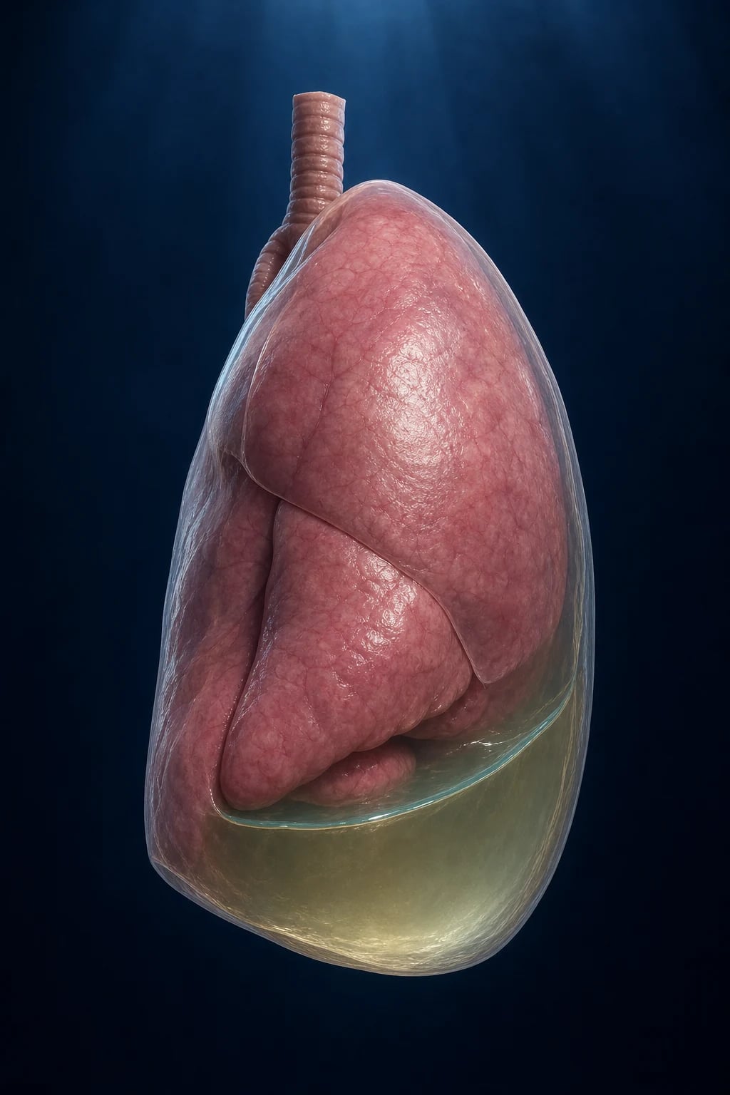
Overview
[1]Pleural effusion is ubiquitous in critical care — roughly 60% of ventilated patients and up to two-thirds of patients in ICU for >1 week develop a clinically significant effusion, and it is often both a diagnostic clue (heart failure, infection, malignancy) and a therapeutic target (the effusion itself worsens gas exchange, prolongs ventilation, and hides an underlying pneumonia). The skill the exam wants is not "how to insert a chest tube" but how to reason from the pleural fluid: transudate vs exudate, when to drain, when to reach for intrapleural fibrinolytics, and when to call the surgeon. The whole topic pivots on Light's criteria and pleural fluid pH, with MIST2 as the keystone trial of modern management. [1]
Why pleural effusion matters in the ICU
[1]Pleural anatomy and pathophysiology — why fluid accumulates
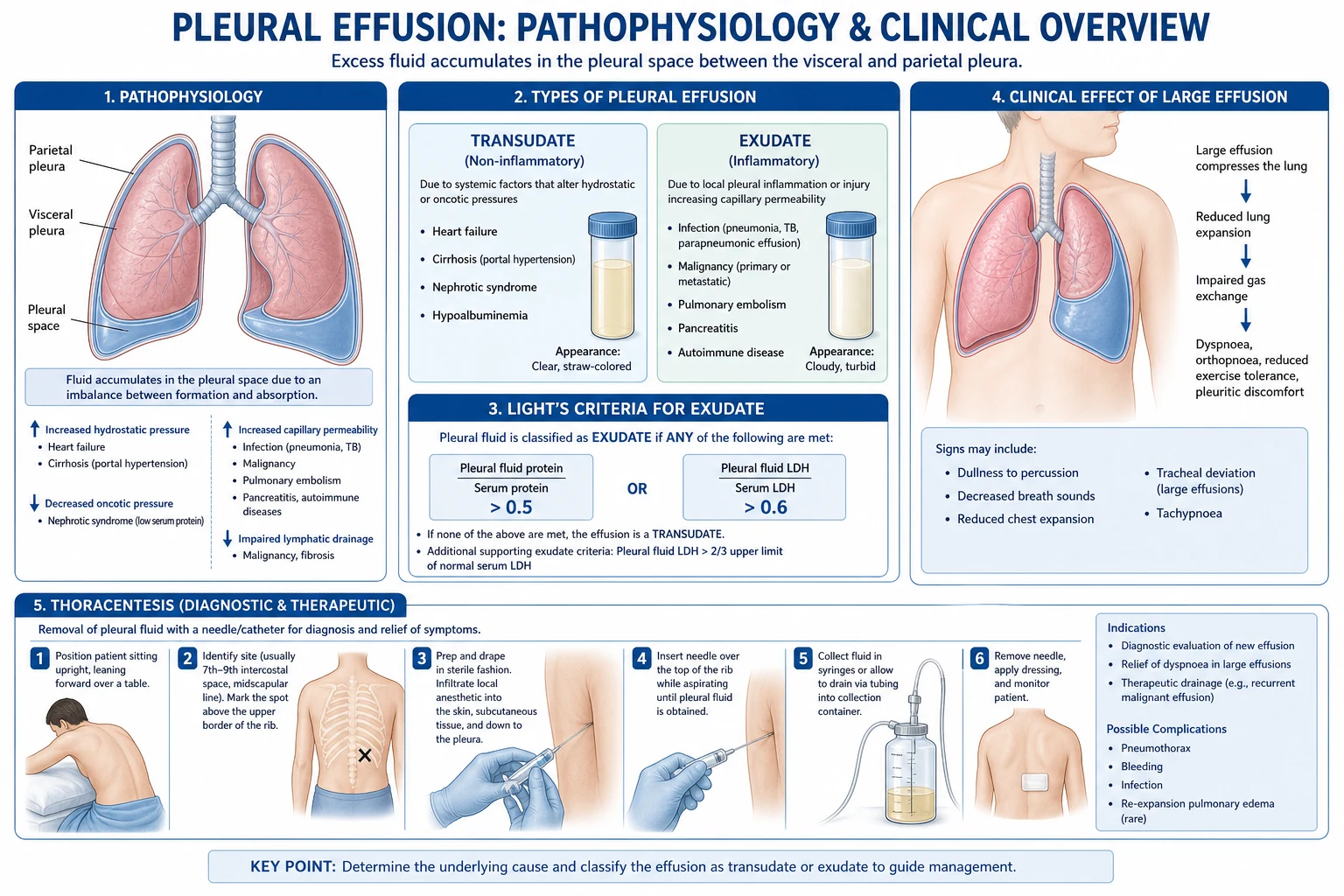
The pleural space is a potential space between the parietal pleura (lining the chest wall, supplied by systemic vessels, with a high-pressure capillary bed) and the visceral pleura (covering the lung, supplied by the low-pressure pulmonary circulation, and partly by systemic bronchial vessels). Normally only 0.1–0.3 mL/kg of hypostatic fluid sits there, lubricating the apposed surfaces. Fluid is filtered from the parietal pleural capillaries and removed predominantly by parietal pleural lymphatic stomata, which have huge reserve capacity (up to ~30-fold increase in flow before an effusion accumulates). [1]
Effusion forms when formation overwhelms removal, by one of four mechanisms: [1]
Mechanisms of pleural fluid accumulation
| Mechanism | Physiology | Typical cause | Fluid type |
|---|---|---|---|
| Increased hydrostatic pressure | Systemic and pulmonary venous pressure rises → transudation across both pleuras | Heart failure (right, left, or biventricular), constrictive pericarditis, superior vena cava obstruction | Transudate |
| Decreased oncotic pressure | Low serum albumin removes the force holding fluid in vessels | Nephrotic syndrome, cirrhosis, severe sepsis/critical illness, protein-losing enteropathy | Transudate |
| Increased capillary permeability | Inflamed pleura leaks protein-rich fluid | Pneumonia (parapneumonic), malignancy, autoimmune (RA, SLE), PE infarction, pancreatitis, TB | Exudate |
| Lymphatic obstruction / impaired drainage | Fluid cannot be cleared from the space | Malignant mediastinal nodes, post-radiation, yellow-nail syndrome, hepatic hydrothorax (diaphragmatic defects + ?impaired lymphatics) | Transudate (hepatic) or exudate (malignant) |
The practical corollary: a transudate tells you the problem is systemic (look at the heart, liver, kidneys, and the fluid balance chart), while an exudate tells you the problem is on the pleural surface or in adjacent lung (look for infection, malignancy, infarction, inflammation). [1]
Light's criteria — the cornerstone of pleural fluid interpretation
Light and colleagues (1972) showed that pleural fluid could be separated into transudate and exudate using the relationships between pleural fluid and serum protein and LDH, rather than absolute values. The criteria are the standard because they are sensitive (identify nearly all exudates) — at the price of over-classifying some heart-failure effusions as exudates.[4][1]
Light's criteria — three tests, any ONE positive = exudate
| Criterion | Threshold | What it measures |
|---|---|---|
| Pleural/serum protein ratio | >0.5 | Protein leak across inflamed pleura (exudate) vs ultrafiltrate (transudate) |
| Pleural/serum LDH ratio | >0.6 | Non-specific marker of pleural inflammation/injury |
| Pleural fluid LDH | >2/3 (two-thirds) of the upper limit of normal for serum LDH (~>200 IU/L if ULN is ~300) | Absolute pleural inflammation |
Interpretation rule: any ONE of the three positive → exudate. All three must be negative to call it a transudate. This is a deliberately sensitive set — it errs toward calling things exudates, because missing an infected or malignant effusion is dangerous. The trade-off is the pseudoexudate problem (below). [1]
Pitfalls of Light's criteria — and the rescue tests
| Problem | Why it happens | Rescue test |
|---|---|---|
| Pseudoexudate (heart failure on diuretics) | Diuresis concentrates pleural protein and LDH, pushing a true transudate over the exudate line | Serum–pleural fluid albumin gradient >12 g/L (= transudate); OR pleural fluid NT-proBNP >1500 ng/L (= heart failure) |
| Pseudoexudate (misclassified on one criterion only) | Borderline values; Light's is sensitive but not specific | Use the albumin gradient / cholesterol (pleural cholesterol <1.55 mmol/L = transudate) as confirmatory |
| Iatrogenic dilution | Diagnostic tap drawn immediately after saline flush through a pleural drain | Interpret with caution; re-sample if unsure |
| Chylous effusion | Triglyceride-rich fluid; LDH/protein may be high or low | Always send triglycerides on milky fluid (>1.1 mmol/L = chylothorax) |
Pleural fluid analysis — the full panel
A diagnostic thoracentesis yields ~20–50 mL. Send it heparinised, on ice, and split it across biochemistry (protein, LDH, glucose, pH, amylase, triglycerides, cholesterol, NT-proBNP as indicated), microbiology (Gram stain, culture, anaerobic culture, AFB/TB PCR if suspected), cytology (cell count and differential, malignant cells), and a reserved aliquot for add-ons. The pH must be measured on a blood-gas analyser in an anaerobic, heparinised syringe — pH meters and dipsticks are unreliable, and leaving the sample in air or filling through a needle without heparin both corrupt the value.[1]
Pleural fluid tests — what each result means
| Test | Transudate / normal | Exudate / abnormal | What it tells you |
|---|---|---|---|
| Protein | <25–30 g/L (and ratio <0.5) | >30 g/L (or ratio >0.5) | Transudate vs exudate split |
| LDH | Low | High (and ratio >0.6 or absolute >2/3 ULN) | Degree of pleural inflammation |
| pH | >7.30 (normal ~7.60) | <7.20 = drain; <7.0 severe | Single best drainage trigger; also low in malignancy, RA, TB, oesophageal rupture |
| Glucose | Same as serum | <2.2 mmol/L (40 mg/dL) = drain; very low in RA, empyema, TB, malignancy | Metabolic consumption by cells/bacteria |
| Cell count | Usually <1000 | Often >1000; neutrophils = acute (parapneumonic, PE); lymphocytes = chronic (TB, malignancy) | Acute vs chronic; infection vs malignancy |
| Gram stain / culture | Negative | Positive in ~60% of empyema | Organism & sensitivities — culture BEFORE antibiotics if possible |
| Cytology | Negative | Malignant cells in ~60% of malignant effusion (sensitivity rises with repeat sampling) | Metastatic adenocarcinoma, mesothelioma, lymphoma |
| Amylase | Normal | High in pancreatic pleural effusion and oesophageal rupture (salivary isoamylase) | Diagnoses pancreatitis-associated & oesophageal perforation |
| Triglycerides | Low | >1.1 mmol/L (110 mg/dL) = chylothorax | Thoracic duct injury; also consider pseudochylothorax (cholesterol crystals) |
| NT-proBNP | Low in non-cardiac | >1500 ng/L = heart failure | Resolves the pseudoexudate problem |
| Adenosine deaminase (ADA) | Low | >40 U/L (sens >90%) = tuberculous pleurisy | TB pleuritis — especially in high-prevalence regions |
Interpreting a pleural fluid sample — the stepwise read
- TRANSUDATE OR EXUDATE? Apply Light's criteria (protein ratio, LDH ratio, LDH). If all three negative → transudate → treat the systemic cause. If on diuretics for heart failure, confirm with albumin gradient / NT-proBNP to exclude pseudoexudate
- DOES IT NEED A DRAIN? Check pH (and glucose, LDH): pH <7.2 (or glucose <2.2 mmol/L, or frank pus, or positive Gram stain) → complicated parapneumonic/empyema → chest tube
- WHAT CELLS? Neutrophil predominance → acute process (parapneumonic, PE, pancreatitis); lymphocyte predominance → chronic (TB, malignancy, chylothorax after days); eosinophilia → often air or blood, less specific than once thought
- IS THERE AN ORGANISM? Gram stain/culture; if negative but high suspicion, send anaerobic cultures and consider TB (AFB, TB PCR, ADA, pleural biopsy)
- IS IT MALIGNANT? Cytology (sensitivity ~60% first tap, rises with repeats); if negative and suspicion persists → CT-guided or thoracoscopic pleural biopsy
- SPECIAL SYNDROMES? Milky fluid → triglycerides (chylothorax); high amylase → pancreatitis or oesophageal rupture; very low glucose/pH with lymphocytes → think RA empyema or TB
Classification
Transudate
Systemic cause
- Light: protein ratio <0.5 AND LDH ratio <0.6 AND LDH <2/3 ULN
- Causes: heart failure (#1), cirrhosis (hepatic hydrothorax), nephrotic syndrome, peritoneal dialysis, myxoedema, atelectasis
- Bilateral, usually symmetric
- Treatment: treat underlying cause (diuretics for HF, etc.)
- Do NOT routinely drain unless symptomatic (large effusion causing dyspnoea)
Exudate
Local cause
- Light: protein ratio >0.5 OR LDH ratio >0.6 OR LDH >2/3 ULN
- Causes: parapneumonic (#1), malignancy (mesothelioma, metastatic), PE, autoimmune (RA, SLE), pancreatitis, TB, chylothorax
- Usually unilateral
- Investigate: pleural fluid analysis (cell count, protein, LDH, pH, glucose, Gram stain, culture, cytology)
- Treatment: cause-specific
Transudate causes — in detail
Causes of a transudative pleural effusion
| Cause | Clues | Approach |
|---|---|---|
| Heart failure (#1) | Bilateral, cardiomegaly, elevated JVP, NT-proBNP >1500; often right > left | Diurese; drain only if symptomatic/refractory or to exclude coexisting infection |
| Hepatic hydrothorax | Cirrhosis, ascites, right-sided in ~70%, can occur without clinically evident ascites | Treat liver disease; salt restriction, spironolactone; avoid chest tube (protein loss, infection, mortality); consider TIPSS |
| Nephrotic syndrome | Heavy proteinuria, hypoalbuminaemia, often bilateral | Treat nephrotic syndrome; thoracentesis mainly to exclude infection/PE |
| Hypoalbuminaemia / capillary leak | Sepsis, critical illness, malnutrition | Fluid balance, nutritional support; resolves with recovery |
| Atelectatic / positional (ventilated) | Dependent, small–moderate, on positive pressure | Lung recruitment, position change; usually no drain |
| Constrictive pericarditis | Bilateral effusions + raised JVP + pericardial calcification/Kussmaul's sign | Echo; pericardiectomy |
| Peritoneal dialysis | Right > left, dialysate tracks through diaphragmatic defects | Reduce volumes, change modality |
| Myxoedema | Hypothyroid, yellow-nail features | Thyroid replacement |
| Superior vena cava obstruction / central line misplacement | Unilateral, venous-pattern swelling | Relieve obstruction, reposition line |
Exudate causes — in detail
Causes of an exudative pleural effusion
| Cause | Clues | Approach |
|---|---|---|
| Parapneumonic / empyema (#1) | Pneumonia/VAP adjacent, fevers, purulent sputum; pH <7.2 | Antibiotics + drain complicated/empyema (see below) |
| Malignancy | Weight loss, smoking, mesothelioma/asbestos; cytology +; lymphocytic | Cytology/biopsy; IPC or talc pleurodesis for recurrence |
| Pulmonary embolism | Sudden dyspnoea, DVT risk, small-moderate effusion, may be transudate or exudate | CTPA; anticoagulate; drain only if large/infarcted |
| Autoimmune (rheumatoid arthritis, SLE, granulomatosis with polyangiitis) | Known disease; RA effusion has very low glucose/pH | Treat underlying disease; RA effusion can be chronic |
| Pancreatitis | Acute pancreatitis, left-sided, high pleural amylase (pancreatic isoamylase) | Treat pancreatitis; drain if large/persistent |
| Tuberculous pleurisy | Young, high-prevalence, lymphocytic, ADA >40, granulomas on biopsy | Anti-TB therapy; often self-limited fluid |
| Chylothorax | Milky, post-surgery/trauma (thoracic duct) or malignancy (lymphoma), triglyceride >1.1 mmol/L | Treat cause; dietary (MCT), octreotide; thoracic duct embolisation/surgery if traumatic |
| Haemothorax | Trauma, post-procedure; haematocrit >50% of serum | Large-bore chest tube; VATS if >1000 mL initial or >200 mL/h |
| Oesophageal rupture (Boerhaave) | Post-emesis, left-sided, high amylase (salivary), pleural pH very low | Surgical emergency; broad-spectrum antibiotics |
| Benign asbestos pleural effusion | Asbestos exposure, benign, exclusion diagnosis | Observation; risk of mesothelioma later |
| Drug-induced (amiodarone, nitrofurantoin, methotrexate, bromocriptine) | Exposure history; eosinophilic | Withdraw drug |
| Uraemic pleuritis | CKD, often bilateral, fibrinous | Dialysis |
Parapneumonic effusion and empyema — natural history and staging
A parapneumonic effusion is fluid in the pleural space associated with pneumonia (or lung abscess, bronchiectasis). It complicates ~20–40% of bacterial pneumonias and ~all cases that progress to empyema. The process evolves through three pathological stages that map onto management decisions; recognising the stage is the central exam skill.[8]
Parapneumonic effusion progression (click each)
Frank pus
Frank pus in pleural space. Treatment: chest tube (large bore, ultrasound-guided) + IV antibiotics (covering anaerobes). Consider intrapleural tPA/DNase (MIST2 trial) if loculated/not draining. VATS (video-assisted thoracoscopic surgery) for failed drainage. Open decortication as last resort.
The three stages of parapneumonic effusion — what to know for the exam
| Feature | Stage 1 — Simple (exudative) | Stage 2 — Complicated (fibrinopurulent) | Stage 3 — Organised (empyema) |
|---|---|---|---|
| Pleural fluid | Thin, sterile, free-flowing | Infected, fibrin deposits form loculations/septations | Frank pus; thick fibrous peel (cortex) forms and entraps the lung |
| pH | >7.2 | <7.2 | <7.2 (often <7.0) |
| Glucose | >2.2 mmol/L (40 mg/dL) | <2.2 mmol/L | Very low |
| LDH | Modestly raised | >1000 IU/L (three times ULN) | Very high |
| Gram stain / culture | Negative | Often positive | Often positive (pus) |
| Imaging | Free-flowing, anechoic on US | Septations/loculations on US; split pleura | Thick peel, trapped lung on CT; rind of enhancing tissue |
| Management | Antibiotics only (treat the pneumonia) | Chest tube + antibiotics ± intrapleural tPA/DNase | Chest tube + antibiotics ± tPA/DNase, and VATS/open decortication to peel off the cortex |
| Drain needed? | No | Yes | Yes (+/- surgery) |
Microbiology of pleural infection
Community-acquired vs hospital-acquired pleural infection — different organisms, different antibiotics
| Setting | Typical organisms | Empiric antibiotics |
|---|---|---|
| Community-acquired | Streptococcus pneumoniae, Streptococcus milleri group, anaerobes (Bacteroides, Peptostreptoccus, Fusobacterium), Staphylococcus aureus (including CA-MRSA in some regions) | Amoxicillin-clavulanate + metronidazole, OR ceftriaxone + metronidazole, OR piperacillin-tazobactam (covers the streptococci AND anaerobes) |
| Hospital-acquired / post-surgical / VAP-associated | Gram-negatives (Klebsiella, Pseudomonas, E. coli), Staph. aureus (MRSA), mixed anaerobes | Anti-pseudomonal beta-lactam (piperacillin-tazobactam or meropenem) + MRSA cover (vancomycin/linezolid) + anaerobe cover (metronidazole unless carbapenem used) |
Management — the complete algorithm
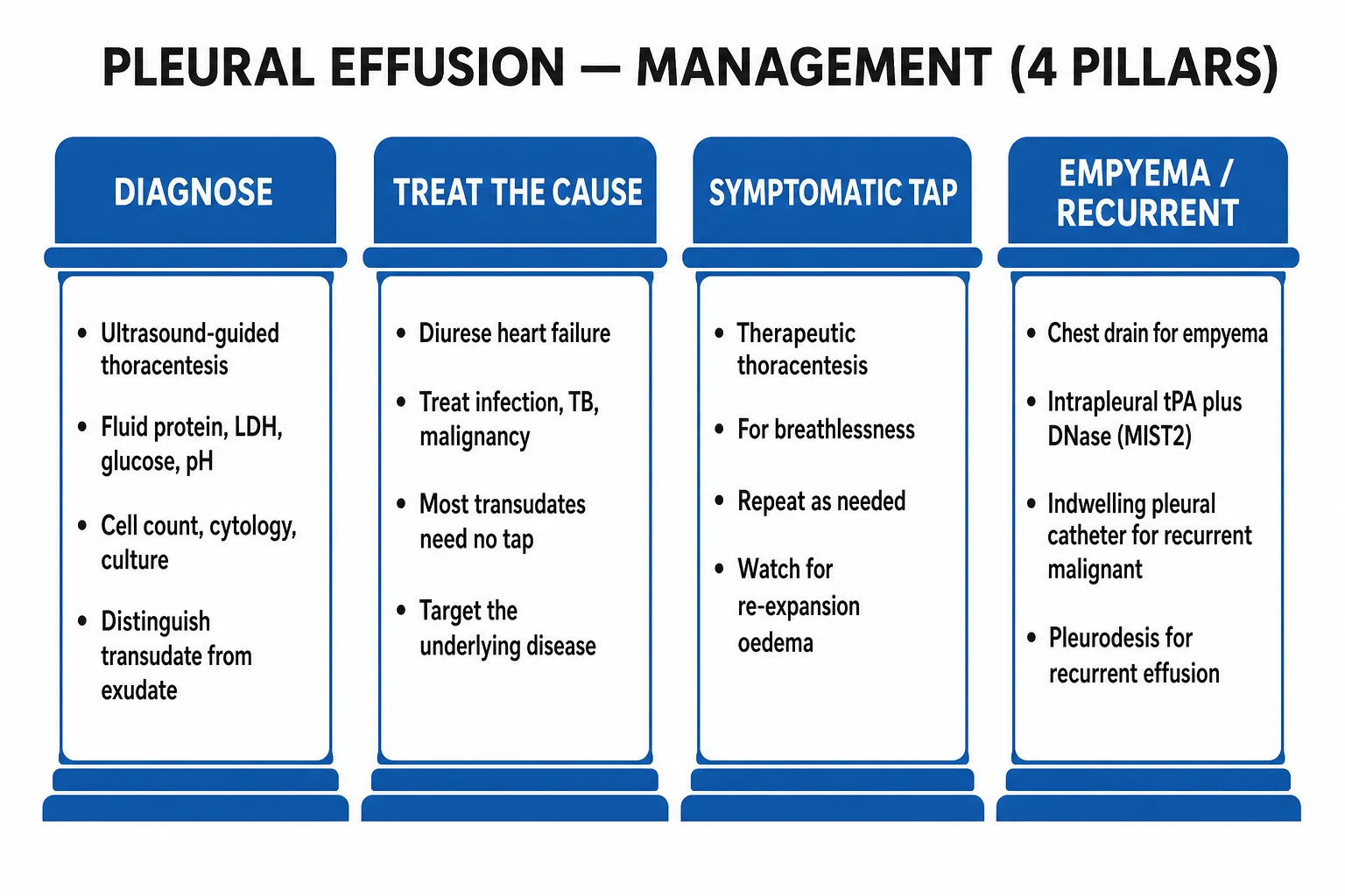
Empyema/complicated effusion management
Ultrasound-guided diagnostic tap
Use ultrasound to identify effusion size and location, mark site. Avoid blind insertion (risk of organ injury — liver, spleen, heart). Diagnostic tap: send fluid for cell count, differential, protein, LDH, glucose, pH, Gram stain, culture (including anaerobes), cytology. pH <7.2 = complicated/empyema → chest tube needed.
Chest tube drainage (large bore)
For complicated parapneumonic (pH <7.2) or empyema: insert large-bore chest tube (28-32 Fr for empyema, smaller pigtail for simple effusion). Ultrasound/CT-guided. Connect to underwater seal drainage. Monitor output daily. Leave until output <50 mL/24h AND no residual collection on imaging.
IV antibiotics
Cover typical pneumonia organisms + anaerobes. Empiric: piperacillin-tazobactam OR ceftriaxone + metronidazole OR amoxicillin-clavulanate + metronidazole. Duration: 2-6 weeks (longer for empyema). Adjust based on cultures.
Intrapleural tPA/DNase (MIST2 trial)
If effusion is loculated and not draining despite chest tube: intrapleural tissue plasminogen activator (tPA 10 mg) + recombinant human DNase (5 mg) daily x 3 days. MIST2 trial: combination improved drainage, reduced surgery, shortened hospital stay. Do NOT give tPA alone (worse outcomes) or DNase alone (no benefit). The COMBINATION is key.
Surgery (VATS)
Video-assisted thoracoscopic surgery (VATS): for failed drainage (persistent collection, multiloculated, thick peel). Breaks down loculations, removes fibrinous peel, drains collection. Preferable to open thoracotomy (less invasive, better outcomes). Open decortication reserved for chronic empyema (organized fibrous peel).
The RAPID score — prognosticate pleural infection at the bedside
[7]The MIST2 trial — the keystone of modern empyema management
Intrapleural fibrinolytics for pleural infection had a troubled history: MIST1 (Maskell 2005) showed that streptokinase was no better than placebo, a negative trial that set the field back. The breakthrough was recognising that pleural pus is held in loculations by fibrin (broken down by a plasminogen activator, tPA) AND rendered viscous by extracellular DNA (broken down by DNase). MIST2 tested the two agents alone and in combination in a 2x2 factorial design.[3][2]
MIST2 2011 — Intrapleural tPA + DNase for pleural infection (PMID 21830966)
Design
Multicentre, double-blind, randomised 2x2 factorial RCT, 210 patients with pleural infection (pus in pleural space, or pH <7.2 with sepsis)
Arms
(1) tPA + DNase; (2) tPA + placebo; (3) DNase + placebo; (4) double placebo — each given intrapleurally twice daily for 3 days via the chest tube
Intervention
Alteplase (tPA) 10 mg + recombinant human DNase 5 mg, intrapleural, twice daily x 3 days
Primary outcome
Change in pleural opacity on chest radiograph (area of effusion) at day 7 — combination group had SIGNIFICANTLY greater reduction (~29.5% vs ~17% placebo)
Secondary
Combination reduced **surgical referral** (~4% vs ~16%), **hospital stay** (mean ~6.7 days shorter), and days of systemic sepsis. tPA alone tended to be WORSE; DNase alone was no different from placebo
Safety
No increase in bleeding, pain, or adverse events. Chest pain and bleeding rates similar across arms
Bottom line
Intrapleural **tPA + DNase together** (NOT either alone) improves drainage, reduces surgery, and shortens hospital stay in loculated pleural infection. It is now standard second-line therapy after a poorly-draining chest tube. Reversed the nihilism from MIST1 (streptokinase)
MIST1 2005 — Intrapleural streptokinase (PMID 15745810, NEGATIVE)
Design
Multicentre, double-blind RCT, 454 patients with pleural infection
Intervention
Intrapleural streptokinase daily x 3 days vs placebo
Primary outcome
No difference in mortality (streptokinase 13.9% vs placebo 13.2%), surgery rate, length of stay, or radiographic change
Bottom line
Streptokinase does NOT work for pleural infection — abandoned. The lesson: not all 'fibrinolytics' are equal; MIST2 (tPA + DNase) succeeded where streptokinase failed
Intrapleural tPA/DNase protocol — practical administration
- CONFIRM INDICATION — loculated/organised pleural infection with inadequate drainage via a well-positioned chest tube (persistent collection on imaging, low output despite radiographic effusion). Exclude contraindication (clotting disorder, recent intracranial/other major bleed)
- PREPARE THE DOSE — alteplase (tPA) 10 mg + recombinant human DNase 5 mg in ~50–100 mL normal saline
- INSTIL VIA THE CHEST TUBE — clamp the tube, instil the mixture, then flush with a small volume of saline to clear the line
- CLAMP & DWELL — keep the tube clamped for 1 hour (some protocols use 2 x 1-hour dwells/day), with the patient turning to distribute the agent
- DECLAMP & MEASURE OUTPUT — release and record drainage; repeat daily (typically up to 3 days, i.e. 6 doses total; some centres extend to 6 days)
- RE-IMAGE & REASSESS — repeat ultrasound/CT; if drainage improves and collection shrinks, continue; if not, refer for VATS
- TIMING OF ESCALATION — if no improvement after 3 days of tPA/DNase, do not persist: surgical drainage (VATS) is the next step
Chest tube insertion — technique, sizes, and safety
Every pleural drainage in the ICU should be image-guided (ultrasound at minimum, CT for complex collections) and the site marked before draping. "Blind" chest-tube insertion in the ventilated ICU patient carries a real risk of liver, splenic, or even cardiac injury, and is no longer acceptable where imaging exists.[1]
The "safe triangle" — where to put the tube
Surface anatomy of the safe triangle (of Less) — borders
| Border | Landmark |
|---|---|
| Anterior | Lateral border of pectoralis major |
| Posterior | Lateral border of latissimus dorsi |
| Inferior | Line of the 5th intercostal space (level of the nipple / inframammary fold) |
| Apex (superior) | Axilla |
| Level of insertion | Usually the 4th–5th intercostal space, anterior axillary line — within the safe triangle, above the diaphragm to avoid intra-abdominal injury |
Seldinger vs blunt dissection
Two insertion techniques — when to use which
| Technique | How it works | Indication | Pros / cons |
|---|---|---|---|
| Seldinger (guidewire) | Needle → guidewire → serial dilators → drain over wire | Small-bore (8–14 Fr) pigtail drains; image-guided drainage of free-flowing or simple loculated effusion; diagnostic-to-therapeutic upgrade | Less traumatic, bedside ultrasound-friendly, less pain/scar; may block with thick pus if too small |
| Blunt dissection | Scalpel incision → blunt clamp through intercostal muscles, over rib, into pleura → finger sweep → tube tract dilated → tube inserted | Large-bore (24–36 Fr) surgical chest tubes; empyema/frank pus, haemothorax; organised collections | Handles thick pus/blood/clot; allows finger sweep to break loculations; more invasive, larger scar, more pain |
Seldinger chest-tube insertion — step by step
- IMAGE & MARK — ultrasound/CT to confirm effusion, depth, and a safe path; mark the site in the safe triangle; confirm position with the patient supine and seated if possible (mark in the position of insertion)
- CONSENT, POSITION, STERILE — explain; patient supine or semi-recumbent, arm behind head; full sterile prep and drape; cap, mask, sterile gown, gloves
- LOCAL ANAESTHESIA — infiltrate skin, intercostal muscle, and parietal pleura with 1% lidocaine (max 3 mg/kg); aspirate at each layer; aspiration of fluid confirms correct depth
- SKIN INCISION — small stab incision at the marked site, along the superior border of the rib below
- NEEDLE & WIRE — advance the needle over the rib, aspirating; on free flow of fluid, pass the guidewire through the needle into the pleural space; remove the needle
- DILATE — serial dilators over the wire to widen the tract (small stabs at the skin edge as needed); maintain control of the wire at all times
- PASS THE DRAIN — thread the pigtail/small-bore drain over the wire into the pleural space; remove the wire
- CONFIRM POSITION — aspirate fluid; connect to underwater seal; confirm with CXR immediately (and ultrasound/CT if complex); suture and secure the drain; document
- CHECK FOR SWINGING/TIDALING & BUBBLING — swinging indicates the tube is in the pleural space; an air leak (bubbling) suggests pneumothorax/bronchopleural fistula
Chest-tube size — the evolving debate
Small-bore pigtail vs large-bore chest tube for empyema
| Aspect | Small-bore pigtail (8–14 Fr) | Large-bore (24–36 Fr) |
|---|---|---|
| Advantages | Less pain, less trauma, bedside ultrasound insertion, easier to manage; modern series show non-inferior drainage when combined with tPA/DNase | Handles thick pus, blood, clot; less likely to block; allows finger sweep at insertion (blunt technique) |
| Disadvantages | Can block with viscous pus; may not drain organised peel | More painful, larger scar, more invasive |
| When preferred | Most non-purulent complicated effusions; loculated effusion with tPA/DNase; first-line in many centres | Frank pus / thick empyema, haemothorax, large organised collections |
| Modern trend | Increasingly first-line (pigtail + tPA/DNase), with escalation to large-bore or VATS only on failure | Reserved for very thick pus or when small-bore fails |
Surgical options — VATS vs open decortication
Surgical approaches for pleural infection
| Approach | Indication | Procedure | Outcomes |
|---|---|---|---|
| VATS (video-assisted thoracoscopic surgery) | Failed tube drainage; multiloculated empyema; organised stage 2–3 with accessible peel; preferred first surgical step | Thoracoscopic: breaks down loculations, debrides fibrin, drains collection, releases trapped lung; minimally invasive, 2–3 ports | Shorter stay, less pain than open; lower mortality than open in many series; first-line surgery for organising empyema |
| Open thoracotomy & decortication | Chronic/organised stage 3 with thick fibrous cortex ("trapped lung"), failed VATS, extensive disease | Removes the fibrous peel off the visceral and parietal pleura, allowing lung re-expansion; major surgery | Higher morbidity (pain, bleeding, longer stay); reserved for chronic/advanced or failed VATS |
| Open drainage (Eloesser flap / rib resection) | Chronic empyema in a patient unfit for definitive surgery; ongoing open drainage | Surgically created stoma allowing long-term open drainage and gradual obliteration of the cavity | Palliative; for the debilitated or chronic-tuberculous empyema |
Antibiotics for pleural infection — duration and regimens
Antibiotic principles in pleural infection
| Principle | Detail |
|---|---|
| Empiric cover | Community: streptococci + anaerobes (amox-clav + metronidazole, or ceftriaxone + metronidazole, or piperacillin-tazobactam). Hospital-acquired: anti-pseudomonal + MRSA + anaerobes (pip-tazo + vancomycin/linezolid ± metronidazole; or meropenem + vancomycin) |
| Anaerobes always | Cover in every regimen; send fluid in anaerobic culture bottles |
| Culture before antibiotics | If feasible, sample pleural fluid before starting/switching antibiotics to maximise yield |
| Duration | Typically 2–6 weeks (commonly 4–6 weeks for empyema); there is no single trial-defined endpoint — guide by clinical response, source control, inflammatory markers. IV → oral switch once afebrile and improving, using an oral agent with anaerobic cover (amox-clav + metronidazole) |
| Intrapleural antibiotics | Not routinely recommended; systemic antibiotics penetrate pus and pleura adequately |
Differentiating the cause of an ICU pleural effusion
The ICU patient commonly has several reasons to accumulate pleural fluid at once — heart failure, hypoalbuminaemia, parapneumonic, and atelectasis may all coexist. The job is to identify the dominant and any actionable cause (above all, infection), and to avoid draining a passive transudate that will recur. [1]
The four big ICU causes — distinguishing features
| Feature | Heart failure | Parapneumonic | Malignancy | Hepatic hydrothorax |
|---|---|---|---|---|
| Laterality | Bilateral (often R>L) | Unilateral (ipsilateral to pneumonia) | Unilateral or bilateral | Right-sided (~70%) |
| Pleural fluid | Transudate; NT-proBNP >1500 | Exudate; pH <7.2 if complicated; pus = empyema | Exudate; cytology + (~60%); lymphocytic | Transudate; low protein |
| Imaging | Cardiomegaly, Kerley B lines, pulmonary venous congestion | Adjacent consolidation, abscess | Nodules, pleural masses, adenopathy | Cirrhotic liver, ascites, small heart |
| Context | Raised JVP, gallop, echo changes | Fever, purulent sputum, sepsis | Weight loss, smoking, primary known | Cirrhosis, low albumin, ascites |
| Key action | Diurese; drain only if refractory/symptomatic or to exclude infection | Antibiotics + drain if complicated/empyema | Cytology/biopsy; IPC or talc pleurodesis for recurrence | Avoid chest tube; salt restriction, spironolactone, consider TIPSS |
| Pitfall | Pseudoexudate on diuretics (use albumin gradient/NT-proBNP) | Don't wait for pus — pH <7.2 drains | Cytology may need repeat sampling; mimic PE | Chest tube = protein loss, infection, high mortality |
Diagnostic approach to a new ICU pleural effusion
- CLINICAL CONTEXT — Is there heart failure, sepsis/pneumonia, cirrhosis, malignancy, hypoalbuminaemia, or recent instrumentation? Bilateral vs unilateral; size and progression
- IMAGE — Bedside ultrasound first (size, anechoic vs septated, safe pocket for sampling); CXR; contrast CT chest if complex/loculated, suspected mass, or diagnosis unclear (pleural phase enhancement helps distinguish simple fluid from empyema/malignancy)
- DIAGNOSTIC THORACENTESIS (ultrasound-guided) — any unexplained exudate, any effusion >10 mm with sepsis, any unilateral effusion of uncertain cause. Send the full panel (above)
- APPLY LIGHT'S CRITERIA + rescue tests (albumin gradient, NT-proBNP) for pseudoexudate
- DECIDE ON DRAINAGE — infected (pH <7.2, pus, positive Gram/culture) → drain; large symptomatic transudate refractory to medical therapy → drain for symptom relief; otherwise treat the cause
- TARGETED INVESTIGATION — malignancy (cytology ± CT/biopsy); PE (CTPA); TB (ADA, AAFB, biopsy); pancreatitis (amylase); chylothorax (triglycerides)
- RE-EVALUATE — repeat sampling if the cause remains obscure or the clinical picture changes; escalate to thoracoscopy/biopsy for undiagnosed exudate
Specific syndromes the ICU candidate must know
Hepatic hydrothorax
[1]Chylothorax and pseudochylothorax
Chylothorax vs pseudochylothorax — both look milky
| Feature | Chylothorax | Pseudochylothorax (cholesterol effusion) |
|---|---|---|
| Appearance | Milky white (chyle) | Milky/turbid |
| Cause | Thoracic duct disruption (trauma, surgery — oesophagectomy, cardiothoracic; or non-traumatic — lymphoma, metastatic malignancy) | Long-standing chronic effusion (chronic RA pleurisy, TB, old empyema) |
| Pleural triglycerides | >1.1 mmol/L (110 mg/dL) | Low triglycerides; high cholesterol with cholesterol crystals |
| Cells | Lymphocyte predominant | Variable |
| Management | Treat cause; dietary (medium-chain triglycerides, NPO + TPN); octreotide; thoracic duct embolisation or surgical ligation for traumatic/non-resolving; pleurodesis/IPC for malignant | Treat the underlying chronic disease; drainage only if symptomatic |
Haemothorax
[1]Malignant pleural effusion
Management of recurrent malignant pleural effusion
| Strategy | When | Notes |
|---|---|---|
| Therapeutic thoracentesis | First episode, prognosis uncertain, palliation | Rapid symptom relief; recurs in most within weeks |
| Talc pleurodesis | Recurrent, expandable lung (no trapped lung), reasonable prognosis | Talc slurry or poudrage; ~70–80% success; requires lung re-expansion and hospital stay |
| Indwelling pleural catheter (IPC / tunneled catheter) | Recurrent, trapped lung, poor prognosis, outpatient preference | Ambulatory drainage; auto-pleurodesis in a proportion; less hospital time (TIME2 / AMPLE trials support IPC) |
| Pleuro-peritoneal shunt / VATS pleurodesis | Selected, fit patients | Less common now IPC is available |
Imaging of pleural effusion and empyema
CXR vs ultrasound vs CT — what each shows
| Modality | Detects | Features of infection/empyema |
|---|---|---|
| CXR (erect PA/lateral) | >200 mL blunts the costophrenic angle; lateral decubitus detects as little as 5 mL; meniscus sign; large effusion → complete opacification with mediastinal shift AWAY | Air-fluid level suggests empyema with gas-former or bronchopleural fistula |
| Bedside ultrasound | Detects small volumes; characterises fluid: anechoic (simple/transudate), echogenic/septated (complicated/empyema, haemorrhage); identifies loculations and a safe window for sampling | Septations, echogenic swirling debris, thickened split pleura = empyema; pleural fluid with moving echoes suggests exudate/pus |
| Contrast-enhanced CT | Best for complex/loculated collections; pleural phase enhancement and thickened, enhancing pleura (split pleura sign) distinguishes empyema from simple transudate; identifies gas, masses, nodes, trapped lung | Split pleura sign (thickened, enhancing visceral and parietal pleura around fluid) = empyema; helps plan drainage/surgery |
SAQ — Parapneumonic effusion that turns out to be a complicated empyema
10 minutes · 10 marks
A 58-year-old man with poorly controlled type 2 diabetes and hazardous alcohol use is admitted with a 6-day history of fever, productive cough and pleuritic chest pain. On examination he is septic: T 39.0 degrees C, HR 128, BP 88/52 (MAP 57) on noradrenaline 0.2 mcg/kg/min, RR 30, SpO2 92 per cent on 15 L oxygen via non-rebreather. WCC 28.4, CRP 340, lactate 3.6 mmol/L, creatinine 145 micromol/L. Chest X-ray shows a right lower lobe consolidation with a moderate right pleural effusion. Bedside ultrasound demonstrates a septated, echogenic collection with debris. Diagnostic thoracentesis returns turbid, foul-smelling fluid: pH 7.08, glucose 1.4 mmol/L, LDH 1850 IU/L, neutrophil predominance, Gram stain showing Gram-positive cocci in chains. Serum protein 68 g/L, serum LDH 220 IU/L.
SAQ — Large unilateral effusion in a cirrhotic patient (hepatic hydrothorax)
10 minutes · 10 marks
A 64-year-old woman with alcohol-related cirrhosis (Child-Pugh C, MELD 24) and ascites is admitted to ICU with worsening dyspnoea and confusion. On examination she is jaundiced, has spider naevi, tense ascites and reduced air entry at the right base. T 37.4 degrees C, HR 102, BP 104/62, RR 26, SpO2 92 per cent on room air. WCC 14.2, CRP 65, albumin 22 g/L. Chest X-ray shows a large right pleural effusion (the left is clear). Diagnostic thoracentesis: straw-coloured fluid, pH 7.42, protein 26 g/L (serum 48), LDH 110 IU/L (serum 200), glucose 5.2 mmol/L, neutrophils 320 x10^6/L, Gram stain and culture negative. Serum-to-pleural albumin gradient 22 g/L.
Clinical pearls
Red flags
Prognosis
Outcomes in pleural infection by stage and intervention
| Scenario | Mortality / outcome | Comment |
|---|---|---|
| Simple parapneumonic effusion | Very low | Resolves with pneumonia treatment |
| Complicated parapneumonic (drained) | ~10–15% | Higher in elderly, comorbid, hospital-acquired |
| Empyema | ~15–20% (up to 30% in high-RAPID) | RAPID high-risk (~36% mortality); prompt drainage + tPA/DNase + early VATS reduce this |
| Hospital-acquired pleural infection | Higher than community-acquired | Resistant organisms, sicker patients |
| tPA/DNase (MIST2) | Reduced surgery referral (~4% vs ~16%) and shorter stay | Standard second-line after failing tube drainage |
| Early VATS | Shorter stay, possibly lower mortality vs continued tube drainage | First surgical step for organising empyema |
| Chronic empyema / open decortication | Higher morbidity | Reserved for organised stage 3 / failed VATS |
The RAPID score is the best validated bedside mortality predictor in pleural infection; high-RAPID patients warrant aggressive, early source control (drainage ± tPA/DNase ± early surgery) and prognostic discussion. Outside infection, prognosis tracks the underlying disease — heart failure, cirrhosis, malignancy — and the effusion itself is usually a marker, not the driver. [1]
Key trials and guidelines summary
The landmark pleural-infection evidence — what each changed
| Study / guideline (year) | Intervention / scope | Key result | What it changed |
|---|---|---|---|
| Light et al. (1972) | Pleural fluid criteria vs pleural biopsy | Protein & LDH ratios separate transudate/exudate | Light's criteria — the standard classification |
| ACCP guideline (2000) | Risk-based parapneumonic management | Categorised effusions by risk; pH <7.2/large/loculated → drain | Category-based drainage decisions |
| MIST1 (2005) | Intrapleural streptokinase vs placebo | No benefit (mortality, surgery, stay identical) | Streptokinase abandoned |
| BTS Pleural Guideline (2010) | Diagnosis of unilateral effusion | Algorithm: ultrasound guidance, full fluid panel, NT-proBNP | Standard of care for workup & procedure safety |
| MIST2 (2011) | Intrapleural tPA + DNase (2x2 factorial) | Combination improved drainage, reduced surgery & stay; either alone no/worse | tPA + DNase = standard for loculated pleural infection |
| RAPID score (2014) | Prognostic score for pleural infection | Stratifies 3-month mortality (low ~3%, high ~36%) | Prognostication & intensity of management |
| TIME2 / AMPLE (2012–2018) | IPC vs talc pleurodesis in malignant effusion | IPC non-inferior, fewer procedures/hospital days | IPC as first-line for many malignant effusions |
Bottom line
Pleural effusion in the ICU is common and is solved by a disciplined, three-step logic. First, classify with Light's criteria (and rescue with the albumin gradient / NT-proBNP when heart failure on diuretics masquerades as an exudate). Second, decide on drainage using pleural fluid pH — <7.2 (or pus, or positive Gram stain/culture) means complicated parapneumonic/empyema and mandates an ultrasound-guided chest tube plus antibiotics covering anaerobes. Third, escalate the failing drain with intrapleural tPA + DNase together (MIST2 — never either agent alone, and never streptokinase) and move to VATS for organised/loculated collections that do not respond. Beware the special syndromes: do not chest-drain hepatic hydrothorax; recognise chylothorax (triglycerides), haemothorax (large-bore, surgery thresholds), and the trapped lung in malignancy (use an IPC, not pleurodesis). Measure pH correctly (heparinised, blood-gas analyser), insert above the rib within the safe triangle, and never persist with a blocked or malpositioned tube. Master Light's criteria, pH <7.2, and MIST2, and you have the bulk of the fellowship answer. [1]
References
- [1]Hooper C, Lee YCG, Maskell N. Investigation of a unilateral pleural effusion in adults: British Thoracic Society Pleural Disease Guideline 2010 Thorax, 2010.PMID 20696692
- [2]Rahman NM, Maskell NA, West A, et al. Intrapleural use of tissue plasminogen activator and DNase in pleural infection N Engl J Med, 2011.PMID 21830966
- [3]Maskell NA, Davies CWH, Nunn AJ, et al. Melanocortin subtype-4 receptor agonists containing a piperazine core with substituted aryl sulfonamides Bioorg Med Chem Lett, 2005.PMID 15745810
- [4]Light RW, Macgregor MI, Luchsinger PC, Ball WC. Syntheses of 1 -tetrahydrocannabinol and related cannabinoids J Am Chem Soc, 1972.PMID 5054408
- [5]Porcel JM. GPs, rather than the government, should be blamed for failures in new NHS, MPs are told BMJ, 2011.PMID 22016455
- [6]Clive AO, Jones HE, Bhatnagar R, et al. (TIME2). Pericardial, But Not Hepatic, Fat by CT Is Associated With CV Outcomes and Structure: The Multi-Ethnic Study of Atherosclerosis JACC Cardiovasc Imaging, 2017.PMID 28330662
- [7]Rahman NM, Kahan BC, Miller RF, et al. Development of Magnetic Probe for Sentinel Lymph Node Detection in Laparoscopic Navigation for Gastric Cancer Patients Sci Rep, 2020.PMID 32019961
- [8]Colice GL, Curtis A, Deslauriers J, et al. Unmasking leprosy: an unusual immune reconstitution inflammatory syndrome in a patient infected with human immunodeficiency virus Am J Trop Med Hyg, 2010.PMID 20595470